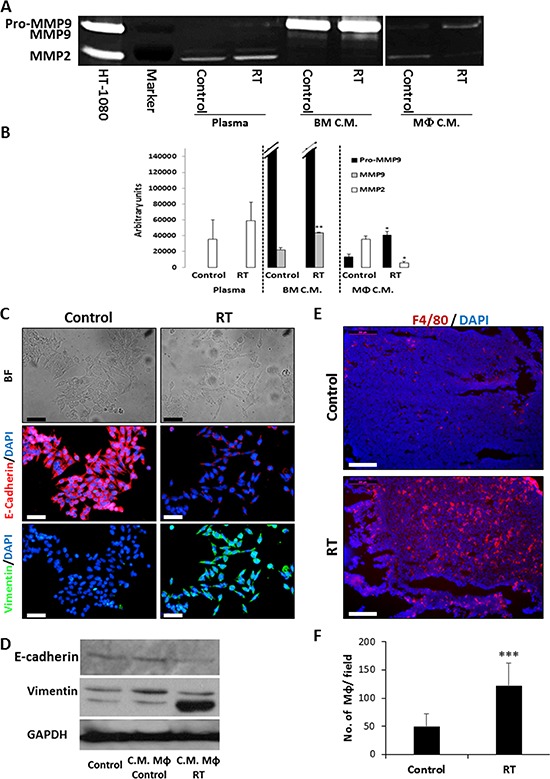
Figure 3. Conditioned medium of macrophages collected from locally irradiated mice promotes metastasis.

A. The activities of MMP2 and MMP9 in plasma extracted from control and 2 Gy locally irradiated (RT) mice as well as in conditioned medium (C.M.) of BMDCs and macrophages derived from these mice were assessed by zymography. Conditioned medium of HT1080 cells was used as a positive control. Experiments were performed in triplicate. B. The expression levels of MMP2, pro-MMP9, and active MMP9 were determined by densitometry analysis of the zymography gels. C-D. SW480 cells were cultured for 24 hours in conditioned medium of macrophages obtained from control or locally-irradiated mice (RT). To determine the extent of epithelial-to-mesenchymal transition (EMT), cells were stained with anti-E-cadherin (red), anti-vimentin (green) antibodies, and counterstained with DAPI (C), or cell lysates were analyzed by Western Blot (D). Images were captured using the Leica CTR 6000 microscope system. Representative bright field (BF) and immunofluorescent images are shown. Scale bars = 50 μm. E. Eight-to-ten week old SCID mice were orthotopically implanted with SW480 tumors and either left untreated (control) or exposed to 2Gy RT. After 72 hours, the tumors were harvested, sectioned and immunostained for macrophages (F4/80, red). Nuclei were stained with DAPI (blue). GPDH served as a loading control. Scale bars = 200 μm. F. The number of macrophages per field were counted (n > 10 fields/group). *p < 0.05; **p < 0.01; ***p < 0.001.
