Abstract
Background:
Although studies have shown that porcelain veneers are very efficient for treating discolored teeth, they did not address in particular the minimum thickness of a multilayer IPS e.max Press (IvoclarVivadent, Schaan, Liechtenstein) restoration required to mask discolored tooth. The aim of this study was to determine the minimum thickness of a multilayer porcelain restoration required for masking severe tooth discoloration.
Materials and Methods:
A total of 24 disk-shaped multilayer specimens were prepared from IPS e.max Press with the diameter of 13 mm and four different thicknesses (core/veneer: 0.4/0.4 mm, 0.5/0.5 mm, 0.6/0.6 mm and 0.8/0.7 mm). Two backgrounds, C4-shade body porcelain and an opaque background from the selected IPS e.max ceramic itself were fabricated to mimic a discolored or stained natural tooth structure and to determine the masking ability. After applying the resin cement layer (Panavia F2.0) with 0.01 mm thickness on each background, all specimens were measured on both background using a spectrophotometer and values of L*, a* and b* were calculated to determine the color differences (ΔE*ab). One-way ANOVA and post-hoc tests of specimen average one-to-one comparison (Tukey HSD) were conducted and P ≤ 0.05 was set as the level of significance.
Results:
ΔE*ab of all groups were within the range of the clinically acceptable color difference (ΔE≤3.3), thus all the groups could mask the C4 background even group 1 with only 0.8 mm thickness. A trend was shown in the results as by increasing the thickness, ΔE*ab is was decreased. The mean ΔE*1*a*b between different thicknesses were statistically significant (P < 0.05) only between group 4 with groups 1 and 2, respectively.
Conclusion:
Within the limitations of this study, all studied thicknesses could mask the C4 background. However, the minimum thickness of a multilayer porcelain restoration (IPS e.max Press) required for masking severe tooth discoloration was 0.8 mm.
Keywords: Color perception, dental porcelain, masking, tooth discoloration
INTRODUCTION
With the increase in the demand for possessing a beautiful smile and white teeth in recent years, management of discolored teeth has high importance in aesthetic dentistry. Depending on the severity of discoloration, there are several treatment options including vital and nonvital bleaching, micro abrasion, composite and porcelain veneers, porcelain crowns and sometimes a combination of them.[1]
All-ceramic restorations are more translucent and thus have more aesthetic properties than restorations with metal substrates and can be used in aesthetic areas properly.[2] It has been proven that porcelain veneers are very efficient for treating discolored teeth, and they last for a long time if they bond properly to the tooth structure. Although limiting the preparation to enamel leads to more efficient bonding, the porcelain restoration should be also thick enough to mask the discoloration. However, in treating a deeply discolored tooth, a full coverage crown might be the ultimate option.[1] Therefore, determining the minimum thickness of a porcelain restoration required for masking heavily discolored teeth can be very useful in clinical treatments.
There are several factors that determine the final aesthetic properties of an all-ceramic restoration in vivo: Color of the ceramic, thickness and the combination of ceramic layers (such as core and veneer with different shade and opacity), the thickness and the color of the luting agent and the color of underlying tooth structure.[3] The ability of an all-ceramic restoration for masking a tooth with severe discoloration can be determined by measuring the color difference (ΔE*ab) when the restoration is placed on two different backgrounds: A dark background and a background fabricated with the same material as the tested restoration but with enough thickness to be completely opaque. The masking ability can be determined using the following formula [Eq. 1].[4,5]

Spectrophotometer is used to determine International Commission on Illumination (French Commission internationale de l’éclairage, hence its CIE initialism) L*a*b* (CIELAB) color coordinates by spectral reflectance measurements. L*, a* and b* are representatives of lightness, greenness-redness and yellowness-blueness respectively.[2] When there is no color difference (ΔE*ab = 0), the masking ability of the system is perfect. However, it has been shown that the visually acceptable color difference, for dental applications, is when ΔE*ab is ≤3.3.[4]
Heat-pressed glass-ceramic lithium disilicate-reinforced ceramics (IPS e.max press, IvoclarVivadent, Schaan, Liechtenstein) is a lithium silicate glass ceramic with proper aesthetic features and strength which can be used as extremely thin anterior veneers.[6]
Dozic et al. concluded that thin porcelain veneers (IPS e.max press, A1, 0.6 mm thick, IvoclarVivadent) cannot mask the underlying tooth color even when different shades of resin cements are used.[7]
According to the results of a study by Zhou et al., high opaque (HO) series of IPS e.max press disks with 0.6 mm, 0.8 mm and 1 mm thicknesses could mask the metal substrate (ΔE*ab <1.5)[8] Shono and Al Nahedh demonstrated that 1.5 mm thickness of IPS e.max (A2-shade) was not able to completely mask the black background.[9]
However, the influence of severe discolored tooth on a multilayer (core and veneer) IPS e.max restoration has not been established previously. Therefore, we investigated this aspect of influence on IPS e.max press by measuring its L*, a* and b* values.
The purpose of this in vitro study was to determine the minimum thickness of a multilayer all-ceramic restoration (IPS e.max press) required for masking severe tooth discoloration (C4-shade) including a HO core for masking the underlying discoloration and an A1 shade veneering layer to provide some translucency simulating tooth enamel. In addition, Panavia F 2.0 light (Kurrary, Kurashiki, Okayama, Japan) resin cement was used to enhance the masking ability.[10] The null hypothesis was that all of the selected thicknesses for the ceramic restoration in corporation with the selected resin cement would mask the C4-shade background.
MATERIALS AND METHODS
Fabrication of ceramic specimens
In this in vitro study, a total of 24 disk-shaped multilayer specimens were prepared from IPS e.max Press (Ivoclar Vivadent, Schaan, Liechtenstein), which is a lithium disilicate glass ceramic in form of ingots for the heat-press technique. Different thicknesses of disks were fabricated as recommended by the manufacturer using lost wax and heat pressed techniques. For IPS e.max press ceramic, the baseline disk thickness was 0.4 mm of core and of 0.4 mm of veneer (group 1). Additional specimens were made with increasing core/veneer thickness (mm) of 0.5/0.5 (group 2), 0.6/0.6 (group 3) and 0.8/0.7 (group 4) according to the manufacturer's instruction. According to a similar study of color analysis on IPS e.max ceramic,[6] sample size was considered six specimens for each group. HO1 shade was selected to fabricate the ceramic core layer according to the manufacturer's recommendation for masking severe tooth discoloration. To obtain the desired thicknesses, cores’ wax patterns were fabricated by PixCera machine (Perfactory, Gladbeck, Germany). Sets containing three wax patterns were invested in an investment ring with a phosphate-bonded investment (Ivoclar Vivadent, Schaan, Liechtenstein) after attachment of a 3 mm-diameter sprue to each of the patterns. The rings were bench set for 60 min and placed into a burn out furnace (Ivoclar Vivadent, Schaan, Liechtenstein) for 120 min. The specimens were then heat-pressed in an EP600 furnace (Ivoclar Vivadent, Schaan, Liechtenstein), air-cooled, divested by blasting with 80 μ glass beads at 4 bar pressure, and ultrasonically treated in an acidic cleaning liquid (Invex, Ivoclar Vivadent, Schaan, Liechtenstein).
The manufacturer's instructions were followed during all procedures. The thickness were measured using a digital caliper with 0.01 mm resolution (Mitutoyo Digimatic, Kawasaki, Japan) and were adjusted if needed with 350, 600 and 1500 grit silicon carbide paper (Matador Wasserfast, Germany) under running water.
According to the manufacturer's instruction a thin layer of IPS e.max deep dentin should be applied on HO cores before appling a more translucent veneering layer. We applied a 0.2 mm thickness of deep dentin (A1 shade) using a digital caliper. In the next step, the core disks were veneered with A1 shade of IPS e.max ceram layering material (mixture of 50% T and 50% TI shades, Ivoclar Vivadent, Schaan, Liechtenstein). To obtain a uniform thickness, the core disks were inserted into custom made brass molds with 13 mm internal diameter and 1, 1.2, 1.4 and 1.7 mm thicknesses to the desired depth. These molds were fabricated 0.2 mm thicker than the desired final thicknesses to compensate for the veneering layers shrinkage. The veneering porcelain was mixed with build-up liquids and introduced to the molds with hand vibration and condensation. Excess moisture was removed with a tissue. After firing, the thickness was adjusted if needed with silicone carbide paper under running water.
Firing was performed according to the manufacturer's recommended procedure. After firing, the disks were ground on the veneer side using the same polishing apparatus with 350, 600 and 1500 grit silicon carbide paper under running water to adjust to the designated total thicknesses and an auto-glazing process was performed at 730°C.
Later, the disks were immersed in IPS e.max Press Invex liquid for 20 min (<1% hydrofluoric acid) and cleaned in an ultrasonic cleaner (DENTSPLY NeyTech, CA, USA) for 10 min and then cleaned with airborne-particle abrasion using 100 μ Al2O3 powder at 2 bar pressure (BEGO, ZiroDent Dentalhandel GbR,. Cologne, Germany). Finally, etching of the disks was done using IPS ceramic etching gel (4.5% hydrofluoric acid) for 20 s. Afterwards, all specimens were cleaned in an ultrasonic bath (Ultrasound Vita-Sonic II, Vita Zahnfabrik, Germany) for 5 min and dried.
Fabrication of backgrounds
Two backgrounds (C4-shade body porcelain [C4] and an opaque background from the selected IPS e.max ceramic itself, and Opaque Ceramic [OC]) were used to mimic a discolored or stained natural tooth structure and to determine the masking ability. For OC background, a 4 mm-thick specimen (core and veneer = 2.2 and 1.8) was fabricated using the waxing machine mentioned before for the core (HO1) and a split brass ring mold (with 4.2 mm thickness) for the veneerig layer (mixture of 50% T and 50% TI shades). Deep dentin (A1: 0.2 mm) was applied between the core and veneering layer. Finally, inherent CIELAB values were measured. The C4 plate (Vita VMK68, Vita Zahnfabrik, BadSackingen, Germany) was fabricated with a 4.2 mm-thick mold and its inherent CIELAB values were measured. The two backgrounds had the same diameter as the specimens.
The thickness sufficient to mask a discolored tooth structure (critical thickness) was determined by calculating the ΔE1*a*b of specimens between the C4 and OC backings.
We selected the Panavia F 2.0 (Kurrary, Kurashiki, Okayama, Japan) light cure resin cement to benefit from its partial masking ability.[10] Since the luting cement was the same for all ceramic specimen in our study, we placed the cement layer on the two backgrounds instead of applying it to the 24 ceramic specimens.
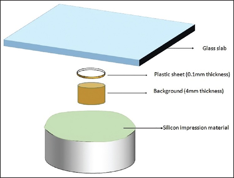
For applying the cement layer on each background, a disc with 13 mm in diameter was obtained from a plastic sheet which had 0.1 mm thickness, placed on the background and silicone mold was prepared. Four gaps were made in the silicon mold for the cement excess. Inside the space, cement was placed. First, a plastic sheet and the glass slab were put on the uncured resin cement and pressed with finger and cured by Quartz-Tungsten-Halogen light curing device for 40 s (Optilux 501, Demetron Kerr, Orange, CA, USA)[11] [Figure 1].
Figure 1.

Schematic figure for the method to prepare constant cement layer thickness.
Spectrophotometric analysis
The color measurements were performed using a Gretag Macbeth ColorEye 7000A spectrophotometer (Color Eye 7000 A, Model C6; Gretag Macbeth, New Windsor, NY, USA). This spectrophotometer with an integrating sphere d/8 geometry has two measuring modes; specular component included and specular components excluded (SCE). In the present study the specular excluded (SCE) configuration was applied to compensate for errors caused by surface glaze. Before each measurement, the spectrophotometer was calibrated using the calibration tile supplied by the manufacturer. The ceramic specimens were placed individually on each of the backgrounds. In Commission Internationale de l’Eclairage colorimetry color is quantified using the parameters of lightness (L*) and chromaticity along the red-green (a*) and yellow-blue (b*) axes. The difference between two colors (ΔE) is determined by measuring CIELAB values by a spectrophotometer. CIELAB coordinates provide a numerical description of the color's position in a three-dimensional color space. The L* color coordinate ranges from 0 to 100 and represents lightness. The a* color usually coordinate ranges from 90 to 70 and represents the greenness on the negative axis and redness on the positive. The b* color coordinate ranges almost from 80 to 100 and represents yellowness (positive b*) and blueness (negative b*). ΔE*ab represents the numerical distance between L*a* b* coordinates of 2 colors using the Eq. 1.[12] The ΔE*ab value (difference between C4 [1] and OC [2]) was evaluated for each thickness. The total color difference ΔE*ab was calculated using the Eq. 1.
A smaller ΔE indicates that the specimen is less sensitive to (as better able to mask) the C4-shade background color. Critical thickness is the minimum ceramic thickness sufficient for masking the C4-background that is determined through the clinically acceptable ΔE range (ΔE*ab ≤ 3.3).
Determining the minimum thickness of ceramic for masking C4-shade background (critical thickness) is a cut-off point, which does not need any statistical analysis. For the four different thicknesses specimen groups, one-way ANOVA and post-hoc tests of specimen (Tukey HSD) were conducted, and P ≤ 0.05 was set as the level of significance.
Mean ΔE values were statistically evaluated. Differences between means at P ≤ 0.05 was considered statistically significant. To make a statistical analysis, SPSS 21 computer program (SPSS Inc., Chicago, Il, USA) was used.
RESULTS
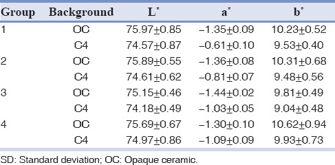
According to the spectrophotometric measurements, L*, a* and b* values of the backgrounds are presented in Table 1. The mean CIELAB color values and ΔE values of IPS e.max ceramic specimens placed on both backgrounds are shown in Tables 2 and 3.
Table 1.
Chromatic values of ceramic backgrounds

Table 2.
Chromatic values of ceramic specimen under different backgrounds (mean ± SD)

Table 3.
ΔE*ab of ceramic specimen over different backgrounds (mean ± SD)

ΔE*ab of all groups were within the range of the clinically acceptable color difference (ΔE ≤3.3), thus all the groups could mask the C4 background. A trend was showing the results as by increasing the thickness, ΔE*ab was decreased.
The mean ΔE*ab between different thicknesses was statistically significant (P < 0.05) only between groups 1 and 4 (mean difference of 0.71) and between 2 and 4 (mean difference of 0.59).
DISCUSSION
According to the results of our study, the minimum thickness of a multilayer porcelain restoration (IPS e.max Press) required for masking severe tooth discoloration was 0.8 mm. Thus, our null hypothesis that all the specimen groups would mask the C4-shade background was supported.
Management of tooth discoloration caused from intrinsic or extrinsic factors is one of the major challenges any dentist may face. Depending on the etiology and severity of the tooth discoloration, treatment options can range from a simple scaling and polishing of the teeth and different bleaching procedures to restorative treatments such as veneers or even crowns.[1]
In treating sever tooth discolorations, restoring an anterior tooth by an all-ceramic restoration with the advantages such as excellent aesthetic properties, biocompatibility and wear resistance can be an appropriate option. But, there is a question that how much tooth reduction is necessary to mask the severe discoloration? Our study was conducted to answer this question.
Although a thin porcelain veneer of an opaque ceramic can mask the underlying discolored tooth, it leads to a nonvital appearance. Therefore, in our study we tried to find the minimum thickness of a multilayer ceramic restoration with an opaque core and a translucent veneer to not only mask the tooth discoloration, but also to have a vital appearance by simulating the translucency of the natural tooth enamel.
Although increasing the thickness of a ceramic restoration improves its masking ability, the increased amount of tooth reduction thereby can jeopardize pulpal health.[13] On the other hand, an efficient bonding of a ceramic restoration can be achieved when the tooth preparation is limited to the enamel. In addition, increasing the opacity of the ceramic restoration adversely affects its aesthetic properties. Therefore, using a multilayer ceramic restoration including an opaque core for masking the underlying discoloration and also a veneering layer to give some translucency in combination with an opaque luting cement would be beneficial.[10]
In the present study, we used a C4-shade dental porcelain to simulate a severely discolored tooth and evaluated the ability of different thicknesses of IPS e.max ceramic specimens for masking this discoloration by measuring their CIELAB values.
Besides color differences, the masking ability of ceramic materials can be evaluated with a spectrophotometric instrument in terms of the opacity or contrast ratio (CR). The (CR = Yb/Yw) is defined as the ratio of illuminance (Y) of the test material when it is placed over a black background (Yb) to the illuminance of the same material when it is placed over a white background (Yw).[5]
According to some studies, when the numerical distance between L*a* b* coordinates of 2 colors (ΔE) is <1 unit, it means that the colors are match and when it is between 1 and 2, the difference is frequently detected by observers and ΔE of higher than 2 is detectable by all observers. However, ΔE of below 3.7 in oral environment is unnoticeable because of the uncontrolled clinical condition.[14,15,16] ΔE values of <1 unit were regarded as not appreciable by the human eye; ΔE values >1 and <3.3 units were considered appreciable by skilled operators, but clinically acceptable; ΔE values >3.3 were considered perceivable by untrained observers (e.g., patients), and for that reason were regarded as not acceptable.[17] In this study, all groups had ΔE*ab of between 1 and 2.
According to some other studies, differences in ΔE*ab lower than 1.1 cannot be detected by the human eye, a ΔE*ab between 1.1 and 3.3 can be detected but is still considered clinically acceptable, while a ΔE*ab higher than 3.3 can be detected and is by an aesthetic point of view considered as clinically not acceptable.[18,19,20] We also considered ΔE*ab < 3.3 as a clinically acceptable color difference in our study.
The overall optical behavior and aesthetic properties of an all-ceramic restoration are determined by the underlying tooth structure color, the thickness of the ceramic layers, and the color of the cement.[11,12,21,22,23]
Vichi et al. investigated the masking ability of ceramic restorations of various thicknesses (1.0, 1.5, and 2.0 mm) of a leucite-reinforced ceramic material (IPS Empress; Ivoclar Vivadent, Schaan, Liechtenstein) over different opaque posts and reported that full masking or acceptable ΔE was achieved only with the 2 mm-thick ceramic material.[11]
Results of a study by Shimada et al. revealed that there were no significant differences in ΔE*ab value at 2.0 mm thickness of IPS Empress2 Ingot-100 under different backgrounds (a composite build-up material, a gold alloy and a silver palladium alloy).[24]
Chu et al. suggested that Procera and Empress 2 had significantly higher CR and masking abilities when compared to Vitadur Alpha. However, when the discoloration is too intense, the application of these two materials may still be limited.[13]
In this study, we investigated the masking ability of IPS e.max Press. By the tremendous advances in the mechanical and optical properties and fabrication methods of ceramic materials, heat-pressed glass-ceramic lithium disilicate-reinforced ceramics (IPS e.max Press) have become popular due to the material's favorable mechanical and aesthetic properties. IPS e.max Press with the compressive strength of 350-450 MPa and fracture toughness of approximately 3 times more that of the Lucite glass ceramic also has the ability of bonding to tooth structure. Its fabrication technique (lost wax) is more practical than layering technique and leads to an excellent adaption of the restoration. It is the optical compatibility between the glassy matrix and the crystalline phase which makes the glass-ceramic lithium disilicate-reinforced ceramics high translucent aesthetic ceramics. Including various shades with different chroma and opacities gives the dentist the opportunity of achieving the desired color even in treating severe discolored teeth.[12,25]
An investigation on masking ability of IPS e.max computer-aided design/computer-aided manufacturing low translucent in a group with a dark-colored abutment tooth revealed that 1.0 mm of this restorative material cemented using either translucent cement or opaque cement and 1.5 mm cemented with translucent cement had ΔE values higher than the clinically unacceptable range (ΔE >3.7).[12] But we concluded that by using a restoration from IPS e.max Press with only 0.8 mm thickness in combination with Panavia resin cement on C4 shade abutment, ΔE*ab would be in clinically acceptable range.
Zhou et al. concluded that IPS e.max Press HO series ceramic specimen thickness of 0.4 mm could not guarantee completely masking the metal substrate background and produced clinically unacceptable color match. While the thicknesses of 0.6 mm, 0.8 mm and 1.0 mm could significantly mask the color of metal substrate disks (ΔE <1.5).[8] In our study, groups 3 (1.2 mm) and 4 (1.5 mm) had ΔE*ab of <1.5.
Studies in this field, however, did not address in particular the minimum thickness of multilayer IPS e.max Press required masking a discolored tooth. The purpose of this in vitro study was to determine the minimum thickness of all-ceramic restoration required to mask a C4-shade background while using Panavia F2 resin cement to enhance the masking ability. To achieve the best aesthetic results in masking a severe discoloration, our ceramic specimen included two ceramic layers with different properties; a HO core layer to mask the underlying discoloration and a translucent veneering layer to replicate the natural tooth vitality appearance. Our results showed that the minimum thickness of the porcelain required for masking severe tooth discoloration was 0.8 mm. By limiting the tooth preparation to only 0.8 mm (in the enamel), we can get a more predictable bonding of the ceramic restoration to the tooth structure.[1]
In our study, ΔE*ab of all groups were lower than 3.3 or 3.7 which are clinically acceptable color difference. Even considering the perceptible difference (ΔE >2),[12] still all groups had the ability of masking the discolored background.
CONCLUSION
The minimum thickness of a multilayer porcelain restoration (IPS e.max Press) required for masking severe tooth discoloration was 0.8 mm including a 0.4 mm core and 0.4 mm veneer.
Financial support and sponsorship
Kerman University of Medical Sciences.
Conflicts of interest
The authors of this manuscript declare that they have no conflicts of interest, real or perceived, financial or non-financial in this article.
ACKNOWLEDGMENTS
The authors thank Kerman University of Medical Sciences for financial support, Dr. Jahani for statistical analysis and Toos Dental laboratory for specimens’ fabrication.
REFERENCES
- 1.Setien VJ, Roshan S, Nelson PW. Clinical management of discolored teeth. Gen Dent. 2008;56:294–300. [PubMed] [Google Scholar]
- 2.Azer SS, Ayash GM, Johnston WM, Khalil MF, Rosenstiel SF. Effect of esthetic core shades on the final color of IPS Empress all-ceramic crowns. J Prosthet Dent. 2006;96:397–401. doi: 10.1016/j.prosdent.2006.09.020. [DOI] [PubMed] [Google Scholar]
- 3.Shokry TE, Shen C, Elhosary MM, Elkhodary AM. Effect of core and veneer thicknesses on the color parameters of two all-ceramic systems. J Prosthet Dent. 2006;95:124–9. doi: 10.1016/j.prosdent.2005.12.001. [DOI] [PubMed] [Google Scholar]
- 4.Dozic A, Kleverlaan CJ, Meegdes M, van der Zel J, Feilzer AJ. The influence of porcelain layer thickness on the final shade of ceramic restorations. J Prosthet Dent. 2003;90:563–70. doi: 10.1016/s0022-3913(03)00517-1. [DOI] [PubMed] [Google Scholar]
- 5.Paris: Bureau Central de la CIE; 2004. Colorimetry: Official recommendations of the International Commission on Illumination, Publication CIE No. 15.3. [Google Scholar]
- 6.Barizon KT. Relative Translucency of Ceramic Systems for Porcelain Veneers. Master's Thesis, University of Iowa. 2011 [Google Scholar]
- 7.Dozic A, Tsagkari M, Khashayar G, Aboushelib M. Color management of porcelain veneers: Influence of dentin and resin cement colors. Quintessence Int. 2010;41:567–73. [PubMed] [Google Scholar]
- 8.Zhou SY, Shao LQ, Wang LL, Yi YF, Deng B, Wen N. Masking Ability of IPS e.max all-ceramics system of HO series. Key Eng Mater. 2012;512(5):1784–7. [Google Scholar]
- 9.Shono NN, Al Nahedh HN. Contrast ratio and masking ability of three ceramic veneering materials. Oper Dent. 2012;37:406–16. doi: 10.2341/10-237-L. [DOI] [PubMed] [Google Scholar]
- 10.Omar H, Atta O, El-Mowafy O, Khan SA. Effect of CAD-CAM porcelain veneers thickness on their cemented color. J Dent. 2010;38(Suppl 2):e95–9. doi: 10.1016/j.jdent.2010.05.006. [DOI] [PubMed] [Google Scholar]
- 11.Vichi A, Ferrari M, Davidson CL. Influence of ceramic and cement thickness on the masking of various types of opaque posts. J Prosthet Dent. 2000;83:412–7. doi: 10.1016/s0022-3913(00)70035-7. [DOI] [PubMed] [Google Scholar]
- 12.Chaiyabutr Y, Kois JC, Lebeau D, Nunokawa G. Effect of abutment tooth color, cement color, and ceramic thickness on the resulting optical color of a CAD/CAM glass-ceramic lithium disilicate-reinforced crown. J Prosthet Dent. 2011;105:83–90. doi: 10.1016/S0022-3913(11)60004-8. [DOI] [PubMed] [Google Scholar]
- 13.Chu FC, Chow TW, Chai J. Contrast ratios and masking ability of three types of ceramic veneers. J Prosthet Dent. 2007;98:359–64. doi: 10.1016/S0022-3913(07)60120-6. [DOI] [PubMed] [Google Scholar]
- 14.Wyszecki G, Stiles WS. 2nd ed. New York: John Wiley & Sons; 1982. Color Science: Concepts and Methods, Quantitative Data and Formulae; pp. 45–7. [Google Scholar]
- 15.Seghi RR, Hewlett ER, Kim J. Visual and instrumental colorimetric assessments of small color differences on translucent dental porcelain. J Dent Res. 1989;68:1760–4. doi: 10.1177/00220345890680120801. [DOI] [PubMed] [Google Scholar]
- 16.Johnston WM, Kao EC. Assessment of appearance match by visual observation and clinical colorimetry. J Dent Res. 1989;68:819–22. doi: 10.1177/00220345890680051301. [DOI] [PubMed] [Google Scholar]
- 17.Vichi A, Louca C, Corciolani G, Ferrari M. Color related to ceramic and zirconia restorations: A review. Dent Mater. 2011;27:97–108. doi: 10.1016/j.dental.2010.10.018. [DOI] [PubMed] [Google Scholar]
- 18.Um CM, Ruyter IE. Staining of resin-based veneering materials with coffee and tea. Quintessence Int. 1991;22:377–86. [PubMed] [Google Scholar]
- 19.Kuenhi RC, Marcus RT. An experimental visual scaling of small color differences. Color. 1979;4:83–91. [Google Scholar]
- 20.Hunter RS. New York: Wiley; 1975. The Measurement of Appearance; pp. 77–80. 151-2, 225, 234. [Google Scholar]
- 21.Nakamura T, Saito O, Fuyikawa J, Ishigaki S. Influence of abutment substrate and ceramic thickness on the colour of heat-pressed ceramic crowns. J Oral Rehabil. 2002;29:805–9. doi: 10.1046/j.1365-2842.2002.00919.x. [DOI] [PubMed] [Google Scholar]
- 22.Li Q, Yu H, Wang YN. Spectrophotometric evaluation of the optical influence of core build-up composites on all-ceramic materials. Dent Mater. 2009;25:158–65. doi: 10.1016/j.dental.2008.05.008. [DOI] [PubMed] [Google Scholar]
- 23.Koutayas SO, Kakaboura A, Hussein A, Strub JR. Colorimetric evaluation of the influence of five different restorative materials on the color of veneered densely sintered alumina. J Esthet Restor Dent. 2003;15:353–60. doi: 10.1111/j.1708-8240.2003.tb00308.x. [DOI] [PubMed] [Google Scholar]
- 24.Shimada K, Nakazawa M, Kakehashi Y, Matsumura H. Influence of abutment materials on the resultant color of heat-pressed lithium disilicate ceramics. Dent Mater J. 2006;25:20–5. doi: 10.4012/dmj.25.20. [DOI] [PubMed] [Google Scholar]
- 25.Shenoy A, Shenoy N. Dental ceramics: An update. J Conserv Dent. 2010;13:195–203. doi: 10.4103/0972-0707.73379. [DOI] [PMC free article] [PubMed] [Google Scholar]


