Figure 3.
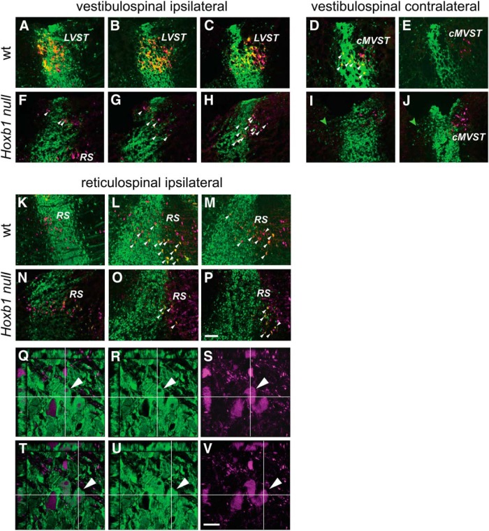
Loss of vestibulospinal neurons and depletion of reticulospinal neurons in the Hoxb1-null mutant. A–J, Loss of vestibulospinal neurons. Comparison of the two vestibulospinal groups that derive wholly or partly from r4 in the wild-type (LVST, A–C; cMVST, D, E) and the Hoxb1-null mutant (F–J) shows a complete absence of r4-derived LVST and cMVST neurons. The white arrowheads in D indicate examples of r4-derived cMVST neurons. Those in F–H indicate examples of non-r4-derived spinally projecting neurons in the Hoxb1-null mutant in the area where the LVST is normally located. The green arrowheads in I and J indicate non-spinally projecting r4-derived cells that migrate into r3 within the vestibular nuclear complex in the Hoxb1-null mutant. K–P, Depletion of r4-derived reticulospinal neurons. Reticulospinal neurons derived from r4 (white arrowheads) are more numerous in the wild-type (K–M) than in the Hoxb1-null mutant (N–P). RDA/BDA labeling is depicted by magenta, YFP immunolabeling is depicted by green, and double labeling appears as varying hues of yellow and orange. Q–V, Examples of confocal z-stacks to demonstrate double labeling, in this case of two reticulospinal neurons. Each panel shows a z-stack viewed in the x–y plane and from the x–z and y–z faces, with x and y transects intersecting at a reticulospinal neuron that is double labeled (one in Q–S, another in T–V). In each row of panels, the right panel shows only RDA (magenta), the middle panel shows only YFP (green), and the left panel shows a merge of the two (note that here the magenta and green combine to create a pale white, as opposed to the yellow/orange that depicts double labeling in the panels above; see Materials and Methods). RS, Reticulospinal neurons. Scale bars: A–P, 200 µm; Q–V, 20 µm.

