Abstract
Objectives:
Over the years, professional dental prophylaxis has involved the use of rubber-cup, bristle brush, and abrasive paste for coronal polishing. Although air polishing is an excellent alternative for removal of tooth stain and dental plaque, very few studies have compared their efficacy in vivo. The present study attempts to evaluate and compare the efficacy of air polishing (test) alone versus rubber-cup polishing (control).
Materials and Methods:
A total of 35 individuals having generalized mild to moderate gingivitis were enrolled as the study population after obtaining their informed consent. Before commencement of the study, all subjects underwent scaling to remove calculus deposits (if any), following which the ipsilateral quadrant of the patient's mouth was randomly assigned as the test side and the contralateral quadrant of the same arch was assigned as the control side for polishing procedures. Time employed for both methods of polishing was held constant at 5 min for each technique. Subjects were assessed before and immediately after polishing and again after 15 days following treatment, for plaque and gingival status along with gingival bleeding.
Results:
Overall, the results of the intra-group comparison of both the polishing procedure sites indicated similar but significant plaque and gingival status changes, whereas the inter-group comparison showed no significant difference between the efficacies of both the groups.
Conclusions:
Air polishing and the rubber-cup, bristle brush with paste polishing demonstrated equivalent efficacy regarding removal of supragingival plaque and in reducing gingival inflammation.
Keywords: Dental polishing, dental prophylaxis, gingivitis, plaque
INTRODUCTION
As accumulation of bacteria on tooth surfaces is the primary cause of gingivitis and periodontitis, regular mechanical removal of bacterial plaque from all non-shedding oral surfaces is considered the primary means to prevent and stop the progression of periodontal disease. In spite of all the potentially harmful effects involving routine scaling and polishing techniques,[1,2,3] it still remains as an integral part of periodontal therapy. The ultimate goal of professional dental polishing is complete removal of plaque and stains; however, the use of traditional rubber-cup and abrasive paste is often laborious, time consuming, and ineffective in completely removing supragingival deposits, particularly in the inaccessible interdental areas and around bonded orthodontic appliances.[4] Also, routine use of rubber-cup with prophylaxis paste has been shown to remove the fluoride-rich outer layer of the enamel and cause significant loss of cementum and dentin over time.[3]
In spite of numerous techniques and materials being available for getting rid of the plaque and extrinsic stains, none has qualified as a gold standard treatment modality till date. Routine home dental polishing done with powered toothbrush[5,6] and whitening paste,[7,8] although effective, still lacks efficiency in inaccessible areas, thus necessitating a more efficient professional polishing other than conventional rubber-cup polishing.
With the growing body of evidence to support alternative tooth polishing methods, air polishing (APP) has shown more promising results not only for supragingival polishing[9,10,11,12,13] but also for effective subgingival plaque removal.[3,14]
Even though there is conclusive evidence that APP can be used on a routine basis to polish teeth, its widespread use has not occurred among clinicians.[15] Failure to adopt this alternative method of polishing teeth may be due to a lack of knowledge regarding the proper use of the device. It may also be due to several unsubstantiated misconceptions surrounding air polishers.
Considering the fact that since 1945, APP has been effective in rapid removal of tooth stains and plaque, and causing less invoked hypersensitivity, less operator fatigue, and improved access to pit and fissures, very few studies have compared the efficacy of traditional rubber-cup bristle brush with prophylaxis paste polishing (RCP) and APP for routine coronal polishing.[11,12,13] In light of all the above aspects, an attempt has been made in this study to compare the efficacy of APP and RCP in a split-mouth design with respect to whole mouth plaque, total marginal and papillary gingivitis, along with bleeding from gingival sulcus.
MATERIALS AND METHODS
A clinical split-mouth study was carried out at the Department of Periodontics to evaluate the comparative effectiveness of APP alone (test side) and rubber-cup, bristle brush with abrasive paste (control side) on supragingival plaque and gingivitis.
The study population consisting of a total of 35 individuals (18 males and 17 females) within the age range of 18–65 years, having all teeth except third molars and a probing depth of not more than 3 mm, and suffering from chronic marginal/papillary gingivitis was selected, among which 5 patients dropped out during the study.
Subjects with history or signs of periodontitis, systemic disorder, or contagious disease, pregnant/lactating women, chronic illness/condition (hypertension, diabetes, respiratory diseases, etc.), and those who had undergone radiotherapy or chemotherapy were excluded. Also, subjects who had taken dental prophylaxis 1 month prior to inclusion in the study or antibiotics for at least 3 months before baseline visit, or having any plaque retentive factors like orthodontic brackets, defective restorations, etc., were not included. The study protocol was approved by the institutional ethical committee and Maharashtra University of Health Sciences, Nashik, Maharashtra (India), and it was explained to each patient. Those who consented and met the selection criteria were enrolled in the study.
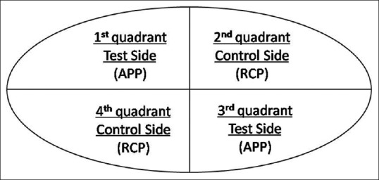
The relevant data pertaining to the case history was recorded in a special proforma. Before commencement of the study, all subjects underwent ultrasonic scaling for removal of calculus deposits (if any), immediately following which each quadrant of the patient's mouth was randomly assigned and polished as TEST SIDE and the other quadrant of the same arch as CONTROL SIDE and vice versa for the opposite arch by coin toss method. In the present study, split-mouth design was used [Figure 1] to assess supragingival plaque, gingival bleeding, and gingivitis.
Figure 1.

Split-mouth design
In the test group, APP system (Air Prophy unit®; Compass international, Guangdong, China) with sodium bicarbonate powder (cleaning powder for Prophy unit; Greeloy, Shanghai, China) was used (particle size standardized up to 250 µm). The technique used for APP involved positioning the nozzle 5–6 mm away from the tooth surface[16] with the spray directed toward the middle third of the crowns of two to three teeth at one time, cleansed with a constant circular motion.[17,18] Since the APP device generates aerosol, a mask and protective eyewear were used.[19] In the control group, the bristle brush followed by rubber-cup with prophylaxis paste (ShineNSmile®; ICPA Health Products Ltd, Gujarat, India) was used in circular motion for polishing. Time employed for both procedures was held constant at 5 min for each technique.[11]
Among the three clinical parameters, for a comprehensive dental plaque assessment with better objectivity, Turesky–Gilmore and Glickman modification of Quigley Hein plaque index (PI)[20] using two-tone plaque disclosing agent (AlphaPlac®; Dental Products of India, Mumbai, India) was used on the facial and lingual surfaces of all teeth (except third molars). Whereas to assess the severity of gingival bleeding and gingivitis, sulcus bleeding index (SBI) by Muhlemann[21] and gingival index (GI) by Loe and Silness[22] were used, respectively. The measurements were assessed thrice for each subject, i.e. at baseline, immediately postoperative, and 15 days postoperatively. All individuals were asked to continue with their own routine oral hygiene procedures.
All the steps in this study were carried out by a single operator. The power of the study was calculated by considering 95% confidence interval. The entire data obtained from the study population at all the three time intervals were put to statistical analysis (SPSS software package 16), and the mean, standard deviation, standard error and paired t-test were calculated to derive an evidence based scientific interpretation.
RESULTS
Out of 35 individuals, a total of 30 individuals including 16 males and 14 females with a mean age of 27 and 24 years, respectively, were assessed in this study. The clinical parameters were compared between the test (APP) sites and the control (RCP) sites [Table 1].
Table 1.
Mean and SD of PI, GI, and SBI at baseline, immediate post-op, and 15 days post-op in test and control group

Furthermore, an intra-group comparison was carried out using t-test (i.e., between baseline, immediately post-op, and 15 days post-op of each group). All 30 patients completed the study with no adverse event reported by any subject during the study.
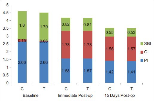
Intra-group comparison within each group with respect to PI, GI, and SBI [Graph 1] at baseline, immediately postoperative, and 15 days postoperatively showed a statistically significant difference (P = 0.000) [Table 2].
Graph 1.

Comparison of mean of PI, GI, and SBI between test (T) and control (C) groups at baseline, immediate post-op, and 15 days post-op
Table 2.
Intra-group comparison PI, GI, and SBI at baseline, immediate post-op, and 15 days post-op in test and control groups

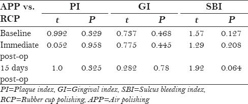
But inter-group comparison between APP and RCP with respect to PI, GI, and SBI [Graph 1] at baseline, immediately postoperative, and 15 days postoperatively did not show any statistical significance (P > 0.01) [Table 3].
Table 3.
Inter-group comparison of PI, GI, and SBI at baseline, immediate post-op, and 15 days post-op in test and control groups

Overall, the results of the intra-group comparison of both the APP and RCP sites indicated similar but significant plaque and gingival status changes, whereas the inter-group comparison showed no significant difference in the efficacy of both the groups.
DISCUSSION
The present study compared the clinical efficiency of APP and RCP in removing supragingival plaque, and also aimed to study their effects on gingival status in a split-mouth design. The results indicated that when comparing the effectiveness of polishing treatments, there was no statistically significant difference in plaque removal or gingival status within the established time interval. Also, both the treatment modalities showed a significant change in plaque accumulation and gingival status from baseline, immediate post-op, to 15 days post-op.
There was a significant reduction in plaque scores from baseline to immediate post-op in both the treatments (APP and RCP), but there was a considerable increase in GI and SBI scores from baseline to immediate post-op period. This finding could be attributed to the increase in gingival bleeding in the immediate post-op period as compared to the baseline scores, due to the therapy performed. At 15 days of follow-up, there was a substantial increase in plaque as compared to the immediate post-op findings, but there was a remarkable reduction in gingival and bleeding scores. This observation could not be attributed to either of the treatment modalities (APP or RCP), as this could entirely be credited to supra- and subgingival scaling alone. But the improvement in the gingival status from immediate post-op period to 15 days postoperatively indicated that although both the polishing methods were traumatic, their effects on the soft tissues were temporary.
The design of the APP system uses a mixture of air, water, and sodium bicarbonate powder to deliver a controlled stream of sodium bicarbonate particles onto the tooth surface. This slurry of powder and water polishes the surface by removing deposits attached to it or smoothing its texture. The APP powder used in this study was the sodium bicarbonate powder, which is readily available, biocompatible, and is relatively soft and only mildly abrasive.[23]
Patients with exposed roots were excluded from this study as the abrasive effect of sodium bicarbonate powder on the root surfaces was observed to be detrimental by previous studies.[24,25,26] Most investigators agree that intact enamel surfaces are not damaged when stain removal is accomplished with an air polisher. Even after exposure to enamel for the equivalent of a 15-year recall program, the surfaces were not altered.[27]
A similar split-mouth study was conducted in 1991[11] to compare the effectiveness of polishing techniques using indices to measure plaque, stain, and gingival trauma prior to and after the polishing treatment in 30 adult subjects, with comparable numbers of teeth contralaterally exhibiting observable plaque and stain. One side of a subjects’ mouth was polished with a rubber-cup and flour of pumice and the other side was polished with the Prophy-jet APP device; plaque, stain, and gingival trauma measurements were assessed prior to and following both polishing treatments. The split-mouth design used in this study accounted for some possible confounding factor/variability due to assignment of one side of patient's mouth to APP treatment and the other side to RCP treatment. In this case, bias may occur on the basis of patient's brushing habit and mastication. For example, if the patient is right handed, it more likely that the left side would present less plaque as compared to the right side; also, if the patient has unilateral mastication, plaque accumulation would differ on either side of the patient's mouth. To eliminate this possible variable in the present study, split-mouth design was modified, i.e. one quadrant of a patient's mouth was assigned to RCP and the other quadrant of the same arch was polished by APP and it was vice versa for the other arch [Figure 1]. The results of the present study are found to be in accordance with those of the 1991 study.[11]
Smoking or tobacco chewing habit was noted in three males in the present study. As smoking has least effect on plaque accumulation, it did not affect the plaque scores. Although the gingival status shows significant changes in smokers as compared to non-smokers, the split-mouth design helps to eliminate the variable. Another observation made in the present study was that in patients with malaligned teeth or teeth exhibiting crowding, the RCP seemed to be less efficient in eradicating plaque and stain from the malaligned teeth surfaces, whereas APP presented as a more efficient modality in such cases due to better accessibility.
Almost all the patients in this clinical trial were comfortable with both the treatment modalities and no patient suffered from any adverse effect of any sort. There was no complaint of any discomfort/sensitivity from any patient after APP or RCP at 15 days post-treatment. Overall, APP has proven to be causing less operator fatigue, is less time consuming, and is equally efficient as the conventional rubber-cup polishing with respect to plaque removing efficacy.[28]
Lack of negative control being a limitation of the current study, future research should include randomized controlled trials that directly compare the effects of air polishers; sonic, ultrasonic, and piezo-scalers; and rubber-cup polishers and hand instruments on all tooth surfaces. In addition, these studies should attempt to control the variables of quantity of stain, abrasiveness of polishing pastes, and amount of pressure applied to each surface.
CONCLUSIONS
Based on the findings of this study, the following conclusions were reached:
APP and the rubber-cup, bristle brush with paste polishing (RCP) demonstrated equivalent efficacy regarding removal of supragingival plaque for a single-visit setting.
Over a period of 15 days, both APP and RCP (along with scaling) demonstrated equivalent efficacy in reducing gingival inflammation. Both APP and RCP exhibited significant increase in bleeding from gingival sulcus immediately after therapy. But after 15 days, a significant reduction in gingival bleeding was presented by both the treatment sites.
It can be concluded that other factors, in addition to effectiveness and time efficiency, should be considered when selecting rubber-cup polishing with paste or an APP device.
Financial support and sponsorship
Nil.
Conflicts of interest
There are no conflicts of interest.
REFERENCES
- 1.Worthington HV, Clarkson JE, Bryan G, Beirne PV. Routine scale and polish for periodontal health in adults. Cochrane Database Syst Rev. 2013;11:CD004625. doi: 10.1002/14651858.CD004625.pub4. [DOI] [PubMed] [Google Scholar]
- 2.Moëne R, Décaillet F, Mombelli A. Subgingival air-polishing: New perspectives for periodontal maintenance? Schweiz Monatsschr Zahnmed. 2010;120:891–911. [PubMed] [Google Scholar]
- 3.Graumann SJ, Sensat ML, Stoltenberg JL. Air polishing: A review of current literature. J Dent Hyg. 2013;87:173–80. [PubMed] [Google Scholar]
- 4.Ramaglia L, Sbordone L, Ciaglia RN, Barone A, Martina R. A clinical comparison of the efficacy and efficiency of two professional prophylaxis procedures in orthodontic patients. Eur J Orthod. 1999;21:423–8. doi: 10.1093/ejo/21.4.423. [DOI] [PubMed] [Google Scholar]
- 5.Terezhalmy GT, Walters PA, Bartizek RD, Grender JM, Biesbrock AR. A clinical evaluation of extrinsic stain removal: A rotation-oscillation power toothbrush versus a dental prophylaxis. J Contemp Dent Pract. 2008;9:1–8. [PubMed] [Google Scholar]
- 6.Maloney VP, Kemp J, Panagakos F, Mateo LR. Laboratory evaluation of extrinsic stain removal by a specially engineered sonic powered toothbrush with unique sensing and control technologies. J Clin Dent. 2012;23:A21–5. [PubMed] [Google Scholar]
- 7.Patil PA, Ankola AV, Hebbal MI, Patil AC. Comparison of effectiveness of abrasive and enzymatic action of whitening toothpastes in removal of extrinsic stains-a clinical trial. Int J Dent Hyg. 2015;13:25–9. doi: 10.1111/idh.12090. [DOI] [PubMed] [Google Scholar]
- 8.Khan MK, Bokhari SA, Haleem A, Kareem A, Khan AA, Hosein T, et al. Extrinsic stain removal with a toothpowder: A randomized controlled trial. Int J Health Sci (Qassim) 2014;8:269–74. doi: 10.12816/0023979. [DOI] [PMC free article] [PubMed] [Google Scholar]
- 9.Gutmann ME. Air polishing: A comprehensive review of the literature. J Dent Hyg. 1998;72:47–56. [PubMed] [Google Scholar]
- 10.Brown DM, Barnhart RC. A scientific foundation for clinical use of air polishing systems. J Pract Hyg. 1995;4:36–40. [Google Scholar]
- 11.Miller DL, Hodges KO. Polishing the surface. A comparison of rubber cup polishing and airpolishing. Probe. 1991;25:103, 105–9. [PubMed] [Google Scholar]
- 12.Mishkin DJ, Engler WO, Javed T, Darby TD, Cobb RL, Coffman MA. A clinical comparison of the effect on the gingiva of the Prophy-Jet and the rubber cup and paste techniques. J Periodontol. 1986;57:151–4. doi: 10.1902/jop.1986.57.3.151. [DOI] [PubMed] [Google Scholar]
- 13.Weaks LM, Lescher NB, Barnes CM, Holroyd SV. Clinical evaluation of the Prophy-Jet as an instrument for routine removal of tooth stain and plaque. J Periodontol. 1984;55:486–8. doi: 10.1902/jop.1984.55.8.486. [DOI] [PubMed] [Google Scholar]
- 14.Müller N, Moëne R, Cancela JA, Mombelli A. Subgingival air-polishing with erythritol during periodontal maintenance: Randomized clinical trial of twelve months. J Clin Periodontol. 2014;41:883–9. doi: 10.1111/jcpe.12289. [DOI] [PMC free article] [PubMed] [Google Scholar]
- 15.White SL, Hoffman LA. A practice survey of hygienists using an air-powder abrasive system-An investigation. J Dent Hyg. 1991;65:433–7. [Google Scholar]
- 16.Barnes CM, Hayes EF, Leinfelder KF. Effects of an airabrasive polishing system on restored surfaces. Gen Dent. 1987;35:186–9. [PubMed] [Google Scholar]
- 17.Petersilka GJ, Bell M, Mehl A, Hickel R, Flemmig TF. Root defects following air polishing. J Clin Periodontol. 2003;30:165–70. doi: 10.1034/j.1600-051x.2003.300204.x. [DOI] [PubMed] [Google Scholar]
- 18.Tada K, Wiroj S, Inatomi M, Sato S. The characterization of dentin defects produced by air polishing. Odontology. 2012;100:41–6. doi: 10.1007/s10266-011-0019-0. [DOI] [PubMed] [Google Scholar]
- 19.Orton GS. Clinical use of an air-powder abrasive system. Dent Hyg (Chic) 1987;61:513–8. [PubMed] [Google Scholar]
- 20.Turesky S, Gilmore ND, Glickman I. Reduced plaque formation by the chloromethyl analogue of victamine C. J Periodontol. 1970;41:41–3. doi: 10.1902/jop.1970.41.41.41. [DOI] [PubMed] [Google Scholar]
- 21.Mühlemann HR, Son S. Gingival sulcus bleeding - a leading symptom in initial gingivitis. Helv Odontol Acta. 1971;15:107–13. [PubMed] [Google Scholar]
- 22.Loe H, Silness J. Periodontal disease in pregnancy. I. Prevalence and severity. Acta Odontol Scand. 1963;21:533–51. doi: 10.3109/00016356309011240. [DOI] [PubMed] [Google Scholar]
- 23.Lehne RK, Winston AE. Abrasivity of sodium bicarbonate. Clin Prev Dent. 1983;5:17–8. [PubMed] [Google Scholar]
- 24.Kontturi-Närhi V, Markkanen S, Markkanen H. Effects of airpolishing on dental plaque removal and hard tissues as evaluated by scanning electron microscopy. J Periodontol. 1990;61:334–8. doi: 10.1902/jop.1990.61.6.334. [DOI] [PubMed] [Google Scholar]
- 25.Bühler J, Schmidli F, Weiger R, Walter C. Analysis of the effects of air polishing powders containing sodium bicarbonate and glycine on human teeth. Clin Oral Investig. 2015;19:877–85. doi: 10.1007/s00784-014-1317-z. [DOI] [PubMed] [Google Scholar]
- 26.Sahrmann P, Ronay V, Schmidlin PR, Attin T, Paqué F. Three-dimensional defect evaluation of air polishing on extracted human roots. J Periodontol. 2014;85:1107–14. doi: 10.1902/jop.2014.130629. [DOI] [PubMed] [Google Scholar]
- 27.Gerbo LR, Lacefield WR, Barnes CM, Russell CM. Enamel roughness after air-powder polishing. Am J Dent. 1993;6:96–8. [PubMed] [Google Scholar]
- 28.Kozlovsky A, Soldinger M, Sperling I. The effectiveness of the air-powder abrasive device on the tooth and periodontium: An overview. Clin Prev Dent. 1989;11:7–11. [PubMed] [Google Scholar]


