Abstract
Background
U.S. incidence estimates of pediatric community-acquired pneumonia hospitalizations based on prospective data collection are limited. Updated estimates with radiographic confirmation and current laboratory diagnostics are needed.
Methods
We conducted active population-based surveillance for community-acquired pneumonia requiring hospitalization among children <18 years in three hospitals in Memphis, Nashville, and Salt Lake City. We excluded children with recent hospitalization and severe immunosuppression. Blood and respiratory specimens were systematically collected for pathogen detection by multiple modalities. Chest radiographs were independently reviewed by study radiologists. We calculated population-based incidence rates of community-acquired pneumonia hospitalizations, overall and by age and pathogen.
Results
From January 2010-June 2012, we enrolled 2638 (69%) of 3803 eligible children; 2358 (89%) had radiographic pneumonia. Median age was 2 years (interquartile range 1-6); 497 (21%) children required intensive care, and three (<1%) died. Among 2222 children with radiographic pneumonia and specimens available for both bacterial and viral testing, a viral and/or bacterial pathogen was detected in 1802 (81%); ≥1 virus in 1472 (66%), bacteria in 175 (8%), and bacterial-viral co-detection in 155 (7%). Annual pneumonia incidence was 15.7/10,000 children [95% confidence interval (CI) 14.9-16.5], with highest rates among children <2 years [62.2/10,000 (CI 57.6-67.1)]. Respiratory syncytial virus (37% vs. 8%), adenovirus (15% vs. 3%), and human metapneumovirus (15% vs. 8%) were more commonly detected in children <5 years compared with older children; Mycoplasma pneumoniae (19% vs. 3%) was more common in children ≥5 years.
Conclusions
Pediatric community-acquired pneumonia hospitalization burden was highest among the very young, with respiratory viruses most commonly detected.
Keywords: pneumonia, etiology, pediatric, epidemiology, incidence
Pneumonia is a leading cause of hospitalization among children in the United States1-3 with medical costs estimated at almost $1 billion in 2009.4 Despite this large disease burden, critical gaps in our knowledge about pediatric pneumonia remain.5
Contemporary estimates of the incidence and etiology of U.S. pediatric community-acquired pneumonia hospitalizations would be of value.5 Most recent published pneumonia incidence estimates have used administrative data which are limited by inability to apply a strict clinical and radiographic definition of community-acquired pneumonia and lack systematic diagnostic testing and detailed etiologic data.6 Other existing U.S. pediatric pneumonia etiology studies are limited to single sites and short duration.5,7 This is a critical time for an etiology study as over the last three decades, pneumococcal conjugate (PCV) and Haemophilus influenzae type b (Hib) vaccines have markedly reduced the incidence of diseases associated with these pathogens.8-11 Improvements in molecular diagnostics also provide new opportunities to improve our knowledge.12,13
The Centers for Disease Control and Prevention (CDC) Etiology of Pneumonia in the Community (EPIC) study is a prospective, multicenter, population-based, active surveillance study. Systematic enrollment and comprehensive diagnostic methods were used to determine incidence and etiology of community-acquired pneumonia requiring hospitalization among U.S. children.
Methods
Active Population-based Surveillance
From January 1, 2010 to June 30, 2012, children <18 years old were enrolled in the EPIC study at Le Bonheur Children's Hospital (Memphis, TN), Monroe Carell Jr. Children's Hospital at Vanderbilt (Nashville, TN), and Primary Children's Hospital (Salt Lake City, UT). We sought to enroll all eligible children; thus trained staff screened for enrollment for at least 18 hours each day, 7 days each week. Written informed consent was obtained before enrollment. The study protocol was approved by the institutional review boards at each institution and the CDC. Weekly study teleconferences, required weekly enrollment reports, data audits, and annual site visits were conducted to ensure uniform procedures among sites.
Children were included if they 1) were admitted to one of the three study hospitals;2) resided in one of the 22 counties in the study catchment areas;3) had evidence of acute infection defined as reported fever or chills, documented fever or hypothermia, or leukocytosis or leukopenia; 4) had evidence of an acute respiratory illness defined as new cough or sputum production, chest pain, dyspnea, tachypnea, abnormal lung examination, or respiratory failure; and 5) had chest radiography consistent with pneumonia ≤72 hours of admission.
Children were excluded if they were recently hospitalized (<7 days for immunocompetent, <90 days for immunosuppressed), enrolled in the EPIC study <28 days earlier, resided in an extended care facility, had an alternative respiratory diagnosis, or were newborns who never left the hospital. Children with the following were excluded: tracheostomy, cystic fibrosis, cancer with neutropenia, solid organ or hematopoietic stem cell transplant ≤90 days earlier, active graft-versus-host-disease or bronchiolitis obliterans, or human immunodeficiency virus infection with CD4 cell count <200 cells/mm3 (or CD4%<14%).
Data and Specimen Collection
Trained staff obtained blood, acute sera, and naso/oropharyngeal (NP/OP) swabs on all enrolled children as soon as possible after presentation. Pleural fluid (PF), endotracheal (ET) aspirates, and bronchoalveolar (BAL) specimens obtained for clinical care were also collected. Only specimens obtained ≤72 hours of admission were included, except for PF (included if collected ≤7 days after admission).
Enrolled children and/or their caregivers were interviewed using a standardized questionnaire and medical charts were abstracted after discharge; demographic, epidemiologic, and clinical data were systematically collected. Children and their caregivers were asked to return 3-10 weeks after enrollment for follow-up interview and convalescent serum collection.
Radiographic Confirmation
Enrollment was based on clinicians' initial interpretation of chest radiographs obtained within 72 hours of admission. However, final inclusion required independent confirmation by the board certified pediatric study radiologist (RAK, JHK, DD) at each study hospital who was blinded to demographic and clinical information. Radiographic evidence of pneumonia (hereafter referred to as radiographic pneumonia) was defined as the presence of consolidation (a dense or fluffy opacity with or without air bronchograms), other infiltrate (linear and patchy alveolar or interstitial densities), or pleural effusion.14 Enrolled children who did not meet these criteria were excluded in the final analyses.
Controls
From February 1, 2011 to June 30, 2012, a weekly convenience sample of asymptomatic children <18 years old without pneumonia were enrolled and provided NP/OP swabs to evaluate the prevalence of respiratory pathogens among asymptomatic children. Eligible controls were undergoing outpatient same-day elective surgery at a study hospital, resided in the study catchment area in Nashville or Salt Lake City, and were willing to consent and be interviewed. Exclusion criteria were the same as for cases; controls were also excluded if they had fever or respiratory symptoms within 14 days before or after enrollment (based on telephone interview), received live attenuated influenza vaccine ≤7 days before enrollment, or were undergoing otolaryngologic surgery.
Laboratory Testing
Gram stain and bacterial culture were performed on blood, PF, ET aspirates, and BAL specimens at each site using standard techniques; only high-quality ET aspirates and quantified BAL specimens were included (Supplementary Appendix).15,16 Real-time polymerase chain reaction (PCR) targeting Streptococcus pneumoniae (lyt-A) and Streptococcus pyogenes (spy) genes was performed on whole blood and PF at CDC.17 PF was also tested at the University of Utah for H. influenzae and other Gram-negative bacteria, Staphylococcus aureus, Streptococcus anginosus/mitis, S. pneumoniae, and S. pyogenes using PCR (Supplementary Appendix).18,19 PCR was performed at the study sites on NP/OP swabs from children with pneumonia and controls using CDC-developed methods for detection of adenovirus (AdV); Chlamydophila pneumoniae; coronaviruses 229E, HKU1, NL63, and OC43 (CoV); human metapneumovirus (HMPV); human rhinovirus (HRV); influenza A/B viruses; Mycoplasma pneumoniae; parainfluenza viruses 1, 2, 3 (PIV); and respiratory syncytial virus (RSV).20-24 Quality assurance and monitoring protocols maintained standardization among sites.25,26 Serology for AdV, HMPV, influenza A/B, PIV, and RSV was performed at CDC on available paired acute and convalescent sera (Supplementary Appendix).27-32
Definition of Pathogen Detection
A bacterial pathogen was defined as detection of H. influenzae and other Gram-negative bacteria, S. aureus, S. anginosus/mitis, S. pneumoniae, or S. pyogenes in blood, ET aspirate, BAL specimen, or PF by culture, or in whole blood or PF by PCR; or C. pneumoniae or M. pneumoniae in a NP/OP swab by PCR. Other bacteria were considered contaminants unless they met specific criteria (Supplementary Appendix).
A viral pathogen was defined as detection of AdV, CoV, HMPV, HRV, influenza, PIV, or RSV in a NP/OP swab by PCR or by a≥4-fold rise in agent-specific antibody titer between acute and convalescent sera for all viruses except HRV and CoV. Determination of influenza serology accounted for influenza vaccination status and timing (Supplementary Appendix).32
Co-detection was defined as detection of ≥2 bacterial or viral pathogens in any combination.
Incidence
Annual incidence rates were calculated from July 1, 2010 to June 30, 2011 and July 1, 2011 to June 30, 2012. To calculate incidence rates, the number of enrolled children with radiographic pneumonia was adjusted by age group for the proportion of eligible children enrolled at each study site and the proportion of pediatric pneumonia admissions to study hospitals in the catchment area (market share) and then divided by the U.S. Census population estimates in the catchment area for the corresponding year.33 Market share was based on discharge diagnosis codes (Supplementary Appendix).
Pathogen-specific rates for pathogens detected in >1% children were calculated by multiplying total pneumonia incidence by the proportion of each pathogen detected among children with radiographic pneumonia who had the opportunity for both bacterial and viral pathogen detection. To calculate 95% confidence intervals (CI), bootstrap methods with 10,000 samples were used. All the authors vouch for the accuracy and completeness of the data presented.
Results
Study Population
Of 3803 eligible children, 2638 (69%) were enrolled; compared with enrolled children, eligible but not enrolled children were less likely to be Hispanic and had shorter length of stay (Supplementary Table S1).
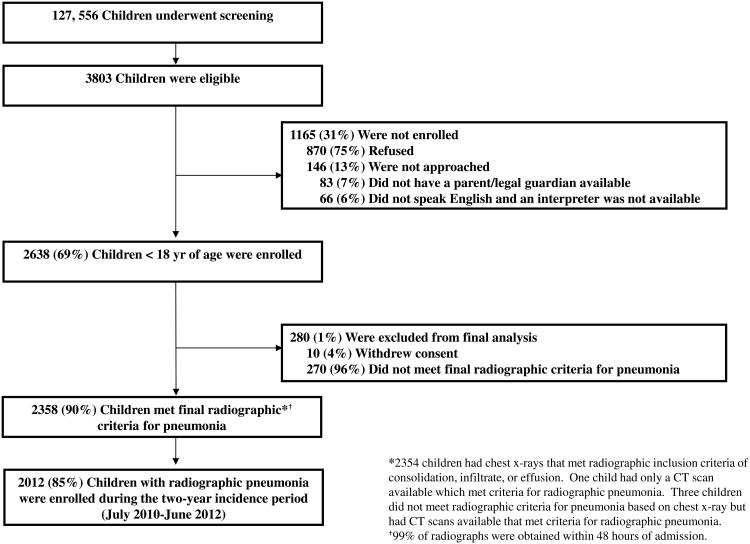
Of 2638 enrolled children, 2358 (89%) had radiographic pneumonia (Figure 1). Inter-rater agreement among the three study radiologists was 84.0% (CI 81.3-86.4) upon review of a 10% random sample of radiographs. Median age was 2 years old (interquartile range [IQR] 1-6); 45% were female; 40% were white, 33% were black, and 19% were Hispanic; 51% had an underlying condition (asthma/reactive airway disease was most common). Median length of stay was 3 days (IQR 2-5); 497 (21%) children required intensive care, and 3 (<1%) died (Table 1, Supplementary Table S1).
Figure 1. Study Enrollment and Final Pneumonia Cases.
Table 1. Characteristics of Children with Community-acquired Pneumonia Requiring Hospitalization.
| Characteristic | Children with radio graphic confirmation of pneumonia (n=2358) |
|---|---|
|
| |
| Age groups – no. (%) | |
| <2 years | 1055 (45) |
| 2-4 years | 595 (25) |
| 5-9 years | 422 (18) |
| 10-17 years | 286 (12) |
|
| |
| Symptoms – no. (%) | |
| Cough | 2230 (95) |
| Fever/feverish | 2155 (91) |
| Anorexia | 1766 (75) |
| Dyspnea | 1657 (70) |
|
| |
| Any underlying condition* – no. (%) | 1197 (51) |
| Asthma/reactive airway disease | 779 (33) |
| Pre-term birth among children <2 yrs. | 218/1055 (21) |
|
| |
| Radiographic findings† – no. (%) | |
| Consolidation | 1376 (58) |
| Alveolar or interstitial infiltrate | 1195 (51) |
| Pleural effusion | 314 (13) |
|
| |
| Hospitalization characteristics – no. (%) | |
| Length of stay – median days, IQR | 3 (2-5) |
| Intensive care unit admission | 497 (21) |
| Mechanical ventilation | 166 (7) |
| Death (in-hospital) | 3 (<1) |
Any underlying medical conditions included asthma/reactive airway disease, chromosomal disorders including Down syndrome, chronic kidney disease, chronic liver disease, congenital heart disease, diabetes mellitus, immunosuppression (either due to chronic condition or medication, malignancy [but not skin cancer], human immunodeficiency virus infection with CD4 count >200 cells/mm3), neurological disorders (including seizure disorder, cerebral palsy, scoliosis), pre-term birth (defined as gestational age <37 weeks at birth for those children who were <2 years old at time of hospitalization), and splenectomy. A more complete list of the prevalence of specific conditions is in Supplementary Table S1.
Findings are not mutually exclusive and therefore do not add to 100%; only 6 children had pleural effusion alone.
Among children with information, 612 of 2053 (30%) children ≥6 months old received ≥1 dose of influenza vaccine for the concurrent season and 1101 (87%) of 1272 children aged 19 months to 12 years old received ≥3 doses of PCV (Supplementary Table S1). Antibiotics were prescribed for 18% of children ≤5 days before hospitalization; 88% received antibiotics during hospitalization.
Pathogen Detection
Among the 2358 children with radiographic pneumonia, 2254 (96%) had a NP/OP swab, 2143 (91%) had blood for culture, 2063 (88%) had whole blood for PCR, 1028 (44%) had paired sera, 86 (4%) had PF, 23 (1%) had a BAL specimen, and 22 (1%) had an ET aspirate. Among those with collection time available, 82% of 2107 blood cultures and 47% of 2022 whole blood PCR samples were collected before inpatient antibiotic administration. To calculate pathogen-specific proportions, only the 2222 (94%) children with radiographic pneumonia for whom blood, PF, ET aspirate, or BAL specimen and a NP/OP swab or paired sera were available were used.
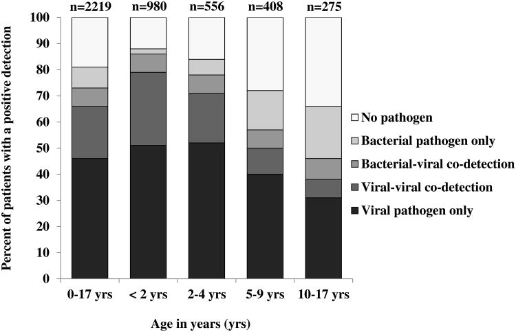
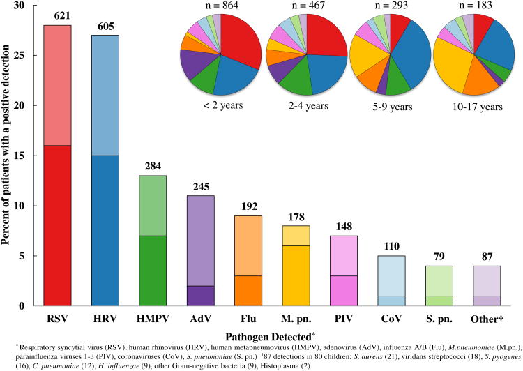
A viral and/or bacterial pathogen was detected in 1802 (81%) of 2222 children; ≥1 virus in 1472 (66%), bacteria in 175 (8%), and bacterial-viral co-detections in 155 (7%). The most commonly detected pathogens were RSV (28%), HRV (27%), HMPV (13%), AdV (11%), M. pneumoniae (8%), PIV (7%), influenza (7%), CoV (5%), S. pneumoniae (4%), S. aureus (1%), and S. pyogenes (<1%) (Figure 2, Supplementary Tables S2, S3). RSV (37% vs. 8%), AdV (15% vs. 3%), and HMPV (15% vs. 8%) were more commonly detected in children <5 years old compared with older children; M. pneumoniae (19% vs. 3%) was more commonly detected in children ≥5 years old compared with younger children (Supplementary Table S4).
Figure 2. A/B: Pathogen Detection among U.S. Children with Community-acquired Pneumonia Requiring Hospitalization, 2010-2012.
Panel A shows the proportion of pathogen types detected from January 1, 2010 through June 30, 2012 among 2222 hospitalized children with radiographic pneumonia who had 1) blood (bacterial culture or real-time polymerase chain reaction [PCR]) or pleural fluid (bacterial culture or PCR), endotracheal aspirate (bacterial culture), or bronchoalveolar lavage (bacterial culture); 2) and naso/oropharyngeal swab (viral and atypical bacterial PCR) or viral serology results available. Panel B shows number and percent of children with specific pathogen detections for all ages in the bar graph. There were 1802 patients who had a viral and/or bacterial pathogen detected among 2222 patients who had available tests for both bacterial and viral detection; there were two patients in whom Histoplasma was detected. Because patients could have more than one pathogen detected, there were a total of 2533 total detections. Darker and lighter shading in the bar graph indicates single and co-pathogen detection, respectively. Proportions of detections (single and co-detection) by age group are depicted on the pie graphs.
Seasonality
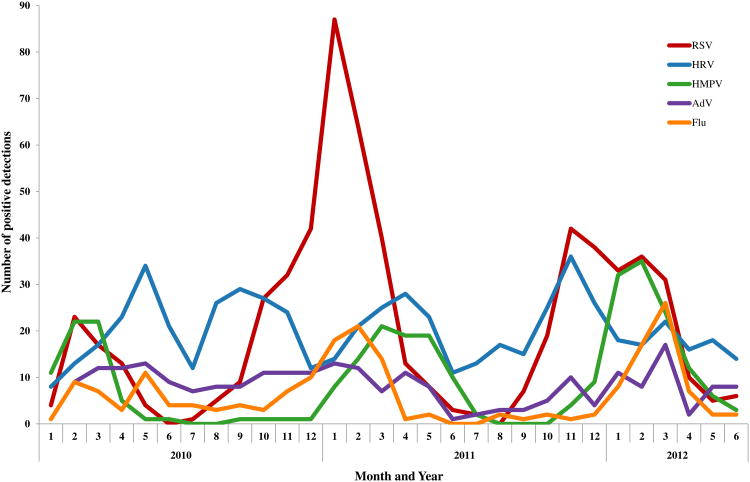
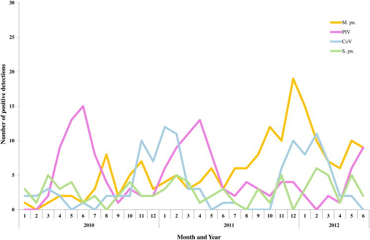
Pneumonia peaked in fall and winter. RSV, influenza, HMPV, and S. pneumoniae increased during winter, whereas HRV was detected year-round (Figure 3). M. pneumoniae rose steadily from summer through fall 2011, peaking that winter.
Figure 3.
A/B: Pathogen Detection by Month and Year among U.S. Children with Community-acquired Pneumonia Requiring Hospitalization, January 1, 2010 through June 30, 2012. Panel A includes respiratory syncytial virus (RSV), human rhinovirus (HRV), human metapneumovirus (HMPV), adenovirus (AdV), and influenza A/B (Flu). Panel B includes M. pneumoniae (M. pn), parainfluenza viruses 1-3 (PIV), coronaviruses (CoV), and S. pneumoniae (S. pn.).
Controls
Of 726 controls, 125 (17%) could not be reached for follow-up and 80 (11%) had fever or respiratory symptoms after surgery, and were excluded. Among 521 remaining asymptomatic controls, 28% were <2 years, 24% were 2-4 years, 24% were 5-9 years, and 25% were 10-17 years old (Supplementary Table S5). Children with radiographic pneumonia (n=832) enrolled during the same period at the same sites were younger; 42% <2 years, 25% 2-4 years, 19% 5-9 years, and 13% 10-17 years. After adjustment for age, HRV was detected in 17% of controls compared with 22% of cases enrolled at the same sites during the same period while all other pathogens were detected in ≤3% of controls.
Overall and Pathogen-specific Incidence
Among 2358 children with radiographic pneumonia, 2012 (85%) were enrolled between July 1, 2010 and June 30, 2012. The annual incidence of pneumonia hospitalization was 15.7/10,000 children (CI 14.9-16.5) (Table 2). Incidence was highest among children <2 years old [62.2/10,000 (CI 57.6-67.1)], lower among those 2-4 years old [23.8/10,000 (CI 21.4-26.3)], and further decreased with increasing age. Incidence of RSV, HRV, HMPV, AdV, influenza, PIV, CoV, and S. pneumoniae was higher among children <5 years old compared with older children, but highest among children <2 years old (Supplementary Table S6). Incidence of M. pneumoniae was similar across age groups.
Table 2. Estimated Annual Incidence Rates* of Community-acquired Pneumonia Hospitalization by Year, Site, Age Group, and Pathogen Detected.
| Variable | Annual no. pneumonia hospitalizations/10,000 children | 95% confidence interval |
|---|---|---|
|
| ||
| Overall incidence rate† | 15.7 | 14.9–16.5 |
| 2010-2011 (year 1) | 16.8 | 15.6-18.0 |
| 2011-2012 (year 2) | 14.6 | 13.5-15.7 |
|
| ||
| Study Site | ||
| Memphis | 19.6 | 18.0–21.3 |
| Nashville | 12.3 | 11.2–13.4 |
| Salt Lake City | 15.2 | 13.8–16.5 |
|
| ||
| Age group | ||
| <2 years | 62.2 | 57.6–67.1 |
| 2-4 years | 23.8 | 21.4–26.3 |
| 5-9 years | 10.1 | 8.9–11.3 |
| 10-17 years | 4.2 | 3.6–4.8 |
|
| ||
| Pathogen-specific incidence‡ | ||
| Respiratory Syncytial Virus | 4.6 | 4.3–5.1 |
| Human Rhinovirus | 4.1 | 3.7–4.4 |
| Human Metapneumovirus | 1.9 | 1.6–2.1 |
| Adenovirus | 1.6 | 1.4–1.8 |
| Mycoplasma pneumoniae | 1.4 | 1.2–1.6 |
| Influenza A/B virus | 1.1 | 0.9–1.3 |
| Parainfluenza viruses 1-3 | 0.9 | 0.8–1.1 |
| Coronaviruses | 0.8 | 0.7–1.0 |
| Streptococcus pneumoniae | 0.5 | 0.4–0.6 |
Based on 2,212,327 person-years of observation
Annual incidence rates were calculated from July 1, 2010 to June 30, 2011 (year 1) and July 1, 2011 to June 30, 2012 (year 2) and represent the 2012 children with radiographic pneumonia enrolled during that time
Pathogen-specific incidence is calculated for the 1899 children with radiographic pneumonia during the incidence period with at least one specimen available for both bacterial and viral testing. Age-specific pathogen-specific incidence is in Supplementary Table S6.
Discussion
The multi-center EPIC study is a prospective, population-based U.S. study of pediatric community-acquired pneumonia. We demonstrated pneumonia hospitalization burden to be highest among children <5 years old. Multi-pathogen diagnostic testing yielded a pathogen in 81% of children with pneumonia; viral and bacterial pathogens were detected in 73% and 15% of children, respectively.
The annual incidence of community-acquired pneumonia hospitalization estimated from our three study hospitals combined was 15.7/10,000 children <18 years old. The estimated pneumonia hospitalization rate, using the 2009 national Kids' Inpatient Database, was 22.5/10,000 children <18 years old2; similar but higher than our rate. These differences might be attributed to differences by year, populations studied, and the strict EPIC study criteria that included standardized clinical and radiological definitions while excluding recently hospitalized or severely immunosuppressed children. Similar to our findings, reports using hospital discharge databases have shown decreasing rates of pneumonia with increasing age of children.1,3,8
RSV was the most common pathogen detected (28%), with greatest burden among children <2 years old with pneumonia. In another study using PCR, RSV was detected among 31% of children <14 years old hospitalized with radiographic pneumonia similar to our results.34
HRV was detected in 27% of children with pneumonia. The literature supports that HRV is associated with pneumonia, either as a sole pathogen, or in synergy with other pathogens.35-37 However, HRV was detected in 17% of controls compared with 22% of cases enrolled at the same sites during the same period. Shedding of HRV can extend >2 weeks after infection,38 making the interpretation of HRV detection among children with pneumonia challenging.
HMPV, AdV, PIV, and CoV accounted for one-third of detections, with highest rates among children <5 years old. In similar pediatric pneumonia studies, these pathogens accounted for 25-40% of detections.12,34 In our study, although PCR was responsible for the majority of viral detections, serology was a useful adjunct.27,28 Our study occurred after the 2009 H1N1 pandemic when the influenza seasons were mild;39 making the influenza burden less than during seasons with more widespread circulation.
Bacterial pathogens were detected in 15% of children with pneumonia. While the incidence of M. pneumoniae was fairly similar across age groups, M. pneumoniae accounted for a steadily increasing proportion of pneumonia with increasing age.40 An earlier U.S. pediatric pneumonia etiology study using PCR targeting pneumolysin, a test with limited specificity,7,41 and conducted before universal Hib and PCV use reported a higher proportion of bacterial detection than we demonstrated.7 While our data partly reflect the substantial reduction of pneumococcal and Hib disease due to conjugate vaccines, bacterial culture-based diagnostics have limited sensitivity and bacteremia is detected in a minority of pneumococcal pneumonias.8-11,41,42 In the absence of a gold standard for bacterial pathogen detection in pneumonia, our findings based on current state-of-the-art diagnostics suggest that the incidence of bacterial pneumonia is lower than previously reported.
In our study, the proportion of pathogen co-detection was 26%. Another U.S. etiology study of 154 children hospitalized with community-acquired pneumonia reported an identical prevalence.7 Given the large proportion and diversity of co-detections, further study is needed.
This study has several limitations. Not every eligible child was enrolled; although incidence calculations accounted for non-enrollment. Among enrolled children, not all specimen types were available, potentially leading to under- or over-estimation of pathogen-specific rates; however, 94% of children with radiographic pneumonia had specimens available for both bacterial and viral detection and no demographic or clinical differences were noted between those with and without specimens available. Despite a comprehensive diagnostic approach, sensitivity of current tests for bacterial pneumonia (particularly in the setting of antibiotic use), is not optimal.43,44 Due to ethical and feasibility considerations, invasive procedures to obtain direct lung samples were not commonly performed. PCR detection of pathogens in NP/OP swabs could represent infection limited to the upper tract or convalescent shedding and thus detection may not denote causation. Our controls were a convenience sample and may not have represented the underlying population; their enrollment was not done for the entire study duration, was restricted to two sites, and focused on the prevalence of pathogens in asymptomatic children thus limiting extrapolations of causality. However, except for HRV, pathogens were uncommonly detected in controls, suggesting that the other viruses and atypical bacteria contribute to pneumonia. We believe control data helped interpret pathogen detections among cases and is an important strength. There is substantial overlap in clinical and radiologic features between bronchiolitis, reactive airway disease, and pneumonia, particularly in young children. Even strict radiographic definitions may not accurately distinguish these entities resulting in potential misclassification.45 Finally, although our multi-center study allowed investigation of diverse populations with standardized procedures, our findings may not be representative of the entire U.S. pediatric population or generalizable to other settings.
In conclusion, the burden of community-acquired pneumonia requiring hospitalization was highest among younger children, with respiratory viruses frequently detected. Effective anti-viral vaccines or treatments, particularly for RSV, could have an impact on pediatric pneumonia. The low prevalence of bacterial detections likely reflects both the effectiveness of bacterial conjugate vaccines and relatively insensitive diagnostics. The pediatric community-acquired pneumonia burden was associated with multiple different and co-detected pathogens, underscoring a need for the enhancement of sensitive, inexpensive, and rapid diagnostics to accurately identify pneumonia pathogens.
Supplementary Material
Acknowledgments
We thank the patients who graciously consented to participate in this study. In addition, we thank the following: Associated Regional and University Pathologists (ARUP) Laboratories: Heather London, Torrance Meyer; BioFire Diagnostics: Mark A. Poritz; CDC: Suzette Bartley, Bernie Beall, Nicole Burcher, Robert Davidson, Michael Dillon, Barry Fields, Phalasy Juieng, Shelley Magill; Le Bonheur Children's Hospital: John Devincenzo, Tonya Galloway, Vivian Lebaroff, Moses Lockhart, Lakesha London, Tekita McKinney, Amanda Nesbit, Chirag Patel, Tina Pitt, Shante Richardson, Naeem Shaikh, Davida Singleton, Mildred Willis; Monroe Carell Jr. Children's Hospital: Thomas Abramo, Gretchen Edwards, Regina Ellis, Angela Harbeson, Deborah Hunter, Romina Libster, Angela Mendoza, Renee Miller, Deborah Myers, Natalee Rathert, Becca Smith, Bob Sparks, Kristy Spilman, Tanya Steinback, Scott Taylor, Sandy Yoder; Primary Children's Hospital: Trenda Barney, Patrick Morris; St. Jude Children's Research Hospital: Edwina Anderson, Nancy Foster, Donna Nance, Ryan Heine, Amanda-Anderson Green, Amy Iverson, Shane Gansebom, Pat Flynn, Randall Hayden, Kim Allison; University of Utah: Fumiko Alger, Alexandra Burringo, Christopher Carlson, Lacey Collom, Gabriel Cortez, Kristina Grim, Keith Gunnerson, David Halladay, Caroline Heyrend, Jarrett Killpack, Kevin Martin, Brittany McDowell, Francesca Nichols, Parker Plant, Margaret Reid, Joshua Shimizu, Luke Schunk, Melanie Sperry, John Sweeley, and Lucy Williams.
Funding/Support: The EPIC study is supported by the Influenza Division in the National Center for Immunizations and Respiratory Diseases at the Centers for Disease Control and Prevention through cooperative agreements with each study site and was based on a competitive research funding opportunity.
Footnotes
Disclaimer: The findings and conclusions in this report are those of the authors and do not necessarily represent the views of the Centers for Disease Control and Prevention.
Disclosure: Disclosure forms provided by the authors are available with the full text of this article at NEJM.org.
References
- 1.Pfuntner A, Wier LM, Stocks C. Rockville, Md.: Agency for Healthcare Research and Quality; 2013. [February 10, 2014]. Most frequent conditions in U.S. hospitals, 2011. HCUP Statistical Brief #162. at http://www.hcup-us.ahrq.gov/reports/statbriefs/sb162.pdf. [PubMed] [Google Scholar]
- 2.Yu H, Wier LM, Elixhauser A. Rockville, Md.: Agency for Healthcare Research and Quality; 2011. [February 10, 2014]. Hospital stays for children, 2009. HCUP Statistical Brief #118. at http://www.hcup-us.ahrq.gov/reports/statbriefs/sb118.pdf. [PubMed] [Google Scholar]
- 3.Lee GE, Lorch SA, Sheffler-Collins S, Kronman MP, Shah SS. National hospitalization trends for pediatric pneumonia and associated complications. Pediatrics. 2010;126:204–12. doi: 10.1542/peds.2009-3109. [DOI] [PMC free article] [PubMed] [Google Scholar]
- 4.Pfuntner A, Wier LM, Steiner C. Rockville, Md.: Agency for Healthcare Research and Quality; 2013. [April 21, 2014]. Costs for Hospital Stays in the United States, 2011. HCUP Statistical Brief #168. at http://www.hcup-us.ahrq.gov/reports/statbriefs/sb168-Hospital-Costs-United-States-2011.pdf. [PubMed] [Google Scholar]
- 5.Bradley JS, Byington CL, Shah SS, et al. The management of community-acquired pneumonia in infants and children older than 3 months of age: clinical practice guidelines by the Pediatric Infectious Diseases Society and the Infectious Diseases Society of America. Clin Infect Dis. 2011;53:617–30. doi: 10.1093/cid/cir625. [DOI] [PMC free article] [PubMed] [Google Scholar]
- 6.Madhi SA, DeWals P, Grijalva CG, et al. The burden of childhood pneumonia in the developed world: a review of the literature. Pediatr Infect Dis J. 2013;32:e119–27. doi: 10.1097/INF.0b013e3182784b26. [DOI] [PubMed] [Google Scholar]
- 7.Michelow IC, Olsen K, Lozano J, et al. Epidemiology and clinical characteristics of community-acquired pneumonia in hospitalized children. Pediatrics. 2004;113:701–7. doi: 10.1542/peds.113.4.701. [DOI] [PubMed] [Google Scholar]
- 8.Grijalva CG, Nuorti JP, Arbogast PG, Martin SW, Edwards KM, Griffin MR. Decline in pneumonia admissions after routine childhood immunisation with pneumococcal conjugate vaccine in the USA: a time-series analysis. Lancet. 2007;369:1179–86. doi: 10.1016/S0140-6736(07)60564-9. [DOI] [PubMed] [Google Scholar]
- 9.Nelson JC, Jackson M, Yu O, et al. Impact of the introduction of pneumococcal conjugate vaccine on rates of community acquired pneumonia in children and adults. Vaccine. 2008;26:4947–54. doi: 10.1016/j.vaccine.2008.07.016. [DOI] [PubMed] [Google Scholar]
- 10.Griffin MR, Zhu Y, Moore MR, Whitney CW, Grijalva CG. U.S. hospitalizations for pneumonia after a decade of pneumococcal vaccination. N Engl J Med. 2013;369:155–63. doi: 10.1056/NEJMoa1209165. [DOI] [PMC free article] [PubMed] [Google Scholar]
- 11.MacNeil JR, Cohn AC, Farley M, et al. Current Epidemiology and Trends in Invasive Haemophilus influenzae Disease – United States, 1989-2008. Clin Infect Dis. 2011;53:1230–6. doi: 10.1093/cid/cir735. [DOI] [PubMed] [Google Scholar]
- 12.Pavia AT. Viral Infections of the Lower Respiratory Tract: Old Viruses, New Viruses, and the Role of Diagnosis. Clin Infect Dis. 2011;52(Suppl):S284–9. doi: 10.1093/cid/cir043. [DOI] [PMC free article] [PubMed] [Google Scholar]
- 13.Caliendo AM. Multiplex PCR and Emerging Technologies for the Detection of Respiratory Pathogens. Clin Infect Dis. 2011;52(Suppl):S326–30. doi: 10.1093/cid/cir047. [DOI] [PMC free article] [PubMed] [Google Scholar]
- 14.Cherian T, Mulholland EK, Carlin JB, et al. Standardized interpretation of paediatric chest radiographs for the diagnosis of pneumonia in epidemiological studies. Bull World Health Organ. 2005;83:353–9. [PMC free article] [PubMed] [Google Scholar]
- 15.Bartlett RC. Medical microbiology: quality, cost and clinical relevance. New York: John Wiley & Sons; 1974. pp. 24–31. [Google Scholar]
- 16.Pollock HM, Hawkins EL, Bonner JR, Sparkman T, Bass JB. Diagnosis of bacterial pulmonary infections with quantitative protected catheter cultures obtained during bronchoscopy. J Clin Microbiol. 1983;17:255–9. doi: 10.1128/jcm.17.2.255-259.1983. [DOI] [PMC free article] [PubMed] [Google Scholar]
- 17.Carvalho MG, Tondella ML, McCaustland K, et al. Evaluation and improvement of real-time PCR assays targeting lytA, ply, and psaA genes for detection of pneumococcal DNA. J Clin Microbiol. 2007;45:2460–6. doi: 10.1128/JCM.02498-06. [DOI] [PMC free article] [PubMed] [Google Scholar]
- 18.Blaschke AJ, Heyrend C, Byington CL, et al. Molecular analysis improves pathogen identification and epidemiologic study of pediatric parapneumonic empyema. Pediatr Infect Dis J. 2010;30:289–94. doi: 10.1097/INF.0b013e3182002d14. [DOI] [PMC free article] [PubMed] [Google Scholar]
- 19.Blaschke AJ, Heyrend C, Byington CL, et al. Rapid identification of pathogens from positive blood cultures by multiplex polymerase chain reaction using the FilmArray system. Diagn Microbiol Infect Dis. 2012;74:349–55. doi: 10.1016/j.diagmicrobio.2012.08.013. [DOI] [PMC free article] [PubMed] [Google Scholar]
- 20.Weinberg GA, Erdman DD, Edwards KM, et al. Superiority of reverse-transcription polymerase chain reaction to conventional viral culture in the diagnosis of acute respiratory tract infections in children. J Infect Dis. 2004;189:706–10. doi: 10.1086/381456. [DOI] [PubMed] [Google Scholar]
- 21.Mullins JA, Erdman DD, Weinberg GA, et al. Human Metapneumovirus infection among children hospitalized with acute respiratory illness. Emerg Infect Dis. 2004;10:700–5. doi: 10.3201/eid1004.030555. [DOI] [PMC free article] [PubMed] [Google Scholar]
- 22.Dare RK, Fry AM, Chittaganpitch M, Sawanpanyalert P, Olsen SJ, Erdman DD. Human coronavirus infections in rural Thailand: a comprehensive study using real-time reverse-transcription polymerase chain reaction assays. J Infect Dis. 2007;196:1321–8. doi: 10.1086/521308. [DOI] [PMC free article] [PubMed] [Google Scholar]
- 23.Lu X, Holloway B, Dare RK, et al. Real-time reverse transcription-PCR assay for comprehensive detection of human rhinoviruses. J Clin Microbiol. 2008;46:533–9. doi: 10.1128/JCM.01739-07. [DOI] [PMC free article] [PubMed] [Google Scholar]
- 24.Thurman KA, Warner AK, Cowart KC, Benitez AJ, Winchell JM. Detection of Mycoplasma pneumoniae, Chlamydia pneumoniae, and Legionella spp in clinical specimens using a single tube multiplex real-time PCR assay. Diagn Microbiol Inf Dis. 2011;70:1–9. doi: 10.1016/j.diagmicrobio.2010.11.014. [DOI] [PMC free article] [PubMed] [Google Scholar]
- 25.Loens K, vanLoon AM, Coenjaerts F, et al. Performance of different mono- and multiplex nucleic acid amplification tests on a multipathogen external quality assessment panel. J Clin Microbiol. 2012;50:977–87. doi: 10.1128/JCM.00200-11. [DOI] [PMC free article] [PubMed] [Google Scholar]
- 26.Wallace PS, MacKay WG. Quality in the molecular microbiology laboratory. Methods Mol Biol. 2013;943:49–79. doi: 10.1007/978-1-60327-353-4_3. [DOI] [PubMed] [Google Scholar]
- 27.Sawatwong P, Chittaganpitch M, Hall H, et al. Serology as an adjunct to polymerase chain reaction assays for surveillance of acute respiratory virus infections. Clin Infect Dis. 2013;54:445–6. doi: 10.1093/cid/cir710. [DOI] [PubMed] [Google Scholar]
- 28.Feiken DR, Njenga MK, Bigogo G, et al. Additional diagnostic yield of adding serology to PCR in diagnosing viral acute respiratory infections in Kenyan patients 5 years of age and older. Clin Vaccine Immunol. 2013;20:113–4. doi: 10.1128/CVI.00325-12. [DOI] [PMC free article] [PubMed] [Google Scholar]
- 29.Geneva: World Health Organization; 2011. [Accessed February 14, 2014]. Manual for the laboratory diagnosis and virological surveillance of influenza; pp. 43–77. at http://www.who.int/influenza/gisrs_laboratory/manual_diagnosis_surveillance_influenza/en/ [Google Scholar]
- 30.Monto A, Maassab HF. Ether treatment of type B influenza virus antigen for the hemagglutination inhibition test. J Clin Microbiol. 1981;13:54–7. doi: 10.1128/jcm.13.1.54-57.1981. [DOI] [PMC free article] [PubMed] [Google Scholar]
- 31.Kendal AP, Cate TR. Increased sensitivity and reduced specificity of hemagglutination inhibition tests with ether-treated influenza B/Singapore/222/79. J Clin Microbiol. 1983;18:930–4. doi: 10.1128/jcm.18.4.930-934.1983. [DOI] [PMC free article] [PubMed] [Google Scholar]
- 32.Fiore AE, Bridges CB, Katz JM, Cox NJ. Inactivate influenza vaccines. In: Plotkin SA, Orenstein WA, Offit PA, editors. Vaccines. 6th. Saunders; 2012. pp. 257–93. [Google Scholar]
- 33.Atlanta, GA: National Center for Health Statistics; 2012. [Accessed February 14, 2014]. Vintage 2012 postcensal estimates of the resident population of the United States by year, county, single-year of age, bridged race, Hispanic origin, and sex. at: http://www.cdc.gov/nchs/nvss/bridged_race.htm. [Google Scholar]
- 34.García-García ML, Calvo C, Pozo F, Villadangos PA, Pérez-Breña P, Casas I. Spectrum of Respiratory Viruses in Children with Community-acquired Pneumonia. Pediatr Infect Dis J. 2012;31:808–13. doi: 10.1097/INF.0b013e3182568c67. [DOI] [PubMed] [Google Scholar]
- 35.Iwane MK, Prill MM, Lu X, et al. Human rhinovirus species associated with hospitalizations for acute respiratory illness in young U.S. children. J Infect Dis. 2011;204:1702–10. doi: 10.1093/infdis/jir634. [DOI] [PubMed] [Google Scholar]
- 36.Fry A, Lu X, Olsen SJ, et al. Human rhinovirus infections in rural Thailand: epidemiological evidence for rhinovirus as both pathogen and bystander. PloS One. 2011:6e17780. doi: 10.1371/journal.pone.0017780. [DOI] [PMC free article] [PubMed] [Google Scholar]
- 37.Hayden FG. Rhinovirus and the lower respiratory tract. Rev Med Virol. 2004;14:17–31. doi: 10.1002/rmv.406. [DOI] [PMC free article] [PubMed] [Google Scholar]
- 38.Jartti T, Lee WM, Pappas T, Evans M, Lemanske RF, Gern JE. Serial viral infections in infants with recurrent respiratory illnesses. Eur Respir J. 2008;32:314–20. doi: 10.1183/09031936.00161907. [DOI] [PMC free article] [PubMed] [Google Scholar]
- 39.Atlanta, GA: Centers for Disease Control and Prevention; [October 23, 2013]. FluView: a weekly influenza surveillance report prepared by the Influenza Division. at http://www.cdc.gov/flu/weekly/ [Google Scholar]
- 40.Waites KB, Talkington DF. Mycoplasma pneumoniae and its role as a human pathogen. Clin Microbiol Rev. 2004;17:697–728. doi: 10.1128/CMR.17.4.697-728.2004. [DOI] [PMC free article] [PubMed] [Google Scholar]
- 41.Werno AM, Murdoch DR. Laboratory diagnosis of invasive pneumococcal disease. Clin Infect Dis. 2008;46:926–32. doi: 10.1086/528798. [DOI] [PubMed] [Google Scholar]
- 42.Madhi SA, Kuwanda L, Cutland C, Klugman KP. The impact of a 9-valent pneumococcal conjugate vaccine on the public health burden of pneumonia in HIV-infected and–uninfected children. Clin Infect Dis. 2005;40:1511–8. doi: 10.1086/429828. [DOI] [PubMed] [Google Scholar]
- 43.Blaschke AJ. Interpreting assays for the detection of Streptococcus pneumoniae. Clin Infect Dis. 2011;52:S331–7. doi: 10.1093/cid/cir048. [DOI] [PMC free article] [PubMed] [Google Scholar]
- 44.Caliendo AM, Gilbert DN, Ginocchio CG, et al. Better tests, better care: improved diagnostics for infectious diseases. Clin Infect Dis. 2013;59:S139–70. doi: 10.1093/cid/cit578. [DOI] [PMC free article] [PubMed] [Google Scholar]
- 45.Scott JAG, Wonodi C, Moïsi, et al. The definition of pneumonia, the assessment of severity, and clinical standardization in the Pneumonia Etiology Research for Child Health Study. Clin Infect Dis. 2012;54:S109–16. doi: 10.1093/cid/cir1065. [DOI] [PMC free article] [PubMed] [Google Scholar]
Associated Data
This section collects any data citations, data availability statements, or supplementary materials included in this article.


