Figure 2.

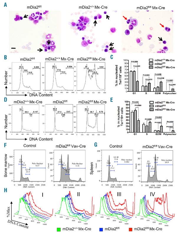
Loss of mDia2 causes binucleated late-stage erythroblasts in adult mice. (A) Wright-Giemsa stains of bone marrow smears from indicated mice. Black and red arrows indicate normal and binucleated erythroblasts, respectively. Scale bars: 15 μm. (B-C) Cell cycle analysis of the nucleated Ter119-positive erythroid cells in bone marrow of the indicated mice. Statistical analysis of each phase of the cell cycle is shown in (C). N=3 in each group. (D-E) Cell cycle analyses as in (B) and (C) except the experiments were performed in the spleen of the indicated mice. N=3 in each group. (F-G) Nucleated Ter119+ erythroblasts from bone marrow (F) and spleen (G) of the mDia2fl/flVav-Cre and control mice were gated for cell cycle analysis as in (B) and (D). Representative plots are presented. (H) Cell cycle analysis was performed on the gated populations from bone marrow, as in Figure 1C. Representative plots of cells from the indicated populations were presented as duplicate in each group. All experiments were repeated at least three times.
