Abstract
Background
Prior exposure to stress is a risk factor for developing post-traumatic stress disorder (PTSD) in response to trauma, yet the mechanisms by which this occurs are unclear. Using a rodent model of stress-based susceptibility to PTSD, we investigated the role of serotonin in this phenomenon.
Methods
Adult mice were exposed to repeated immobilization stress or handling, and the role of serotonin in subsequent fear learning was assessed using pharmacological manipulation and western blot detection of serotonin receptors, measurements of serotonin, high-speed optogenetic silencing, and behavior.
Results
Both dorsal raphe serotonergic activity during aversive reinforcement and amygdala serotonin 2c receptor (5-HT2CR) activity during memory consolidation are necessary for stress enhancement of fear memory, but neither process affects fear memory in unstressed mice. Additionally, prior stress increases amygdala sensitivity to serotonin by promoting surface expression of 5-HT2CR without affecting tissue levels of serotonin in the amygdala. We also show that the serotonin that drives stress enhancement of associative cued fear memory can arise from paired or unpaired footshock, an effect not predicted by theoretical models of associative learning.
Conclusion
Stress bolsters the consequences of aversive reinforcement, not by simply enhancing the neurobiological signals used to encode fear in unstressed animals, but rather by engaging distinct mechanistic pathways. These results reveal that predictions from classical associative learning models do not always hold for stressed animals, and suggest that 5-HT2CR blockade may represent a promising therapeutic target for psychiatric disorders characterized by excessive fear responses such as that observed in PTSD.
Keywords: serotonin, optogenetics, fear, 5-HT2C receptor, PTSD, amygdala
INTRODUCTION
Stress exposure is a risk factor for the development of post-traumatic stress disorder (PTSD) in humans [1,2]. Humans with PTSD often have strong memories for the traumatic experiences that underlie their disorder [3], but also exhibit heightened fear conditioning in laboratory settings [4,5]. In preclinical studies the relationship between stress exposure and subsequent trauma-related memory can be studied by exposing rodents to stressors and examining the impact on Pavlovian fear conditioning. In this model, fear conditioning itself does not lead to PTSD; only stress-exposed animals display the excessively strong fear memories that are also observed in humans with PTSD. The exaggerated fear response typically observed in stress-exposed animals [6] is often attributed to either strengthened encoding [7] or consolidation processes [8].
Serotonin plays a critical role in the regulation of emotion, and dysregulation of serotonergic systems is associated with stress-related affective disorders [9], including PTSD. Multiple lines of evidence suggest that excess serotonin is linked to altered threat processing. For instance, individuals that carry the short variant of the gene encoding the serotonin transporter (SLC6A4), which is thought to impair synaptic serotonin uptake, display increased amygdala reactivity to briefly presented (phasic) aversive stimuli [10]. In rodent studies, during aversive learning, serotonin is released into projection regions of the dorsal raphe nucleus (DRN) via phasic firing changes in response to discrete stimuli [11–13]. The extracellular serotonin levels in downstream DRN targets, like the basolateral amygdala (BLA), can remain elevated for at least an hour after learning is completed [14,15]. Although serotonin acts through several receptor subtypes in the BLA, the serotonin 2c receptor (5-HT2CR) is of interest because these receptors are heavily expressed in BLA neurons that regulate anxiety [16] and 5-HT2CR agonists promote anxiety in humans [17]. Furthermore, viral-mediated overexpression of 5-HT2CR in amygdala produces anxiogenic effects [18] while pharmacological blockade of amygdala 5-HT2CR prevents stress-induced anxiety-like behaviors [19].
Here, we examine behavior in a rodent paradigm in which repeated exposure to stress produces a vulnerability to heightened fear learning [6] and demonstrate that this vulnerability emerges from a serotonergic fear memory consolidation process that is not present in unstressed mice. This consolidation process requires serotonergic activity in the DRN during aversive reinforcement and 5-HT2CR signaling in the BLA, a major target structure of the DRN [20–24], after aversive learning. Interestingly, we also show that serotonin activation by either signaled or unsignaled footshocks is sufficient to enhance associative fear memory in stressed animals, an effect not predicted by classic theoretical models of associative learning. We show that stress enhances cell surface expression of 5-HT2CRs in the amygdala without affecting total serotonin levels during fear conditioning. Thus, aversive reinforcement is processed differently in the brain of a stress-exposed animal, and this profoundly impacts memory for later aversive experiences. These findings reveal fundamental mechanisms underlying the operation of a critical neural system in affective processing, and provide new principles both for associative learning theory and the prevention of stress-related psychiatric disorders.
METHODS AND MATERIALS
Subjects
Adult male C57BL/6 mice (Taconic, Germantown, NY) or transgenic mice [25] were used in all experiments. All procedures were approved by the Committee on Animal Care at the Massachusetts Institute of Technology and the Animal Care and Use Review Office at the U.S. Army Medical Research and Materiel Command.
Virus
AAV vectors were serotyped with AAV 2/8 capsids and packaged by the Vector Core at The University of North Carolina at Chapel Hill. The final viral concentration was approximately 1.0 – 2.0 × 10¹¹ infectious particles/mL.
Surgical Procedures
For some experiments, mice received cannulae implants, optical fiber implants, or virus infusions, as described in Supplemental Materials.
In vivo recording
Single-unit recordings were conducted in anesthetized SERT-Cre mice weeks after stereotactic delivery of virus to the DRN. Cell-attached recordings, which enabled well isolated single-unit recordings, were obtained by using standard blind in vivo patching technique [26]. See Supplemental Materials for details.
Drugs
The selective 5-HT2CR antagonist 6-chloro-2,3-dihydro-5-methyl-N-[6-[(2-methyl-3-pyridinyl) oxy]-3-pyridinyl]-1H-indole-1-carboxyamide dihydrochloride (SB242084, Tocris Bioscience, Minneapolis, MN) was dissolved in 0.9% sterile saline.
Immobilization stress
Mice were transferred to an experimental room and placed in ventilated plastic Decapicone bags (Braintree Scientific, Braintree, MA) for one hour on each of two consecutive days. While fear conditioning is also a type of stress exposure, here we use the term “stress” to exclusively refer to immobilization stress.
See Supplemental Materials for additional procedures and assays.
RESULTS
Repeated stress enhances the consolidation of fear memories established under degraded contingency
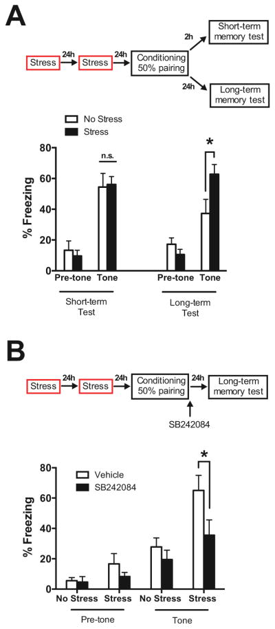
Stress exposure can enhance learned fear memories [6,27,28], modeling the way in which a history of stress exposure can predispose humans to disorders of fear or anxiety [1,29]. Here, we exposed mice to either two days of immobilization stress (Stress; 1 h/day) or handling (No Stress) followed by auditory fear conditioning (Figure 1). Unlike previous studies that examined the relationship between stress and subsequent auditory fear memory [6,27], we used an auditory fear conditioning protocol in which two of four tone and footshock presentations were explicitly unpaired (50% pairing), thereby reducing the tone-footshock contingency. Such a paradigm may be more sensitive to the effects of stress than a conventional protocol where the pairing is 100% [30]. Conditional fear to the tone was assessed in a novel environment either 2 h (short-term memory) or 24 h (long-term memory) after fear conditioning (Figure 1A).
Figure 1. Stress recruits serotonergic fear memory consolidation.
(A) Prior immobilization stress did not impact short-term (2 h) fear memory (left), but increased long-term (24 h) fear memory (right) to the tone. (B) Post-conditioning infusion of the serotonin 2C receptor antagonist SB242084 into the lateral/basolateral amygdala [24] blocked the immobilization stress-induced enhancement of fear consolidation. Data are means ± s.e.m. Fisher’s PLSD comparisons during auditory fear test: * P < 0.05 and n.s. = not significant for Stress versus No Stress.
Prior stress did not impact the amount of conditional freezing to the tone during fear acquisition (Supplemental Figure S1A) or the short-term memory test (Stress: F (1,19) = 0.020, Stress X Tone interaction: F (1,19) = 0.384, Ps = n.s., n = 10–11/group, Figure 1A, left), but did enhance tone-elicited freezing in mice tested 24 h later (Stress: F (1,18) = 1.64, P = n.s, Stress X Tone interaction: F (1,18) = 11.790, P < 0.01; Fisher’s PLSD comparing No Stress = 37.22 ± 9.22% and Stress = 62.78 ± 6.26%, P < 0.05, n = 10/group, Figure 1A, right). All groups exhibited comparable, low levels of freezing during the 3 min baseline period of the auditory fear test (Fisher’s PLSD comparing No Stress to Stress, Figure 1A, left and right, Ps > 0.230), indicating no generalization between the conditioning and testing contexts. Stress did not enhance fear memory via changes in pain processing, general motor activity, or memory retrieval (Supplemental Figures S1B–D, S2). Enhanced fear memory was also observed only after repeated stress (Supplemental Figure S3). The findings that repeated stress enhances long-term but not short-term fear memory when given before fear conditioning suggests that immobilization stress enhances fear responses by strengthening fear memory consolidation.
Serotonergic fear memory consolidation is selectively enabled by stress
Because our stress paradigm enhanced fear memory consolidation and serotonin is also implicated in the consolidation of memories [31–34], we determined whether stress-related enhancement of long-term fear memory consolidation is mediated by serotonin signaling in the BLA. Mice were implanted with bilateral cannulae in the BLA prior to stress or handling. Intra-BLA administration of the highly selective 5-HT2CR antagonist SB242084 (0.4 μg/0.4 μl) [24] immediately following fear conditioning completely blocked stress-induced enhancement of fear when mice were tested for long-term fear memory 24 h later (Stress: F (1,24) = 6.83, Stress X Tone interaction: F (1,24) = 4.277, Ps < 0.05; Fisher’s PLSD comparing Stress-Vehicle = 65.08 ± 9.90% and Stress-SB242084 = 35.56 ± 10.01%, P < 0.05, n = 6–10/group, Figure 1B), but did not affect fear levels in the absence of prior stress (Fisher’s PLSD comparing No Stress-Vehicle = 27.78 ± 5.91% and No Stress-SB242084 = 19.44 ± 6.21%, P = n.s.). These findings reveal that serotonin-mediated consolidation of fear memory occurs through amygdalar 5-HT2CRs and is selectively enabled by a prior history of immobilization stress exposure.
Stress enhances amygdala sensitivity to serotonin
There are at least two possible mechanisms by which repeated stress may selectively engage serotonergic consolidation of fear memory through 5-HT2CR. One possibility is that stress enhances the release of serotonin from DRN afferents to the BLA during fear conditioning. As an alternative or concurrent change, it is possible that stress may increase the membrane expression of postsynaptic serotonin receptors in BLA neurons [35,36], leading to enhanced postsynaptic sensitivity to serotonin release by the DRN.
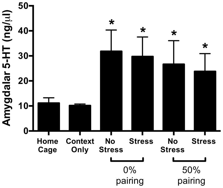
First we determined whether prior stress impacts BLA serotonin levels during conditioning. In addition to the 50% pairing fear conditioning protocol (two tone-shock pairings with two unpaired tones and two unpaired footshocks), a 0% pairing protocol was used (four unpaired tones and four unpaired footshocks). This allowed us to determine whether BLA serotonin levels differ when negative reinforcement is uncoupled from the auditory cue. Two control groups were included: one remained in the home cage (Home Cage group) and the other was placed in the conditioning context without tones or footshocks (Context Only group). Mice were sacrificed 30 min following fear conditioning, a time point where extracellular serotonin in the amygdala is maximally elevated by the conditioning procedure [14,15].
The serotonin content of the BLA was increased by fear conditioning (Conditioning: F (1,36) = 4.381, P < 0.05, n = 4–8/group; Fisher’s PLSD comparing all Stress and No Stress groups with control groups, P < 0.05, Figure 2), but not exposure to the novel context (Fisher’s PLSD comparing Context Only and Home Cage groups, P = n.s., Figure 2), consistent with other studies showing that fear conditioning and other stressors increase extracellular serotonin in the amygdala [14,37]. Within the groups that received fear conditioning, there was no effect of pairing on serotonin levels (Pairing: F (1,27) = 0.46, P = n.s., Figure 2), and, most critically, serotonin was similarly elevated in Stress and No Stress mice (Pairing X Stress: F (1,27) = 0.002, P = n.s., n = 7–8/group, Figure 2).
Figure 2. Stress does not affect conditioning-related increases in amygdalar serotonin.
Fear conditioning produced a significant elevation in serotonin (5-HT) in the BLA, but this was not altered by previous immobilization stress exposure. Data are means ± s.e.m. Fisher’s PLSD comparisons to the Home Cage group: * P < 0.05.
Because the BLA homogenates contain both extracellular and vesicular serotonin content in the BLA, it is possible that the lower levels of serotonin observed in the control groups reflect greater release of serotonin. To clarify this, we used high-pressure liquid chromatography (HPLC) to measure the primary serotonin metabolite 5-hydroxyindoleacetic acid (5-HIAA) in a subset of the homogenates (Supplemental Figure S4). We found that 5-HIAA levels were low in the Context Only control group and significantly increased by fear conditioning (Conditioning: F (1,14) = 10.54, P < 0.01; Fisher’s PLSD comparing all Stress and No Stress groups with Context Only, P < 0.05), but similarly elevated in the Stress and No Stress groups (Pairing X Stress: F (1,8) = 0.558, P = n.s., n = 3–4/group, Supplemental Figure S4). This suggests that the changes we observed in serotonin may predominantly reflect extracellular release, even though our method of detection is not specific for extracellular serotonin. Conservatively, our results show that BLA serotonin content is elevated by fear conditioning, but this is not influenced by the prior immobilization stress history of the animals. The similar post-conditioning levels of serotonin and its metabolite 5-HIAA when comparing subjects receiving the 0% pairing to the 50% pairing paradigm also suggests that footshock is the primary factor in determining conditioning-related increases in BLA serotonin.
We next examined the postsynaptic sensitivity of BLA neurons to serotonin following stress by measuring the surface expression of 5-HT2CR in the BLA. Mice received either two days of immobilization stress (Stress groups) or handling (No Stress groups), followed by auditory fear conditioning with 50% pairing. Mice were sacrificed 10 min after fear conditioning ended. 5-HT2CR density was assessed at this post-conditioning time point because it corresponds roughly to both the time when serotonin is first significantly elevated by fear conditioning [14,15] and a time when cellular consolidation of fear memory is occurring [38].
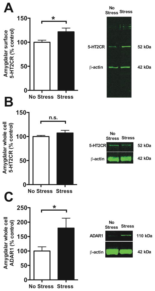
We found that repeated stress produced a significant increase in surface membrane expression of the 5-HT2CR in the amygdala measured shortly following fear conditioning (Stress: F (1,26) = 4.887, P < 0.05, n = 12–16/group, Figure 3A), without affecting the total pool of 5-HT2CR (Stress: F (1,10) = 1.504, P = n.s., n = 6/group, Figure 3B). This finding suggests that repeated stress alters trafficking of 5-HT2CR, as opposed to an upregulation of gene transcription or protein translation. Stress is known to trigger editing of the pre-messenger mRNA for the 5-HT2CR [39] through adenosine deaminase acting on RNA 1 (ADAR1) [40]. Because edited forms of the 5-HT2CR are known to have less internalization from the membrane surface [41], we also examined expression of ADAR1 protein. We found that repeated stress significantly enhances total levels of this protein in the BLA (Stress: F (1,10) = 4.975, P < 0.05, n = 6/group, Figure 3C). Such a finding is consistent with other reports showing that edited 5-HT2CR is more prevalent in the membrane of amygdala cells in mice that display increased anxiety and responsiveness to stress [42]. Together, these data show that the amygdala exhibits an enhanced membrane presence of 5-HT2CR following repeated stress.
Figure 3. Stress enhances surface expression of 5-HT2C receptors in BLA.
Immobilization stress enhanced membrane expression of the 5-HT2C receptor in the BLA (A) without affecting the total levels of 5-HT2C receptors (B), suggesting a change in trafficking of the receptor. (C) Stress also produced a concurrent increase in the whole cell levels of the mRNA editing enzyme ADAR1 in the BLA. Images on the right depict all bands detected in representative samples. Data are means ± s.e.m. Fisher’s PLSD comparisons: * P < 0.05
Serotonergic DRN activity during aversive reinforcement is required for stress facilitation of fear memory
Our data reveal that stress recruits a serotonergic consolidation mechanism for BLA-dependent fear memory, but the conditions during fear learning that lead to serotonin release into the BLA are unclear. Serotonergic DRN neurons exhibit heterogeneous, transient responses to a wide variety of discrete stimuli [11], including footshock [12]. Most DRN neurons are unresponsive to acoustic stimuli [43], but excitation is observed in a very small population of cells [13]. Thus, it is possible that stress could enhance fear memory by altering BLA responses to serotonin released by the auditory or shock stimuli or their contingent pairing during fear conditioning.
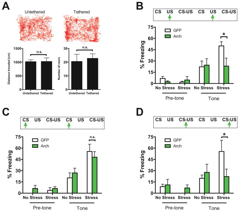
The DRN of Sert-Cre mice was transduced with the light-driven opsin Arch (“Arch-GFP” groups), which enables rapid and reliable large hyperpolarizing currents in neurons in response to pulses of green-yellow light (Supplemental Figures S5,S6) [44]. Control groups received a virus expressing only GFP (“GFP” groups). The lightweight optical fiber system used for light delivery did not impair movement or exploration within the conditioning chamber (Group: F (1,6) = 0.002 and 0.131; P = n.s., n = 4/group, Figure 4A).
Figure 4. Dorsal raphe serotonergic activity is required for the stress-enhancement of fear in a stimulus-dependent manner.
(A) Upper, representative traces of mice tethered or untethered to the fiber-optic patch cord freely exploring the fear conditioning apparatus. Lower, integration of fiber optic cable system with the fear conditioning apparatus does not interfere with voluntary motor behavior in the conditioning chamber. In three separate sets of mice, light was delivered to the dorsal raphe nucleus (DRN) for 30 sec periods encompassing noncontingent footshocks, noncontingent tones, or contingent tones and footshocks. (B) Arch-mediated silencing of serotonin activity in the DRN during unpaired footshocks blocked stress-induced facilitation of fear. (C) Photoinhibition of DRN serotonin activity during unpaired tone presentation failed to affect stress-enhancement of fear. (D) Photoinhibition of DRN serotonin activity during tone-shock pairings completely prevented stress-related enhancement of fear. Green arrows indicate green light delivery (30 sec). T = unpaired tone; S = unpaired footshock; T-S = paired tone and footshock. Data are means ± s.e.m. Group comparisons during auditory fear test: * P < 0.05 and n.s. = not significant for Stress-Arch versus Stress-GFP.
Typically a conditional fear response is established by pairing 100% of neutral tones with aversive footshock. Stress does enhance long-term fear memory (Group: F (1,15) = 6.581, P < 0.05; Fisher’s PLSD comparing No Stress = 54.94 ± 6.83%, Stress = 79.86 ± 6.38%, P < 0.05, n = 8–9/group, Supplemental Figure S7A), without potentiating fear retrieval or performance (Group: F (1,17) = 3.274, P = n.s., n = 9–10/group, Supplemental Figure S7B), when such a paradigm is used. However, consistent with the prior experiments, we used 50% pairing to enable selective silencing of DRN serotonergic activity during the presentation of noncontingent or contingent cues and reinforcers, appropriate for parsing the relationship between serotonergic activity and temporally-limited stimulus presentation during auditory fear conditioning. This also insured that unstressed animals would exhibit moderate levels of conditional freezing in the long-term memory test, with ample room for potentiation by stress. Continuous light was applied for 30 sec periods, a duration corresponding to the length of the tone used, across the three experimental conditions (Figure 4), equating the length of silencing across the different groups.
Photoinhibition during noncontingent footshocks (Figure 4B), noncontingent tones (Figure 4C), or contingent tones and footshocks (Figure 4D) produced different effects on fear memory. Notably, silencing serotonin activity during unpaired footshocks blocked stress-related enhancement of freezing in Stress-Arch relative to Stress-GFP mice (Stress: F (1,18) = 1.39, P = n.s., Stress X Tone X Virus interaction: F (1,18) = 5.904; P < 0.05; Fisher’s PLSD, P < 0.05, n = 5–6/group, Figure 4B). In contrast, despite an enhancement of fear memory by stress exposure (Stress: F (1,20) = 9.08, P < 0.01, Stress X Tone interaction: F (1,20) = 12.285, P < 0.001, n = 6–8/group, Figure 4C), photoinhibition of serotonergic activity during unpaired tones did not result in a difference in freezing levels between Stress-Arch and Stress-GFP controls (P = 0.551, Fisher’s PLSD). As might be expected if the shock were the salient stimulus for the release of serotonin into its efferents, photoinhibition of DRN serotonin neurons during paired tones and footshocks prevented the stress-enhancement of fear (Stress: F (1,16) = 0.498, P = n.s., Stress X Tone X Virus interaction: F (1,16) = 7.228, P < 0.05, n = 4–7/group, Figure 4D). Similar to the effect observed following silencing of DRN neurons during the footshock alone, conditional freezing to the tone in Stress-Arch mice was reduced compared to Stress-GFP (P < 0.05, Fisher’s PLSD).
DISCUSSION
Perhaps the most surprising finding of our study was that serotonergic fear memory consolidation was only engaged in mice with a history of repeated stress exposure. This was demonstrated by the selective reduction of fear in stressed, but not unstressed, mice by post-conditioning intra-BLA infusion of a 5-HT2CR antagonist (Figure 1B) and the lack of effect of DRN photoinhibition on long-term fear memory in unstressed animals under any conditions (Figures 4B–D). This cannot be attributed to “floor” levels of tone-induced freezing in the long-term fear memory test for the unstressed animals: post-tone freezing levels were significantly higher than pre-tone freezing levels for most conditions (Figure 1, Figures 4B–D). Rather, repeated immobilization stress increases the expression of 5-HT2CR membrane receptors in the BLA measured in the post-conditioning consolidation period, and this illuminates a mechanism by which 5-HT2CR-dependent fear memory consolidation is engaged following stress exposure. We measured 5-HT2CR density in the BLA after conditioning because this time point falls within the consolidation window that we identified as critical for stress-related enhancement of fear memory (Figure 1B). We do not know whether immobilization stress altered surface 5-HT2CR expression after stress exposure, or whether it altered trafficking of these receptors after fear conditioning; this issue remains an important open question for future studies. Previous studies reported that lesions or pharmacological inactivation of the DRN did not alter fear conditioning processes in unstressed animals, but did block potentiation of fear produced by a prior stressor [45,46], consistent with our finding that serotonin signaling through the 5-HT2CR has a non-essential role in fear learning in animals lacking a history of stress exposure.
A second surprising finding from our study relates to the observation that DRN serotonin activity during unpaired footshocks regulates the associative memory strength of the tone-footshock pairings (Figure 4B). Typically, when unsignaled reinforcement is given between cue-reinforcer pairings, as in a degraded contingency paradigm (cue-reinforcer pairings held constant, and extra reinforcers given) or a reduced temporal overlap paradigm as used here (total number of cue and reinforcer presentations held constant, but number of pairings reduced), it reduces the overall level of associative learning for the cue-reinforcer pairing [47–49]. Such a finding is accounted for in associative learning theory by positing that a context-reinforcer association competes with the cue-reinforcer association either at the time of encoding or the time of retrieval [50,51]. Thus, one might predict that if stress enhances signaling of the neurotransmitter released by unpaired reinforcement, it should be augmenting the reduction of the associative learning for the cue-reinforcer pairing, and thus blockade of this signaling should actually enhance learning for the cue-reinforcer association. Here, we show that, contrary to this prediction, eliminating serotonergic activity during the unpaired footshocks reduces associative memory strength (Figure 4B), revealing that the serotonergic activity driven by unsignaled footshock enhances associative memory strength for the tone-footshock pairings, but only in mice with a history of stress. The ability of the unsignaled footshocks to affect associative learning for the tone-footshock pairing occurs, in part, because the relevant biochemical signal for consolidation (serotonin, persisting for tens of minutes post-conditioning, Figure 2) greatly outlasts the trigger for a necessary signal (aversive footshocks, persisting for seconds; Figure 4), an effect which is typically not accounted for in classical associative learning models that explain learning through variation in the ability of the aversive reinforcer to support learning [52]. Our results reveal a novel mechanism by which unsignaled aversive reinforcers modulate associative aversive learning, and also reveal a specific set of circumstances in which the “rules” of learning theory are affected by state variables, such as stress. While many associative learning theories have been criticized for a failure to specifically account for the influence of state variables [53], there has been little consideration of this issue by neurobiologists who study stress and other state variables (see Supplemental Discussion). Given the importance of associative learning theory for motivating both behavioral and computational approaches to learning [54], we argue that thoughtful consideration of how experience influences learning theory is worthwhile.
Our observation that repeated stress administered after fear learning during a presumed consolidation window does not enhance fear memory (Supplemental Figure S2) may seem to conflict with our claim that stress enhances fear memory by augmenting consolidation (Figures 1A,B). Additionally, the observation that optogenetic inhibition of the serotonergic dorsal raphe during conditioning is sufficient to prevent stress-related enhancement of fear (Figure 4) also may appear at odds with the claim that serotonin is important for consolidation. However, there are at least two viable resolutions for this apparent conflict. First, while aversive reinforcement triggers activity in serotonergic neurons [12] (Figure 4), it is clear that synaptic serotonin can remain elevated in projection regions such as the BLA for at least an hour following conditioning [14]. Thus, serotonin may bind to its receptors during both fear learning and a brief (~hours) post-training consolidation window. Our finding that stress enhances long-term, but not short-term, fear memory (Figure 1A, Supplemental Figure S1A) via post-conditioning activity at 5-HT2CRs in the BLA (Figure 1B) is consistent with this. Given this temporal constraint, repeated stress started 24 h after fear learning does not alter fear memory strength for prior learning (Supplemental Figure S2) because it cannot alter either the critical time period for serotonergic consolidation shortly following fear learning, or the release of serotonin, most likely triggered by footshock during fear learning. Alternatively, our data are also consistent with a model in which serotonin release by aversive footshocks “prepares” the amygdala for a prolonged period of enhanced consolidation by acting at 5-HT2CRs shortly following fear learning. The prolonged elevation of extracellular serotonin observed after fear conditioning [14,15] may then act through 5-HT2CRs or other serotonin receptors to further stabilize fear memories.
In summary, during fear learning, serotonergic neurons make a critical contribution to the fear-enhancing effect of stress (see Supplemental Discussion), elicited by the presentation of aversive stimuli during fear conditioning. Furthermore, this effect is mediated by postsynaptic actions at 5-HT2CRs in the BLA, which enhance fear memory consolidation, though additional mechanisms may contribute (Supplemental Discussion). These results show that while the triggers leading to serotonin release (i.e., presentation of aversive stimuli) are temporally delimited, the effects of serotonin on downstream targets like the BLA are persistent. This mechanism may explain why polymorphisms in human serotonergic genes are often associated with enhanced aversive processing, especially following a history of traumatic life events [10,55,56].
While our rodent model of PTSD is simple, it does capture critical features of the disorder. The strong fear memory of the fear conditioning experience in stressed animals mirrors the strong memories for traumatic events often observed in humans with PTSD [57]. While PTSD involves additional symptoms, the intrusive nature of the traumatic memory may contribute to some other symptoms, such as hypervigilance or sleep disturbance [3,58]. Also, the dose-response relationship between stress exposure and enhancement of fear in our model (Figure 1 and Supplemental Figure S3) parallels the relationship between stress exposure and vulnerability to PTSD in humans [59]. Our demonstration that pharmacological and optogenetic inhibition of a serotonergic subcircuit selectively reduces fear in stressed animals with “pathological” (exaggerated) fear levels, without affecting fear responding in unstressed animals, overcomes a critical barrier to the successful treatment of stress-induced anxiety disorders such as PTSD. The benchmark for the successful treatment of PTSD should not be the elimination of fear, but simply its reduction to normal, adaptive levels. Our results suggest that administration of a 5-HT2CR antagonist, such as agomelatine, already FDA-approved for human use, might prevent or treat of PTSD by reducing the consolidation or reconsolidation of traumatic memories. One case report has shown that agomelatine produced full remittance of PTSD in one patient [60]; clearly, additional studies are warranted.
Supplementary Material
Acknowledgments
The authors thank Matthew B. Pomrenze and Philip H. Siebler for technical assistance and Elia S. Harmatz and Drs. Susana Correia and Liang Liang for comments on the manuscript. We also thank Dr. Steven F. Maier for providing equipment and laboratory space to perform western blot and HPLC analysis of samples. E.S.B. acknowledges funding by Jerry and Marge Burnett; Department of Defense CDMRP PTSD Program; Human Frontiers Science Program; MIT Intelligence Initiative; MIT McGovern Institute and McGovern Institute Neurotechnology (MINT) Program; MIT Media Lab; MIT Mind-Machine Project; NARSAD; NIH Director’s New Innovator Award (1DP2OD002002), NIH Grants 1R01DA029639, 1R43NS070453, 1RC1MH088182; and the Alfred P. Sloan Foundation. K.A.G. acknowledges funding by NIMH R01 MH084966 and the U.S. Army Research Laboratory and the U.S. Army Research Officer under grant 58076-LS-DRP.
Footnotes
FINANCIAL DISCLOSURES
The authors report no biomedical financial interests or potential conflicts of interest.
Publisher's Disclaimer: This is a PDF file of an unedited manuscript that has been accepted for publication. As a service to our customers we are providing this early version of the manuscript. The manuscript will undergo copyediting, typesetting, and review of the resulting proof before it is published in its final citable form. Please note that during the production process errors may be discovered which could affect the content, and all legal disclaimers that apply to the journal pertain.
References
- 1.Mazure C. Does Stress Cause Psychiatric Illness? Washington, D.C: American Psychiatric Press, Inc; 1995. [Google Scholar]
- 2.Schwartz AC, Bradley RL, Sexton M, Sherry A, Ressler KJ. Posttraumatic stress disorder among African Americans in an inner city mental health clinic. Psychiatr Serv. 2005;56:212–215. doi: 10.1176/appi.ps.56.2.212. [DOI] [PubMed] [Google Scholar]
- 3.Diamond DM, Campbell AM, Park CR, Halonen J, Zoladz PR. The temporal dynamics model of emotional memory processing: a synthesis on the neurobiological basis of stress-induced amnesia, flashbulb and traumatic memories, and the Yerkes-Dodson law. Neural Plast. 2007;2007:60803. doi: 10.1155/2007/60803. [DOI] [PMC free article] [PubMed] [Google Scholar]
- 4.Glover EM, Phifer JE, Crain DF, Norrholm SD, Davis M, Bradley B, et al. Tools for translational neuroscience: PTSD is associated with heightened fear responses using acoustic startle but not skin conductance measures. Depress Anxiety. 2011;28:1058–1066. doi: 10.1002/da.20880. [DOI] [PMC free article] [PubMed] [Google Scholar]
- 5.Roy MJ, Costanzo ME, Jovanovic T, Leaman S, Taylor P, Norrholm SD, et al. Heart rate response to fear conditioning and virtual reality in subthreshold PTSD. Stud Health Technol Inform. 2013;191:115–119. [PubMed] [Google Scholar]
- 6.Meyer RM, Burgos-Robles A, Liu E, Correia SS, Goosens KA. A ghrelin-growth hormone axis drives stress-induced vulnerability to enhanced fear. Mol Psychiatry. 2013 doi: 10.1038/mp.2013.135. [DOI] [PMC free article] [PubMed] [Google Scholar]
- 7.Suvrathan A, Bennur S, Ghosh S, Tomar A, Anilkumar S, Chattarji S. Stress enhances fear by forming new synapses with greater capacity for long-term potentiation in the amygdala. Philos Trans R Soc Lond B Biol Sci. 2014;369:20130151. doi: 10.1098/rstb.2013.0151. [DOI] [PMC free article] [PubMed] [Google Scholar]
- 8.Yehuda R. Post-traumatic stress disorder. N Engl J Med. 2002;346:108–114. doi: 10.1056/NEJMra012941. [DOI] [PubMed] [Google Scholar]
- 9.Lowry CA, Hale MW, Evans AK, Heerkens J, Staub DR, Gasser PJ, et al. Serotonergic systems, anxiety, and affective disorder: focus on the dorsomedial part of the dorsal raphe nucleus. Ann N Y Acad Sci. 2008;1148:86–94. doi: 10.1196/annals.1410.004. [DOI] [PubMed] [Google Scholar]
- 10.Armbruster D, Moser DA, Strobel A, Hensch T, Kirschbaum C, Lesch KP, et al. Serotonin transporter gene variation and stressful life events impact processing of fear and anxiety. Int J Neuropsychopharmacol. 2009;12:393–401. doi: 10.1017/S1461145708009565. [DOI] [PubMed] [Google Scholar]
- 11.Ranade SP, Mainen ZF. Transient firing of dorsal raphe neurons encodes diverse and specific sensory, motor, and reward events. J Neurophysiol. 2009;102:3026–3037. doi: 10.1152/jn.00507.2009. [DOI] [PubMed] [Google Scholar]
- 12.Schweimer JV, Ungless MA. Phasic responses in dorsal raphe serotonin neurons to noxious stimuli. Neuroscience. 2010;171:1209–1215. doi: 10.1016/j.neuroscience.2010.09.058. [DOI] [PubMed] [Google Scholar]
- 13.Waterhouse BD, Devilbiss D, Seiple S, Markowitz R. Sensorimotor-related discharge of simultaneously recorded, single neurons in the dorsal raphe nucleus of the awake, unrestrained rat. Brain Res. 2004;1000:183–191. doi: 10.1016/j.brainres.2003.11.030. [DOI] [PubMed] [Google Scholar]
- 14.Yokoyama M, Suzuki E, Sato T, Maruta S, Watanabe S, Miyaoka H. Amygdalic levels of dopamine and serotonin rise upon exposure to conditioned fear stress without elevation of glutamate. Neurosci Lett. 2005;379:37–41. doi: 10.1016/j.neulet.2004.12.047. [DOI] [PubMed] [Google Scholar]
- 15.Zanoveli JM, Carvalho MC, Cunha JM, Brandao ML. Extracellular serotonin level in the basolateral nucleus of the amygdala and dorsal periaqueductal gray under unconditioned and conditioned fear states: an in vivo microdialysis study. Brain Res. 2009;1294:106–115. doi: 10.1016/j.brainres.2009.07.074. [DOI] [PubMed] [Google Scholar]
- 16.Bonn M, Schmitt A, Lesch KP, Van Bockstaele EJ, Asan E. Serotonergic innervation and serotonin receptor expression of NPY-producing neurons in the rat lateral and basolateral amygdaloid nuclei. Brain Struct Funct. 2013;218:421–435. doi: 10.1007/s00429-012-0406-5. [DOI] [PMC free article] [PubMed] [Google Scholar]
- 17.Van Veen JF, Van der Wee NJ, Fiselier J, Van Vliet IM, Westenberg HG. Behavioural effects of rapid intravenous administration of meta-chlorophenylpiperazine (m-CPP) in patients with generalized social anxiety disorder, panic disorder and healthy controls. Eur Neuropsychopharmacol. 2007;17:637–642. doi: 10.1016/j.euroneuro.2007.03.005. [DOI] [PubMed] [Google Scholar]
- 18.Li Q, Luo T, Jiang X, Wang J. Anxiolytic effects of 5-HT(1)A receptors and anxiogenic effects of 5-HT(2)C receptors in the amygdala of mice. Neuropharmacology. 2012;62:474–484. doi: 10.1016/j.neuropharm.2011.09.002. [DOI] [PMC free article] [PubMed] [Google Scholar]
- 19.Christianson JP, Ragole T, Amat J, Greenwood BN, Strong PV, Paul ED, et al. 5-hydroxytryptamine 2C receptors in the basolateral amygdala are involved in the expression of anxiety after uncontrollable traumatic stress. Biol Psychiatry. 2010;67:339–345. doi: 10.1016/j.biopsych.2009.09.011. [DOI] [PMC free article] [PubMed] [Google Scholar]
- 20.Michelsen KA, Schmitz C, Steinbusch HW. The dorsal raphe nucleus--from silver stainings to a role in depression. Brain Res Rev. 2007;55:329–342. doi: 10.1016/j.brainresrev.2007.01.002. [DOI] [PubMed] [Google Scholar]
- 21.Vertes RP. A PHA-L analysis of ascending projections of the dorsal raphe nucleus in the rat. J Comp Neurol. 1991;313:643–668. doi: 10.1002/cne.903130409. [DOI] [PubMed] [Google Scholar]
- 22.Hale MW, Shekhar A, Lowry CA. Stress-related serotonergic systems: implications for symptomatology of anxiety and affective disorders. Cell Mol Neurobiol. 2012;32:695–708. doi: 10.1007/s10571-012-9827-1. [DOI] [PMC free article] [PubMed] [Google Scholar]
- 23.Cools R, Roberts AC, Robbins TW. Serotoninergic regulation of emotional and behavioural control processes. Trends Cogn Sci. 2008;12:31–40. doi: 10.1016/j.tics.2007.10.011. [DOI] [PubMed] [Google Scholar]
- 24.Kennett GA, Wood MD, Bright F, Trail B, Riley G, Holland V, et al. SB 242084, a selective and brain penetrant 5-HT2C receptor antagonist. Neuropharmacology. 1997;36:609–620. doi: 10.1016/s0028-3908(97)00038-5. [DOI] [PubMed] [Google Scholar]
- 25.Zhuang X, Masson J, Gingrich JA, Rayport S, Hen R. Targeted gene expression in dopamine and serotonin neurons of the mouse brain. J Neurosci Methods. 2005;143:27–32. doi: 10.1016/j.jneumeth.2004.09.020. [DOI] [PubMed] [Google Scholar]
- 26.Margrie TW, Brecht M, Sakmann B. In vivo, low-resistance, whole-cell recordings from neurons in the anaesthetized and awake mammalian brain. Pflugers Arch. 2002;444:491–498. doi: 10.1007/s00424-002-0831-z. [DOI] [PubMed] [Google Scholar]
- 27.Rau V, DeCola JP, Fanselow MS. Stress-induced enhancement of fear learning: an animal model of posttraumatic stress disorder. Neurosci Biobehav Rev. 2005;29:1207–1223. doi: 10.1016/j.neubiorev.2005.04.010. [DOI] [PubMed] [Google Scholar]
- 28.Rau V, Fanselow MS. Exposure to a stressor produces a long lasting enhancement of fear learning in rats. Stress. 2009;12:125–133. doi: 10.1080/10253890802137320. [DOI] [PubMed] [Google Scholar]
- 29.Lederbogen F, Kirsch P, Haddad L, Streit F, Tost H, Schuch P, et al. City living and urban upbringing affect neural social stress processing in humans. Nature. 2011;474:498–501. doi: 10.1038/nature10190. [DOI] [PubMed] [Google Scholar]
- 30.Tsetsenis T, Ma XH, Lo Iacono L, Beck SG, Gross C. Suppression of conditioning to ambiguous cues by pharmacogenetic inhibition of the dentate gyrus. Nat Neurosci. 2007;10:896–902. doi: 10.1038/nn1919. [DOI] [PMC free article] [PubMed] [Google Scholar]
- 31.Liu RY, Cleary LJ, Byrne JH. The requirement for enhanced CREB1 expression in consolidation of long-term synaptic facilitation and long-term excitability in sensory neurons of Aplysia. The Journal of neuroscience : the official journal of the Society for Neuroscience. 2011;31:6871–6879. doi: 10.1523/JNEUROSCI.5071-10.2011. [DOI] [PMC free article] [PubMed] [Google Scholar]
- 32.Meneses A. Do serotonin(1–7) receptors modulate short and long-term memory? Neurobiol Learn Mem. 2007;87:561–572. doi: 10.1016/j.nlm.2006.12.005. [DOI] [PubMed] [Google Scholar]
- 33.Zhang G, Asgeirsdottir HN, Cohen SJ, Munchow AH, Barrera MP, Stackman RW., Jr Stimulation of serotonin 2A receptors facilitates consolidation and extinction of fear memory in C57BL/6J mice. Neuropharmacology. 2013;64:403–413. doi: 10.1016/j.neuropharm.2012.06.007. [DOI] [PMC free article] [PubMed] [Google Scholar]
- 34.Hart AK, Fioravante D, Liu RY, Phares GA, Cleary LJ, Byrne JH. Serotonin-mediated synapsin expression is necessary for long-term facilitation of the Aplysia sensorimotor synapse. The Journal of neuroscience : the official journal of the Society for Neuroscience. 2011;31:18401–18411. doi: 10.1523/JNEUROSCI.2816-11.2011. [DOI] [PMC free article] [PubMed] [Google Scholar]
- 35.Magalhaes AC, Holmes KD, Dale LB, Comps-Agrar L, Lee D, Yadav PN, et al. CRF receptor 1 regulates anxiety behavior via sensitization of 5-HT2 receptor signaling. Nat Neurosci. 13:622–629. doi: 10.1038/nn.2529. [DOI] [PMC free article] [PubMed] [Google Scholar]
- 36.Flugge G. Regulation of monoamine receptors in the brain: dynamic changes during stress. Int Rev Cytol. 2000;195:145–213. doi: 10.1016/s0074-7696(08)62705-9. [DOI] [PubMed] [Google Scholar]
- 37.Bouchez G, Millan MJ, Rivet JM, Billiras R, Boulanger R, Gobert A. Quantification of extracellular levels of corticosterone in the basolateral amygdaloid complex of freely-moving rats: a dialysis study of circadian variation and stress-induced modulation. Brain Res. 2012;1452:47–60. doi: 10.1016/j.brainres.2012.01.010. [DOI] [PubMed] [Google Scholar]
- 38.Abel T, Lattal KM. Molecular mechanisms of memory acquisition, consolidation and retrieval. Curr Opin Neurobiol. 2001;11:180–187. doi: 10.1016/s0959-4388(00)00194-x. [DOI] [PubMed] [Google Scholar]
- 39.Englander MT, Dulawa SC, Bhansali P, Schmauss C. How stress and fluoxetine modulate serotonin 2C receptor pre-mRNA editing. The Journal of neuroscience : the official journal of the Society for Neuroscience. 2005;25:648–651. doi: 10.1523/JNEUROSCI.3895-04.2005. [DOI] [PMC free article] [PubMed] [Google Scholar]
- 40.Liu Y, Emeson RB, Samuel CE. Serotonin-2C receptor pre-mRNA editing in rat brain and in vitro by splice site variants of the interferon-inducible double-stranded RNA-specific adenosine deaminase ADAR1. J Biol Chem. 1999;274:18351–18358. doi: 10.1074/jbc.274.26.18351. [DOI] [PubMed] [Google Scholar]
- 41.Marion S, Weiner DM, Caron MG. RNA editing induces variation in desensitization and trafficking of 5-hydroxytryptamine 2c receptor isoforms. J Biol Chem. 2004;279:2945–2954. doi: 10.1074/jbc.M308742200. [DOI] [PubMed] [Google Scholar]
- 42.Moya PR, Fox MA, Jensen CL, Laporte JL, French HT, Wendland JR, et al. Altered 5-HT2C receptor agonist-induced responses and 5-HT2C receptor RNA editing in the amygdala of serotonin transporter knockout mice. BMC Pharmacol. 2011;11:3. doi: 10.1186/1471-2210-11-3. [DOI] [PMC free article] [PubMed] [Google Scholar]
- 43.Wilkinson LO, Jacobs BL. Lack of response of serotonergic neurons in the dorsal raphe nucleus of freely moving cats to stressful stimuli. Exp Neurol. 1988;101:445–457. doi: 10.1016/0014-4886(88)90055-6. [DOI] [PubMed] [Google Scholar]
- 44.Chow BY, Han X, Dobry AS, Qian X, Chuong AS, Li M, et al. High-performance genetically targetable optical neural silencing by light-driven proton pumps. Nature. 2010;463:98–102. doi: 10.1038/nature08652. [DOI] [PMC free article] [PubMed] [Google Scholar]
- 45.Davis M, Cassella JV, Kehne JH. Serotonin does not mediate anxiolytic effects of buspirone in the fear-potentiated startle paradigm: comparison with 8-OH-DPAT and ipsapirone. Psychopharmacology (Berl) 1988;94:14–20. doi: 10.1007/BF00735873. [DOI] [PubMed] [Google Scholar]
- 46.Maier SF, Grahn RE, Kalman BA, Sutton LC, Wiertelak EP, Watkins LR. The role of the amygdala and dorsal raphe nucleus in mediating the behavioral consequences of inescapable shock. Behav Neurosci. 1993;107:377–388. doi: 10.1037//0735-7044.107.2.377. [DOI] [PubMed] [Google Scholar]
- 47.Rescorla RA. Probability of shock in the presence and absence of CS in fear conditioning. Journal of comparative and physiological psychology. 1968;66:1–5. doi: 10.1037/h0025984. [DOI] [PubMed] [Google Scholar]
- 48.Rescorla RA. Predictability and number pairings in Pavlovian fear conditioning. Psychonomic Science. 1966;4:383–384. [Google Scholar]
- 49.Cain CK, Godsil BP, Jami S, Barad M. The L-type calcium channel blocker nifedipine impairs extinction, but not reduced contingency effects, in mice. Learn Mem. 2005;12:277–284. doi: 10.1101/lm.88805. [DOI] [PMC free article] [PubMed] [Google Scholar]
- 50.Witnauer JE, Miller RR. Degraded contingency revisited: posttraining extinction of a cover stimulus attenuates a target cue’s behavioral control. J Exp Psychol Anim Behav Process. 2007;33:440–450. doi: 10.1037/0097-7403.33.4.440. [DOI] [PMC free article] [PubMed] [Google Scholar]
- 51.Mackintosh NJ. A theory of attention: Variations in the associability of stimuli with reinforcements. Psychol Rev. 1975;82:276–298. [Google Scholar]
- 52.Rescorla RA, Wagner AR. A theory of Pavlovian conditioning: Variations in the effectiveness of reinforcement and nonreinforcement. In: Prokasy AHBWF, editor. Classical Conditioning II: Current Research and Theory. New York: Appleton-Century-Crofts; 1972. pp. 64–99. [Google Scholar]
- 53.Miller RR, Barnet RC, Grahame NJ. Assessment of the Rescorla-Wagner model. Psychol Bull. 1995;117:363–386. doi: 10.1037/0033-2909.117.3.363. [DOI] [PubMed] [Google Scholar]
- 54.Li SS, McNally GP. The conditions that promote fear learning: prediction error and Pavlovian fear conditioning. Neurobiol Learn Mem. 2014;108:14–21. doi: 10.1016/j.nlm.2013.05.002. [DOI] [PubMed] [Google Scholar]
- 55.Hermann A, Kupper Y, Schmitz A, Walter B, Vaitl D, Hennig J, et al. Functional gene polymorphisms in the serotonin system and traumatic life events modulate the neural basis of fear acquisition and extinction. PLoS One. 2012;7:e44352. doi: 10.1371/journal.pone.0044352. [DOI] [PMC free article] [PubMed] [Google Scholar]
- 56.Mekli K, Payton A, Miyajima F, Platt H, Thomas E, Downey D, et al. The HTR1A and HTR1B receptor genes influence stress-related information processing. Eur Neuropsychopharmacol. 2011;21:129–139. doi: 10.1016/j.euroneuro.2010.06.013. [DOI] [PubMed] [Google Scholar]
- 57.American Psychiatric Association. Diagnostic and statistical manual of mental disorders. 5. Arlington, VA: American Psychiatric Publishing; 2013. [Google Scholar]
- 58.Hackmann A, Ehlers A, Speckens A, Clark DM. Characteristics and content of intrusive memories in PTSD and their changes with treatment. J Trauma Stress. 2004;17:231–240. doi: 10.1023/B:JOTS.0000029266.88369.fd. [DOI] [PubMed] [Google Scholar]
- 59.Catani C, Jacob N, Schauer E, Kohila M, Neuner F. Family violence, war, and natural disasters: a study of the effect of extreme stress on children’s mental health in Sri Lanka. BMC Psychiatry. 2008;8:33. doi: 10.1186/1471-244X-8-33. [DOI] [PMC free article] [PubMed] [Google Scholar]
- 60.De Berardis D, Serroni N, Marini S, Moschetta FS, Martinotti G, Di Giannantonio M. Agomelatine for the treatment of posttraumatic stress disorder: a case report. Ann Clin Psychiatry. 2012;24:241–242. [PubMed] [Google Scholar]
Associated Data
This section collects any data citations, data availability statements, or supplementary materials included in this article.


