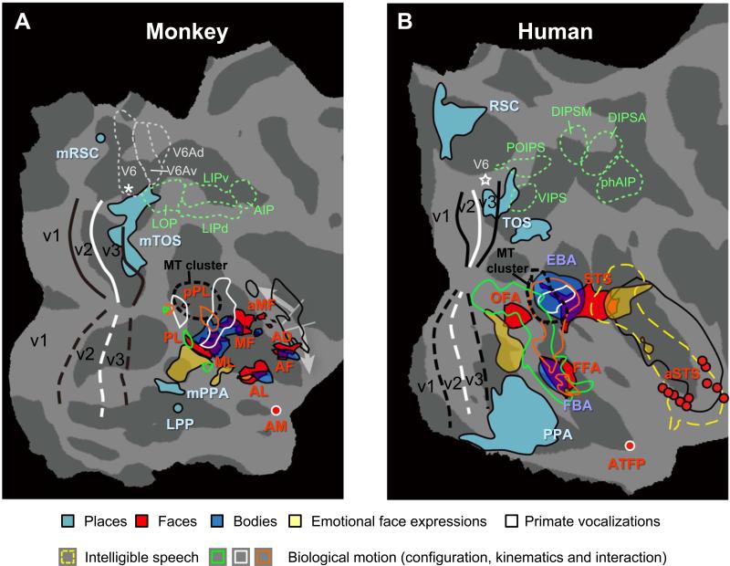
Figure 3.
Comparison of face, place and body patches in monkey and human posterior occipital cortex. (A) Monkey: Activations (real data combined with approximate locations of local maxima) from different studies are projected onto the flattened right hemisphere of F99 atlas. Place-selective patches: real data from Nasr et al. (2011) and approximate data (LPP and mRSC) from Kornblith et al. (2013). Faces-selective regions: real probabilistic data from Janssens et al. (2014); AM: approximate data from Tsao et al. (2008a). Dynamic facial expressions (fear vs. chewing controlled for scrambles): real data from Zhu et al. (2012). Body-selective regions: real group data from Popivanov et al. (2012) (same as in Fig. 1C). Biological motion sensitivity (main effect of configuration and kinematics, as well as the interaction): approximate data from Jastorff et al. (2012). Regions sensitive for primate vocalizations (black outlines): real data from Joly et al. (2012). Retinotopy: real probabilistic data from Janssens et al. (2014). LOP, LIP and AIP from Lewis and Van Essen (2000). V6, V6A and V6Ad are approximate data from Fattori et al. (2009) and Pitzalis et al. (2013). The white arrows indicate regions in the monkey STS which are vastly expanded in the human for auditory processing (including speech).
(B) Human: activations are projected onto flattened right hemisphere of fsaverage atlas. Faces- and body selective regions: approximate probabilistic data of Engell and McCarthy (2013); Dynamic facial expressions: real data from Zhu et al. (2012); ATFP: approximate data from Rajimehr et al. (2009); aSTS: approximate data from Pitcher et al., (2011). Biological motion sensitivity: approximate data from Jastorff and Orban (2009). Regions sensitive for primate vocalizations (black outlines) and intelligible speech (dashed yellow outlines): real data from Joly et al. (2012). Retinotopy: approximate probabilistic data from PALS-B12 atlas; VIPS, POIPS, DIPSM, DIPSA and phAIP: approximate locations from Jastorff et al. (2010). White stars are foveal representations in V6

