

Abstract
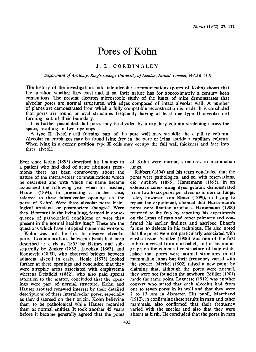
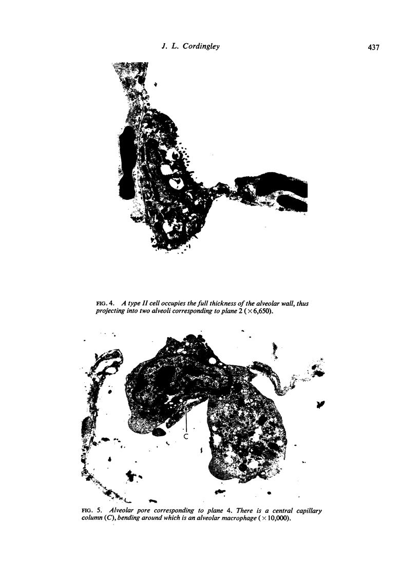
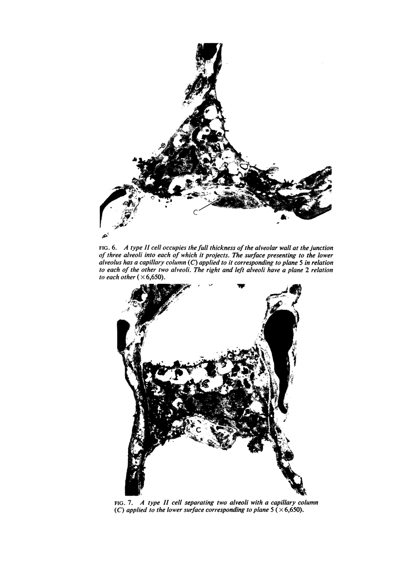
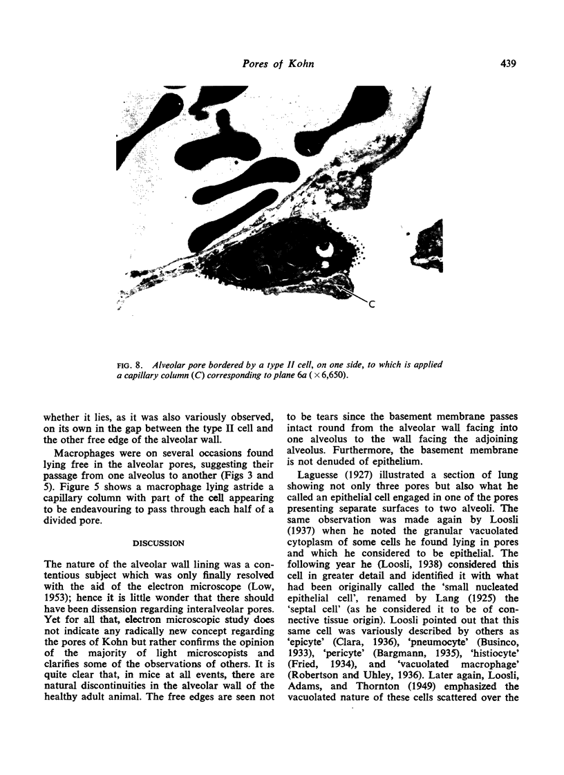
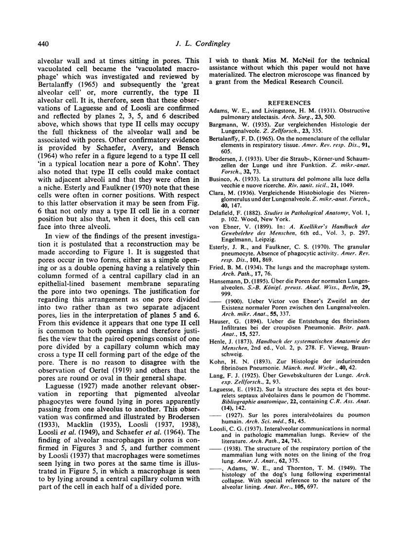
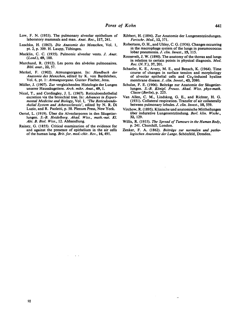
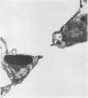
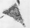
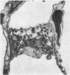
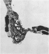
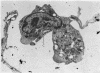
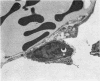
The history of the investigations into interalveolar communications (pores of Kohn) shows that the question whether they exist and, if so, their nature has for approximately a century been contentious. The present electron microscopic study of the lungs of mice demonstrates that alveolar pores are normal structures, with edges composed of intact alveolar wall. A number of planes are demonstrated from which a fully compatible reconstruction is made. It is concluded that pores are round or oval structures frequently having at least one type II alveolar cell forming part of their boundary.
It is further postulated that pores may be divided by a capillary column stretching across the space, resulting in two openings.
A type II alveolar cell forming part of the pore wall may straddle the capillary column. Alveolar macrophages may be found lying free in the pore or lying astride a capillary column. When lying in a corner position type II cells may occupy the full wall thickness and face into three alveoli.
Full text
PDF
Images in this article
Selected References
These references are in PubMed. This may not be the complete list of references from this article.
- BERTALANFFY F. D. ON THE NOMENCLATURE OF THE CELLULAR ELEMENTS IN RESPIRATORY TISSUE. Am Rev Respir Dis. 1965 Apr;91:605–609. doi: 10.1164/arrd.1965.91.4.605. [DOI] [PubMed] [Google Scholar]
- Esterly J. R., Faulkner C. S., 2nd The granular pneumonocyte. Absence of phagocytic activity. Am Rev Respir Dis. 1970 Jun;101(6):869–876. doi: 10.1164/arrd.1970.101.6.869. [DOI] [PubMed] [Google Scholar]
- LOOSLI C. G., ADAMS W. E., THORNTON T. M., Jr The histology of the dog's lung following experimental collapse with special reference to the nature of the alveolar lining. Anat Rec. 1949 Dec;105(4):697–721. doi: 10.1002/ar.1091050406. [DOI] [PubMed] [Google Scholar]
- LOW F. N. The pulmonary alveolar epithelium of laboratory mammals and man. Anat Rec. 1953 Oct;117(2):241–263. doi: 10.1002/ar.1091170208. [DOI] [PubMed] [Google Scholar]
- Macklin C. C. Pulmonic Alveolar Vents. J Anat. 1935 Jan;69(Pt 2):188–192.1. [PMC free article] [PubMed] [Google Scholar]
- Robertson O. H., Uhley C. G. CHANGES OCCURRING IN THE MACROPHAGE SYSTEM OF THE LUNGS IN PNEUMOCOCCUS LOBAR PNEUMONIA. J Clin Invest. 1936 Jan;15(1):115–130. doi: 10.1172/JCI100750. [DOI] [PMC free article] [PubMed] [Google Scholar]
- SCHAEFER K. E., AVERY M. E., BENSCH K. TIME COURSE OF CHANGES IN SURFACE TENSION AND MORPHOLOGY OF ALVEOLAR EPITHELIAL CELLS IN CO2-INDUCED HYALINE MEMBRANE DISEASE. J Clin Invest. 1964 Nov;43:2080–2093. doi: 10.1172/JCI105082. [DOI] [PMC free article] [PubMed] [Google Scholar]
- Van Allen C. M., Lindskog G. E., Richter H. G. COLLATERAL RESPIRATION. TRANSFER OF AIR COLLATERALLY BETWEEN PULMONARY LOBULES. J Clin Invest. 1931 Aug;10(3):559–590. doi: 10.1172/JCI100371. [DOI] [PMC free article] [PubMed] [Google Scholar]