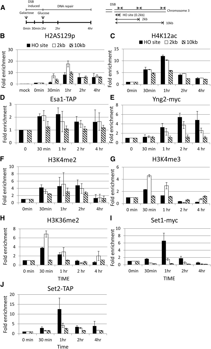
Figure 4.
Histones are specifically modified at the HO DSB site. (A) Schematic of the ChIP experiment. Cells were cultured in SC −Ura −Leu −Trp + 2% glycerol and 1% succinate. The 2% galactose was then added to induce the expression of HO endonuclease at 0-min time point, thus generating a single DSB at the MAT HO locus of chromosome III. One hour later, 2% glucose was added to repress the expression of HO endonuclease, and thus the DSB were repaired. Samples were collected at the indicated time points for the ChIP assay. Specific primers located at HO, 2 kb, and 10 kb from the HO cutting site are indicated as arrows. The ChIP experiments were performed with specific anti-H2A-S129p (B), H4K12ac (C), IgG sepharose beads (to pull down Esa1) (D), myc antibody (to pull down Yng2-myc) (E), H3K4me2 (F), H3K4me3 (G), H3K36me2 (H), myc (to pull down Set1) (I) antibodies, and IgG sepharose beads (to pull down Set2) (J) as indicated.

