

Abstract
Introduction:
Fracture-dislocations of tarsometatarsal joints are rare. Association of these fractures with ankle injuries is very rare; to our knowledge not reported in the literature. Here, we report a rare case of Tarso-metatarsal (Lisfranc) dislocation with bimalleolar fracture with syndesmotic injury.
Case report:
Patient was treated with closed reduction and fixation of Lisfranc injury with combination of screws and k wires and also fixation of bimalleolar and syndesmotic injury. At follow up patient achieved excellent function as assessed by AOFAS (American Orthopaedic Society Foot and Ankle Society Midfoot Score).
Conclusion:
Even though extremely uncommon, early recognition of ankle injuries with uncommon lisfranc fractures & dislocations is important as prompt and simultaneous treatment of both injuries results in excellent clinical outcome.
Keywords: Lisfracn fracture dislocation, ankle fracture
INTRODUCTION:
Injuries to Taro metatarsal joints are rare occurrences, account for 0.2% of all injuries and results of treatment are unsatisfactory [1,2]. These estimates are too low because 20% to 40% of these injuries are overlooked or misdiagnosed as foot sprain or isolated fractures of tarsals or metatarsals at first presentation [2,3]. If overlooked or not treated properly. Lisfranc fracture dislocation frequently results in painful mal-union and impaired function [4].
Here, we report a case of tarso-metatarsal (lisfranc) dislocation with bimalleolar fracture with syndesmotic injury. Purpose of this case is to emphasize importance of ankle injuries in foot fractures, to identify the factors associated with good or poor functional outcome and to explain lack of consensus regarding treatment of this uncommon injury with review of the literature. This combination of injuries has not been reported in the literature to our knowledge.
CASE REPORT:
Forty year old driver had history of road traffic accident with twisting injury to left foot.
Patient was brought to emergency department where Radiograph of the affected foot was done with standard antero posterior, oblique views and lateral views. Ankle Radiograph were done In view of ankle injury. Radiograph of the foot (Fig. 1, 2) showed isfranc dislocation with homolateral type of injury with lateral dislocation of metatarsals. This is the lateral divergent type of injury caused due to violent plantar flexion of forefoot an indirect type of injury [5]. Radiograph of the ankle (Fig. 3, 4) showed transverse fracture of medial malleolus with oblique fracture of fibula above syndesmosis - pronation external rotation injury [6]. There was no associated injury or any neurovascular deficit and patient was stable. Patient was taken to operation theatre after ten hours of injury, closed reduction of lisfranc dislocation done, confirmed under fluoroscopy and fixed with percutaneous 4mm screws to stabilize the medial column. Screws achieving excellent hold in medial and middle column. Lateral column was stabilized by K wires passing from metatarsals to tarsals. (Fig. 5)
Figure 1 and 2.

Pre-operative radiographs of foot showing Lisfranc dislocation
Figure 3, 4.

Pre-operative radiographs of ankle showing bimalleolar fracture
Figure 5.

Immediate post operative radiographs of foot
Open reduction and internal fixation of medial malleolus was done with two 4mm screws and fibula fracture fixed with one third tubular plate. Intraoperative radiograph (Fig. 6) was done showing syndesmotic injury which was confirmed by giving lateral traction with bone hook and looking for displacement of fibula laterally. This was fixed with syndesmotic screw.
Figure 6.
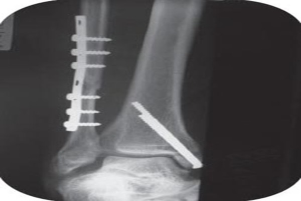
Intra-operative radiographs of ankle showing Syndesmotic injury
(Fig. 7, 8) Patient was given below knee plaster cast. At three weeks follow up, cast was Loosened and one K wire from fourth metatarsal backed out (Fig. 9,10). Reduction was checked again under fluoroscopy and patient was given below knee cast. Partial weight bearing was started at six weeks with gradual full weight bearing. At follow up, distance between plantar surfaces of medial cuneiform and fifth metatarsal were measured on weight bearing lateral radiograph of the foot to rule out flattening of longitudinal arch of foot which was found to be equal [7]. Implant removal was done at four months with removal of syndesmotic screw which backed off at follow up radiograph. AOFAS (American Orthopaedic Society Foot and Ankle Society Midfoot Score) [8] was 90 points (on a scale of 0 to 100 points, with 100 points indicating excellent outcome). At two and half years follow up patient does not have any complaints.
Figure 7 and 8.

Post operative radiographs of bimalleolar fracture with syndesmotic fixation
Figure 9 and 10.

Radiographs at follow up of 3 weeks; K wire backed out
DISCUSSION:
Fractures and dislocations of tarso-metatarsal joint are frequently overlooked or misdiagnosed [3]. This is because of variation in the pattern of injury and clinical presentation. There is lack of knowledge of specific clinical signs and radiological projection such as plantar ecchymosis sign [9], linked toe dislocation and fleck sign at the base of second metatarsal [10]. Road accidents are the most common cause of lisfranc fracture dislocation while twisting injury to the foot is the commonest cause for simple dislocation without fracture [10]. Incomplete reduction of dislocation or redislocation frequently results in permanent disability due to chronic pain, deformity and inability to wear shoes [1,10].
Tarso-metatarsal joint was named after Lisfranc a French surgeon serving in napoleonic army described amputation through that joint [1]. The factors maintaining stability of the normal tarsometatarsal joints must be considered first in order to understand lisfranc fracture dislocations. The capsule of the first metatarso-cuneiform joint is reinforced by dorsal and by plantar ligaments. On the medial side, added support is provided by the insertion of tibialis anterior into the base of the first metatarsal bone and the medial cuneiform bone. There is no interosseous ligament between the bases of the first and second metatarsal bones but there may be an intervening bursa. Bony stability is increased by the interosseous ligaments, the one between the medial cuneiform and the metatarsal being well developed and known as Lisfranc’s ligament [10]. Because of proximal recess of middle cuneiform, second metatarsal is firmly locked in mortice formed by all other metatarsals [5]. Most injuries are produced in plantar flexion leading to dorsal dislocation, this is due to relatively weak toe extensors and weak dorsal ligaments as compared to strong plantar ligaments and fascia [5,10]. Position and shape of tarso metatarsal joints is such that it tends to compress foot in longitudinal axis and bow foot dorsally at tarso metatarsal joint and displace metatarsals laterally once lateral displacement occurs [10].
Previous classification was based on mode of injury classifying lisfranc injury into direct and indirect forces [5,10]. Direct forces may crush the metatarsals and displace them plantarwards while secondary displacements may be lateral and medial. With indirect rotational forces, forefoot must be in the plantar flexion for dislocation to occur. Dorsal aspect of lisfranc joint is unable to resist the forces in contrast to much stronger soft tissue support on the plantar aspect [10]. As per classification described by Quene and Kuss, three patterns of displacement have been described: Homolateral (all metatarsals displaced in coronal plane), Isolated (only one or two metatarsals are displaced in coronal plane) & Divergent (displacement in sagittal and coronal plane). Additional pattern includes complex type which includes interposition of tibialis anterior tendon between medial and middle cuneiform [11]. Radiographs in three planes (Antero posterior, Lateral, 30 deg oblique view) to know the exact anatomy of fracture and to assess the accuracy of reduction. Standard radiographs may not show subtle instability [12] so stress radiographs are must be done to find out gross instability. Careful screening of ankle injuries and injuries to rest of the foot needs to be done. Possibility of vascular injury is to be kept in mind even though rare and uncommon when lateral plantar artery is intact. To reduce the risk of vascular compromise, reduction should be done as early as possible [13].
Lisfranc dislocations are the major injuries associated with high potential for chronic disability so precise anatomical reduction is a must either by closed or open method [1,10,14].
Closed reduction of dislocation is to be done by longitudinal traction in line of foot and checked radiologically for adequacy then it can be fixed preferably with screws for medial columns and K wire for lateral column. Stabilization most commonly done by smooth K wires [1,10,13,14] has high failure rate due to loss of reduction which is not recognized until cast is removed and then it is difficult to restore anatomical reduction again. Also there are chances of infection and migration of K wires. When only K wires are used, the chances of redisplacement are higher due to loss of swelling & bowstring action of strong plantar tendons [1]. So recent literature supports temporary fixation with AO screws for both stable and unstable injuries. Screw fixation for medial and middle columns provide greater biomechanical stability than pinning alone [2,14]. Whereas lateral column can be stabilized by pins as this type of fixation preserves the motion that is normally present in lateral column [2,15] ;as it was done in this case. Stable internal fixation provided by screws permit early and safe weight bearing so stimulate healing and reducing swelling. Closed reduction is unlikely to be successful in cases having severe comminution, large articular fragment and interposition of soft tissues [1] In these cases open reduction should be performed as soon as possible. For open reduction, single or two longitudinal incisions one along lateral border of first metatarsal and second along the long axis of fifth metatarsal can be used to avoid ischemia [1].
Conclusion: Injury to lisfranc joint is one of the rare injuries and association of this injury with ankle injuries is even more rare. But early recognition and fixation of these associated injuries, prompt treatment with accurate reduction and stable internal fixation with screws for medial column and K wires for lateral column for lisfranc fracture dislocation results in good clinical outcome.
CLINICAL MESSAGE:
Lisfracns fracture dislocation may be associated with ankle fractures and simultaneous internal fixation of both the injuries gives good clinical and functional results
Footnotes
Conflict of Interest : NONE
Source of Funding : NONE
REFERENCES
- 1.Hardcastle P, Reschauer R, Kutscha-Lissberg E, Schoffmann W. Injuries to the tarsometatarsal joint. Incidence, classification and treatment. J Bone Joint Surg Br. 1982 Jun;64:349–56. doi: 10.1302/0301-620X.64B3.7096403. [DOI] [PubMed] [Google Scholar]
- 2.Panchbhavi V, Vellurupalli S, Yang J, Anderson C. Screw fixation compared with Suture button fixation of isolated lisfranc ligament injuries. J Bone Joint Surg Am. 2009;91:1143–8. doi: 10.2106/JBJS.H.00162. [DOI] [PubMed] [Google Scholar]
- 3.Ramemelt S, Schneders W, Schikore H, Holch M, Heineck J, Zwipp H. Primary open reduction and fixation compared with delayed corrective arthrodesis in the treatment of tarsometatarsal (Lisfranc) dislocation injuries. J Bone Joint Surg Br. 2008 Nov;11:1499–1506. doi: 10.1302/0301-620X.90B11.20695. [DOI] [PubMed] [Google Scholar]
- 4.Mann R, Prieskorn D, Sobel M. Mid-Tarsal and Tarsometatarsal Arthrodesis for Primary Degenerative Osteoarthrosis or Osteoarthrosis after Trauma J Bone. Joint Surg Am. 1996 Sep;78:1376–85. doi: 10.2106/00004623-199609000-00013. [DOI] [PubMed] [Google Scholar]
- 5.James J, Wiley, Ottawa The Mechanism of Tarso-metatarsal joint injuries. J Bone Joint Surg Br. 1971 Aug;53:474–82. [PubMed] [Google Scholar]
- 6.March J, Saltzman C. fractures in adults by Rockwood & Green's. 5th edition. Philadelphia: Lippincott Williams & Wilkins; Ankle fractures classification. 2009-17. [Google Scholar]
- 7.Faciszewski T, Burks R, Manaster B. Subtle injuries of the Lisfranc joint. J Bone Joint Surg Am. 1990;72:1519–22. [PubMed] [Google Scholar]
- 8.Kitaoka H, Alexander I, Adelaar R, et al. Clinical rating system for the ankle, hindfoot, midffoot, hallux and Lesser toes. Foot Ankle Int. 1994;15:349–53. doi: 10.1177/107110079401500701. [DOI] [PubMed] [Google Scholar]
- 9.Ross G, Cronin R, Hauzenblas J, Juliano P. Plantar ecchymosis sign: A clinical aid to diagnosis of occult lisfranc tarsometatarsal injuries. J Orthop Trauma. 1996;10:119–22. doi: 10.1097/00005131-199602000-00008. [DOI] [PubMed] [Google Scholar]
- 10.Jeffreys T. Lisfranc's Fracture-Dislocation. J Bone Joint Surg Br. Aug. 1963;45:546–51. [PubMed] [Google Scholar]
- 11.Efstathopoulos N, Papachristou G, Agoropoulos Z, Karachalios G, Kokorogiannis K, Vlachou C, Karayiani Al. Open fractures-dislocations of the tarsometatarsal (Lisfranc) joints. Report of three cases. European J of Orthopaedic Surg & Traumatol. 1997 Feb;7(1):41–3. [Google Scholar]
- 12.Norfray J, Geline R, Steinberg R. Subtleties of Lisfranc fracture-dislocations. American Journal of Roentgen. 1981 Dec;:1151–6. doi: 10.2214/ajr.137.6.1151. [DOI] [PubMed] [Google Scholar]
- 13.Willim G. A dangerous type of fracture of foot. J Bone Joint Surg Br. 1951 Nov;33:535–8. doi: 10.1302/0301-620X.33B4.535. [DOI] [PubMed] [Google Scholar]
- 14.Lee C, Birkedal J, Dickerson E, Vieta P, Webb L, Teasdall R. Stabilization of lisfranc injuries; A biomechanical study. Foot Ankle Int. 2004 May;25:365–70. doi: 10.1177/107110070402500515. [DOI] [PubMed] [Google Scholar]
- 15.Desmond E, Chou L. Current concepts review: Lisfranc injuries. Foot Ankle Int. 2006 Aug;27:653–70. doi: 10.1177/107110070602700819. [DOI] [PubMed] [Google Scholar]