Abstract
Negative allosteric modulators (NAMs) of metabotropic glutamate receptor subtype 5 (mGlu5) have potential applications in the treatment of fragile X syndrome, levodopa-induced dyskinesia in Parkinson disease, Alzheimer disease, addiction, and anxiety; however, clinical and preclinical studies raise concerns that complete blockade of mGlu5 and inverse agonist activity of current mGlu5 NAMs contribute to adverse effects that limit the therapeutic use of these compounds. We report the discovery and characterization of a novel mGlu5 NAM, N,N-diethyl-5-((3-fluorophenyl)ethynyl)picolinamide (VU0477573) that binds to the same allosteric site as the prototypical mGlu5 NAM MPEP but displays weak negative cooperativity. Because of this weak cooperativity, VU0477573 acts as a “partial NAM” so that full occupancy of the MPEP site does not completely inhibit maximal effects of mGlu5 agonists on intracellular calcium mobilization, inositol phosphate (IP) accumulation, or inhibition of synaptic transmission at the hippocampal Schaffer collateral-CA1 synapse. Unlike previous mGlu5 NAMs, VU0477573 displays no inverse agonist activity assessed using measures of effects on basal [3H]inositol phosphate (IP) accumulation. VU0477573 acts as a full NAM when measuring effects on mGlu5-mediated extracellular signal-related kinases 1/2 phosphorylation, which may indicate functional bias. VU0477573 exhibits an excellent pharmacokinetic profile and good brain penetration in rodents and provides dose-dependent full mGlu5 occupancy in the central nervous system (CNS) with systemic administration. Interestingly, VU0477573 shows robust efficacy, comparable to the mGlu5 NAM MTEP, in models of anxiolytic activity at doses that provide full CNS occupancy of mGlu5 and demonstrate an excellent CNS occupancy-efficacy relationship. VU0477573 provides an exciting new tool to investigate the efficacy of partial NAMs in animal models.
Introduction
Glutamate signaling in the central nervous system (CNS) is mediated by activation of both ionotropic and metabotropic glutamate receptors. Metabotropic glutamate (mGlu) receptors are G protein–coupled receptors (GPCRs) that belong to the GPCR class C/family 3 group. The mGlu receptor family contains eight members: mGlu1–mGlu8 (Niswender and Conn, 2010). The receptors are grouped according to their structure, signaling partners, and pharmacology. Group 1 receptors (mGlu1, mGlu5) signal through the G protein Gq/11 and mediate inositol phosphate (IP3)/calcium (Ca2+) signal transduction (Abe et al., 1992). Group 1 receptors activate additional signaling pathways, including extracellular signal-related kinases (Mao et al., 2005). The mGlu5 receptors are widely expressed in the CNS and also are expressed in the periphery (Julio-Pieper et al., 2011). In neurons, mGlu5 is primarily expressed postsynaptically, modulating cell excitability and postsynaptic efficacy (Conn and Pin, 1997). Within the CNS, mGlu5 receptor expression levels are high in the hippocampus, nucleus accumbens, striatum, globus pallidus, and substantia nigra pars reticulata (Ferraguti and Shigemoto, 2006). CNS therapeutic indications for which mGlu5 receptors are potential targets include Parkinson disease, fragile X syndrome/autism spectrum disorders, schizophrenia, Alzheimer disease, addiction, depression, anxiety, and pain (Nickols and Conn, 2014). In addition, mGlu5 NAMs have potential in the treatment of gastroesophageal reflux disease (Niswender and Conn, 2010; Zerbib et al., 2011).
The GPCRs contain seven transmembrane regions, with class C GPCRs possessing a large extracellular N-terminal region that contains the endogenous ligand (orthosteric) binding site. Receptor subgroups show abundant sequence homology at the orthosteric binding site, making the synthesis of subtype selective agents challenging. Allosteric ligands bind to a topographically distinct site within the seven transmembrane-spanning domains; this site contains a higher level of sequence diversity between receptor subtypes compared with the orthosteric site (Conn et al., 2009). This sequence diversity allows allosteric ligands to exhibit greater subtype selectivity than can be achieved with orthosteric ligands of mGlu receptors (Christopoulos and Kenakin, 2002; Conn et al., 2009). Allosteric modulators act in concert with the endogenous orthosteric ligand by changing the affinity and/or efficacy of the orthosteric ligand. Alteration of the orthosteric ligand affinity and/or efficacy is defined as cooperativity. Positive allosteric modulators (PAMs) display positive cooperativity and potentiate responses to orthosteric agonists, whereas negative allosteric modulators (NAMs) display negative cooperativity and block orthosteric agonist responses. In addition, neutral allosteric ligands (NALs) bind to the allosteric site and show neutral cooperativity with orthosteric agonists (Christopoulos, 2014). In traditional allosteric models, the cooperativity of allosteric ligands is a saturable effect, with allosteric ligands acting in the presence of the orthosteric agonist, thereby preserving the temporal and spatial activation of the receptor.
The prototypical mGlu5 NAM is MPEP; others include MTEP and fenobam (Gasparini et al., 1999, 2002; Cosford et al., 2003b). Preclinical and clinical studies suggest that mGlu5 NAMs have efficacy in animal models of anxiety disorders (Porter et al., 2005; Swanson et al., 2005; Felts et al., 2013), fragile X syndrome (Bear et al., 2004; Gasparini et al., 2008), Parkinson disease (Marino and Conn, 2002; Marino et al., 2002), Parkinson disease l-DOPA–induced dyskinesia (Dekundy et al., 2006; Mela et al., 2007; Levandis et al., 2008; Rylander et al., 2009, 2010; Morin et al., 2010, 2013), depression (Pecknold et al., 1982; Porter et al., 2005; Li et al., 2006; Chaki et al., 2013; Hughes et al., 2013; Pilc et al., 2013), neuroprotection (Bruno et al., 2000; Bao et al., 2001), addictive disorders (Bird and Lawrence, 2009; Lindsley et al., 2011; Achat-Mendes et al., 2012; Amato et al., 2013), and gastroesophageal reflux disease (Frisby et al., 2005; Jensen et al., 2005). The mGlu5 NAMs have reached or are now in active clinical studies to evaluate clinical efficacy for these disorders (Campbell et al., 2004; McGeehan et al., 2004; Pietraszek et al., 2004; Rodriguez et al., 2010; Zerbib et al., 2011). Despite these encouraging results, both clinical and preclinical studies suggest that mGlu5 NAMs may induce on-target adverse effects, including cognitive and memory impairments and psychotomimetic effects (Campbell et al., 2004; Porter et al., 2005; Rodriguez et al., 2010; Abou Farha et al., 2014), raising the question of whether complete blockade of mGlu5 may contribute to these deleterious effects, thereby limiting the therapeutic utility of mGlu5 NAMs.
The degree of cooperativity of allosteric modulators varies from strong positive cooperativity (PAMs) to strong negative cooperativity (NAMs), with a range of values between these extremes (Conn et al., 2009; Sams et al., 2011; Nickols and Conn, 2014; Gregory et al., 2015). Of note, we and others previously reported discovery of mGlu5 NALs that have no measurable cooperativity with glutamate in the in vitro assays used for compound assessment (O’Brien et al., 2003; Rodriguez et al., 2005; Hammond et al., 2010; Sams et al., 2011; Gregory and Conn, 2015; Gregory et al., 2015). In addition, mGlu5 “partial NAMs” have also been identified that display weak cooperativity, only partially blocking maximal mGlu5-mediated responses at concentrations that fully occupy the allosteric site on mGlu5 (Rodriguez et al., 2005; Felts et al., 2010; Bradley et al., 2011; Gregory et al., 2012). This finding provides a potential breakthrough and raises the possibility that partial NAMs could be developed that provide in vivo efficacy without completely inhibiting mGlu5 function; however, previous mGlu5 partial NAMs had low potencies relative to MPEP and other full NAMs and were not highly optimized for use in in vivo studies (Rodriguez et al., 2005; Gregory et al., 2012). We now report optimization of mGlu5 NAMs belonging to a picolinamide biaryl acetylene series and discovery of VU0477573 as a novel mGlu5 selective partial NAM that displays high affinity and induces a partial inhibition of the maximal response of mGlu5 to glutamate. VU0477573 has an excellent in vivo pharmacokinetic profile and achieves high brain exposure, and systemic administration of VU0477573 provides full CNS occupancy of the receptor in vivo and corresponding efficacy in a model of anxiolytic activity known to be responsive to prototypical mGlu5 NAMs. These results confirm the potential utility of partial GPCR NAMs as a novel approach to reducing receptor signaling and achieving in vivo efficacy without completely eliminating receptor signaling.
Materials and Methods
Reagents
Ammonium formate was purchased from Fisher Scientific (Fair Lawn, NJ). Dulbecco’s modified Eagle’s medium (DMEM), fetal bovine serum (FBS), and antibiotics were obtained from Life Technologies (Carlsbad, CA). MPEP (Gasparini et al., 1999), MTEP (Cosford et al., 2003b), and VU0477573 (Jaeschke et al., 2012) were synthesized inhouse. [3H]myo-inositol was obtained from PerkinElmer (Boston, MA), and l-glutamic acid was obtained from Tocris Bioscience (Ellisville, MO). [3H]-methoxyPEPy (76.3Ci/mmol) was custom synthesized by PerkinElmer Life and Analytical Sciences (Waltham, MA). All other reagents were purchased from Sigma-Aldrich (St. Louis, MO) and were analytical grade.
Cell Culture
Human embryonic kidney cell lines (HEK293) were maintained in complete DMEM supplemented with 10% FBS, 2 mM glutamine, 20 mM HEPES, 0.1 mM nonessential amino acids, 1 mM sodium pyruvate, and antibiotic/antimycotic (Life Technologies), and G418 (500 μg/ml; Mediatech, Manassas, VA) at 37°C in 5% CO2 in a humidified cell incubator. Cell lines used include HEK293A cells expressing low levels of rat mGlu5 (HEK293A-rat-mGlu5-low cells). The cell lines used for selectivity studies are all HEK293A cells. Those used in the calcium release assays included tREX human mGlu1 (induced overnight with 50 ng/ml tetracycline) and the HEK293A-rat-mGlu5-low cells. Additional cell lines used in selectivity studies express G protein inwardly rectifying potassium (GIRK) channels. These include rat mGlu2 GIRK, rat mGlu3 GIRK, rat mGlu4 GIRK, human mGlu6 GIRK, rat mGlu7 GIRK (L-AP4 was used as the agonist in mGlu7 studies; all other selectivity profiling used glutamate as the orthosteric agonist), and rat mGlu8 GIRK (Niswender et al., 2008). The HA-tagged N-terminally truncated rat mGlu5 cell line was used for [3H]IP accumulation studies (Goudet et al., 2004; Noetzel et al., 2013).
Rat Cortical Astrocytes
Rat cortical astrocytes from Lonza (Basel, Switzerland) were received, stored in liquid nitrogen, and thawed according to manufacturer’s instructions. The astrocytes were plated on BD Falcon Primaria dishes in assay growth media (AGM) from Lonza, which consists of the assay basal media plus AGM SingleQuot supplements from Lonza. Approximately 4 hours after plating, the cells were fed with AGM according to the protocol and every 3 days until the cells reached confluent growth. The cells were plated in 96-well poly-d-lysine coated plates in 100μl of the AGM. Each well contained approximately 5 × 10⁴ cells. The following day, the media were supplemented with G5 at a dilution of 1:100 in AGM. On the subsequent day, the fluorescence-based calcium assay was performed using the compound dilutions and conditions described for the HEK293A-rat-mGlu5-low cells.
Fluorescence-Based Calcium Assays
Mobilization of intracellular calcium in response to drug treatment was assessed in HEK293A-rat-mGlu5-low cells. Approximately 24 hours before the assay, cells were plated at a density of 50,000 cells/well in poly-d-lysine-coated clear-bottom, black-walled 96-well plates in assay buffer (DMEM with 10% dialyzed fetal bovine serum, 20 mM HEPES, and 1 mM sodium pyruvate). Cells were grown at 37°C and 5% CO2 overnight. The day of the assay, Fluo4-AM dye (Invitrogen, Carlsbad, CA) was used as a cell-permeant calcium indicator dye to assess receptor-mediated intracellular calcium mobilization on the Flexstation II microplate reader (Molecular Devices, Sunnyvale, CA). Test compounds were diluted into dimethylsulfoxide (DMSO) for a final DMSO concentration of 0.3% at a 3× concentration. Compounds were assayed against an EC80 concentration of glutamate (350–400 nM). Compound alone was added at 20 seconds followed by an EC80 concentration of glutamate added at 80 seconds to detect NAM activity; total read time was 120 seconds. A 5-point smoothing function was applied to the raw calcium fluorescent traces. The basal fluorescence of each well was assessed during the first 20 seconds of the assay. The peak increase in fluorescence over basal was calculated, and the values were normalized to the maximal peak response due to glutamate stimulation (Gregory et al., 2012; Noetzel et al., 2013).
For fold-shift assays, calcium flux was measured using the Functional Drug Screening System (FDSS 6000; Hamamatsu Corporation, Bridgewater, NJ.). Cells were plated at 15,000 cells/well in black-walled poly(d)lysine-coated, 384-well plates (Grenier Bio-One, Monroe, NC) in assay medium. Calcium assay buffer was used in the experiment [Hanks’ balanced saline solution (HBSS), Invitrogen; HBSS containing 2.5 mM probenecid and 20 mM HEPES). On the day of the assay, cells were washed on a BioTek ELx 405 plate washer (Biotek, Winooski, VT), and the cells were incubated with 1 μl Fluo-4 AM prepared as a 2.3 mM stock in DMSO and mixed 1:1 with 10% (w/v) Pluronic F-127 and diluted in assay buffer for 1 hour at 37°C and 5% CO2. Unincorporated dye was removed, cells washed on the BioTek ELx washer, and 20 μl of assay buffer was added to each well. Compounds were diluted to 2× in assay buffer. After 10 minutes, cells were preincubated with test compounds for 5 minutes and stimulated with glutamate for 2 minutes. Data were collected at 0.25 Hz during the preincubation and at 1 Hz during the glutamate addition. Raw data were normalized as follows: based on the initial readings in the well, both cell number and nonuniform illumination/imaging were controlled; signal amplitude using the data point immediately preceding the addition of agonist was subtracted from each point on the trace; and data were normalized to the maximal response for each experiment. Concentration response curves were created using the Prism 5.0 (GraphPad Software Inc., San Diego, CA).
Selectivity Profiling.
mGlu1.
The HEK293A tREX human mGlu1 cell line was used to assess the activity of compounds against mGlu1. Fluorescence-based calcium assays were performed on the FDSS as described already herein for the fold-shift protocol. Cells were plated and induced overnight with 50 ng/ml tetracycline. Screening was performed against a concentration response curve (CRC) of glutamate. Either vehicle or a 10 μM final concentration of compound was added and followed 140 seconds later by a CRC of glutamate.
Group 2 and group 3 mGlus.
To assess the effect of test compounds at the group 2I and group 3 mGlu receptors, a thallium flux assay was used on the FDSS as previously described (Niswender et al., 2008). Briefly, cells were plated at 15,000 cells/well in black-walled poly(d)lysine-coated, 384-well plates (Grenier Bio-One) in assay medium. FluoZin-2 AM dye (Invitrogen) was used as a cell-permeant indicator dye to assess receptor-mediated thallium flux on the FDSS. Screening was performed against a CRC of glutamate or (l-AP4 for mGlu7) diluted in thallium buffer (125 mM NaHCO3, 1 mM MgSO4, 1.8 mM CaSO4, 5 mM glucose, 12 mM Tl2SO4, 10 mM HEPES). Either vehicle or a 10 μM final concentration of compound was added and followed 140 seconds later by a CRC of agonist. Fluorescence was measured using an FDSS 6000, and data were analyzed as described previously (Niswender et al., 2008).
Radioligand Binding
Membranes were prepared from HEK293A-rat-mGlu5-low cells as follows. Cells were placed on ice and washed twice with ice-cold phosphate-buffered saline and harvested by scraping into ice-cold homogenization buffer (50 mM Tris-HCl, 10 mM EDTA, 0.9% NaCl, pH 7.4). Cells were homogenized using a Tekmar TP-18/10S1 homogenizer (Teledyne; Tekmar, Cincinnati, OH) using three 10-second bursts, interspersed with 30-second incubations on ice. The cell fractions were separated by centrifugation at 1000g for 10 minutes at 4°C. The supernatant was subsequently centrifuged at 30,000g for 1 hour at 4°C. The pellet was resuspended in ice-cold binding buffer (50 mM Tris-HCl, 0.9% NaCl, pH 7.4). For competition binding, 50μg/well of membranes were incubated with shaking for 1 hour at room temperature with ∼3nM of [3H]-methoxyPEPy and a range of concentrations of test ligand in binding buffer; 10 μM MPEP was used to determine nonspecific binding. The assay was terminated by rapid filtration through GF/B Unifilter plates (PerkinElmer Life and Analytical Sciences) using a Brandel 96-well plate harvester (Brandel, Inc., Gaithersburg, MD) with three washes using ice-cold binding buffer to separate bound from free radioligand. Plates were dried overnight at room temperature. Microscint20 scintillation fluid was added at 40 μl/well to the plate (PerkinElmer Life and Analytical Sciences), and the radioactivity was counted on a TopCount scintillation counter (PerkinElmer Life and Analytical Sciences) after a 2-hour incubation period at room temperature. Concentration-response curves were generated with a four-parameter logistical equation in GraphPad Prism5.
ERK1/2 Phosphorylation Assay
Receptor-mediated extracellular signal-regulated kinases 1 and 2 (ERK1/2) phosphorylation was assessed using the AlphaScreen SureFire phosphoERK1/2 assay kit (PerkinElmer, Waltham, MA). HEK293A-rat-mGlu5-low cells were plated on poly-d-lysine-coated, clear 96-well plates at a density of 40,000 cells/well, in assay media, 16–24 hours before assay. The next day, media were aspirated and cells washed once with serum/glutamate-free (SF) media by gentle flicking and adding 100 µl/well of SF media followed by gentle flicking again. Then 100 µl/well of SF media was added to the cells and starved for 5 or 6 hours at 37°C before the assay. After the incubation period, 50 µl/well of fresh SF media was added to the cells and equilibrated at room temperature for 15–20 minutes, followed by pretreating the cells, first with 40 µl/well of 2× NAMs (compounds) in SF media for 1 minute and, second, with 10 μl of 10× agonist (glutamate) or FBS (positive control) in SF media for 7 minutes. Immediately after the agonist treatment, the assay was stopped by removal of media by flicking the plate, and 40 µl/well of 1× lysis buffer (from kit) was added to the cells and kept on a shaker for 10 minutes. Cell lysates were stored for 1–4 days at −20°C before carrying out the detection assay using the AlphaScreen SureFire phosphoERK1/2 assay kit. After thawing with shaking for 10 minutes, 4μl of lysate was transferred to a white 384-well plate. (Costar; Corning Life Sciences, Tewksbury, MA). Under dim light, 7 μl of the reaction buffer mix was added to each well. The mixture was incubated at 37°C for 90 minutes in the dark. The AlphaScreen signal was detected using an H4 synergy reader (BioTek Instruments) with standard AlphaScreen settings. The data are expressed as the -fold increase over the basal level of detected phosphoERK1/2.
Inositol Phosphate Accumulation
We used anion-exchange chromatography to assess either [3H]inositol phosphate (IP) accumulation in HEK293A-rat-mGlu5-low cells or to assess the basal level of IP accumulation to determine inverse agonist activity in HEK293 cells stably expressing HA-tagged N-terminally truncated rat mGlu5 (Goudet et al., 2004; Noetzel et al., 2013). Cells were seeded at a density of 100,000 cells/ml per well on poly-d-lysine–coated 24-well plates (Fisher Scientific Co LLC, Suwanee, GA) in the presence of 1µCi of [3H]myo-inositol/well. Cell plates were incubated at 37°C, 5% CO2 for approximately 24 hours for HEK293A-rat-mGlu5-low cells, and for approximately 48 hours for HEK293A cells stably expressing HA-tagged N-terminally truncated rat mGlu5. On the day of the assay, the media were aspirated from the cells, and 1 ml of 10 µM compound (1×) or dimethylsulfoxide (DMSO)-matched vehicle in phosphotidylinositol (PI) assay buffer (10× HBSS, 20 mM HEPES, 4.16 mM NaHCO3, 30 mM LiCl) was added to the cells and incubated at 37°C, 5% CO2 for 1 hour. After compound treatment, buffer was removed from cells and reaction stopped with 1 ml of 10 mM formic acid (prepared fresh) per well. Cells were incubated in formic acid at room temperature for 40 minutes and stored overnight at 4°C. IP accumulation was analyzed by anion-exchange chromatography as follows. Cell lysates were transferred to freshly prepared AG 1-X8 anion-exchange resin-packed (100 g of resin/400 ml of H20) 10-ml poly-prep chromatography columns (Bio-Rad Laboratories, Hercules, CA). The columns were washed with 9 ml of water, 9 ml of 5 mM myo-inositol, and 9 ml of water. [3H]IP was eluted with 8 ml of PI eluent (200 mM ammonium formate, 100 mM formic acid) into 20-ml scintillation vials (RPI, Mt. Prospect, IL). Scintillation fluid (14 ml of Ecoscint Ultra; National Diagnostics, Atlanta, GA) was added to each tube and mixed by vigorous shaking for 30 seconds. The vials were counted on a Packard Instruments Tri-Carb 2900TR Liquid Scintillation Analyzer (PerkinElmer, Waltham, MA). The portion of the protocol including column preparation, washes, and elution averaged approximately 4.5 hours (for 100 columns). Columns were regenerated with PI regeneration solution (1M ammonium formate, 0.1 M formic acid) followed by two water washes. Columns were stored at 4°C and used up to four times.
Extracellular Field Potential Recordings from Brain Slices
All animal studies and experiment procedures were approved by the Vanderbilt University Institutional Animal Care and Use Committee in accordance with the National Institutes of Health’s Guide for the Care and Use of Laboratory Animals. Transverse hippocampal slices were prepared from adult (7–11 weeks old) male C57BL/6 mice (The Jackson Laboratory, Bar Harbor, ME). In brief, mice were anesthetized with isoflurane and decapitated, and the brains were quickly removed from the skull and submerged in oxygenated (95% O2/5% CO2), ice-cold cutting solution (in mM: 210 sucrose, 3 KCl, 1.25 NaH2PO4, 26 NaHCO3, 10 d-glucose, 0.5 CaCl2, 8 MgCl2). Transverse brain slices (400 µm) containing the hippocampus were cut using a Leica VT1200S microtome (Leica Microsystems Inc., Buffalo Grove, IL), incubated in oxygenated artificial cerebrospinal fluid (ACSF) for 30 minutes at 32°C, and then maintained at room temperature for at least 30 minutes until being transferred to a submersion recording chamber. The chamber was continuously perfused with oxygenated ACSF at a flow rate of 2 ml/min. The ACSF contained (in mM): 126 NaCl, 2.5 KCl, 2.0 CaCl2, 1.3 MgSO4, 1.25 NaH2PO4, 26 NaHCO3, and 10 d-glucose. A concentric bipolar-stimulating electrode was placed in the stratum radiatum near the CA3–CA1 border to stimulate the Schaffer collaterals. Recording electrodes were fabricated from borosilicate glass (Sutter Instruments, Novato, CA) using a Narishige micropipette puller (PP-830; Narishige, Japan), filled with ACSF, and placed in the stratum radiatum of area CA1. Extracellular field excitatory postsynaptic potentials (fEPSPs) were evoked every 20 seconds and acquired using a MultiClamp 700B amplifier (Molecular Devices) and pClamp 10.4 software (Molecular Devices). Input-output curves were generated to determine the stimulus intensity that produced 50%–60% of the maximum fEPSP slope before each experiment. This submaximal fEPSP slope was used as a baseline for the remainder of the individual experiment. Stock solutions of mGlu compounds were made using DMSO and diluted to the appropriate concentrations in ACSF with the final DMSO concentration of 0.1%. All drugs were bath applied. Three sequential fEPSP slopes were averaged. The time courses were normalized to the average slope calculated during the predrug period (percent of baseline). Data were analyzed offline using Clampfit 10.4 (Molecular Devices) and GraphPad Prism 5.0 (GraphPad Software), and presented as mean ± S.E.M.
Animals
CD-1 mice (Harlan Laboratories, Indianapolis, IN) were used in pharmacokinetic, in vivo occupancy, and behavioral studies. All animals were group housed (four animals per cage), in polycarbonate cages with corncob bedding in a colony room maintained at 21 ± 2°C with 45% ± 10% relative humidity on a 12-hour light/dark cycle (lights on at 0600 hour Central Standard Time). Standard rodent chow (Purina Mills, Richmond, IN) and water were provided ad libitum. Animals ranged in weight from 30 to 60 g. This study was carried out in accordance with the Institutional Animal Care and Use Committee of Vanderbilt University and the guidelines of the Committee on Care and Use of Laboratory Animal Resources, as adopted and promulgated by the U.S. National Institutes of Health.
In Vivo Pharmacokinetic Study
Compound was formulated in 10% tween 80 in water, and CD-1 male mice (30–40 g) were administered VU0477573 i.p. For the dose-exposure study, mice were dosed 1, 3, 10, and 30 mg/kg of VU0477573 (10 ml/kg, i.p., and samples were obtained at 15 and 45 minutes. For the time-course study, mice were dosed with 10 mg/kg of VU0477573 i.p. and samples obtained at 15, 45, 60, 180, and 360 minutes. At the indicated time points, mice were deeply anesthetized with isoflurane and rapidly decapitated, followed by collection of trunk blood and whole brain. The brains were washed thoroughly in cold phosphate-buffered saline and frozen immediately on dry ice. Plasma was separated by centrifugation (4000 relative centrifugal force (rcf), 4°C) and stored at −80°C until analysis. On the day of analysis, frozen whole mouse brains were weighed and diluted with 1:3 (w/w) parts of 70:30 isopropanol:water. The mixture was then subjected to mechanical homogenization using a Mini-Beadbeater and 1.0-mm zirconia/silica beads (BioSpec Products, Bartlesville, OK) followed by centrifugation. The sample extraction of plasma (20 μl) or brain homogenate (20 μl) was performed by a method based on protein precipitation using three volumes of acetonitrile containing an internal standard (50 ng/ml carbamazepine). The samples were centrifuged (3000 rcf, 5 minutes), and supernatants were transferred and diluted 1:1 (supernatant: water) into a new 96-well plate, which was then sealed in preparation for liquid chromatography-tandem mass spectrometry analysis.
Liquid Chromatography-Tandem Mass Spectrometry Bioanalysis of Samples from In Vivo Studies.
The in vivo exposure study was performed as a satellite study to the marble-burying behavioral assay. All in vivo samples were analyzed via electrospray ionization on an AB Sciex API-5500 QTrap (Foster City, CA) triple-quadrupole instrument that was coupled with Shimadzu LC-20AD pumps (Columbia, MD) and a Leap Technologies CTC PAL autosampler (Carrboro, NC). Analytes were separated by gradient elution using a Fortis C18 3.0 × 50 mm, 3-µm column (Fortis Technologies Ltd, Cheshire, UK) thermostated at 40°C. High-performance liquid chromatography mobile-phase A was 0.1% formic acid in water; mobile phase B was 0.1% formic acid in acetonitrile. The gradient started at 10% B after a 0.2-minute hold and was linearly increased to 90% B over 0.8 minute, held at 90% B for 0.5 minute, and returned to 10% B in 0.1 minute followed by a re-equilibration (0.9 minute). The total run time was 2.5 minutes, and the high-performance liquid chromatography flow rate was 0.5 ml/min. The source temperature was set at 500°C, and mass spectral analyses were performed using multiple reaction monitoring (MRM), with transitions specific for each compound using a Turbo-Ionspray source in positive ionization mode (5.0-kV spray voltage). The calibration curves were constructed, and linear response was obtained in the range of 5–10,000 ng/ml by spiking known amounts of VU0477573 in blank plasma. All data were analyzed using AB Sciex Analyst software v1.5.1.
Marble-Burying Assay
Harlan CD-1 male mice weighing between 30 and 35 g were administered drug or vehicle (10% Twee 80) i.p. 15 minutes before start of the assay. Test sessions were performed between 10:00 AM and 4:00 PM. All dose groups consisted of five to eight mice. Eight small Plexiglass cages (32 × 17 × 14 cm) were arranged in two rows of four cages on top of a large round table. Mice were transported from the colony room to the testing room and were allowed to habituate for 30 minutes. Mice were pretreated with a dose of MTEP or VU0477573 15 minutes before initiation of the assay. Mice were treated with increasing doses of VU0477573 compared with vehicle alone and 15 mg/kg MTEP. The mice were placed individually into cages in which 12 black glass marbles 14 mm in diameter were evenly spaced: 6.4 cm vertically and 4.25 cm horizontally from each other and from the walls of the cage. The marbles were placed on top of 2.5 cm of Diamond Soft Bedding (Harlan Teklad, Madison, WI). The compound and comparator were evaluated in a counterbalanced design, in which all doses of compounds were tested in each session. Mice receiving the same dose were placed in cages on opposite sides of the table to control for context and lighting variations. Perforated clear plastic lids were placed on top of each cage. The amount of marble burying was recorded over a 30-minute interval. The mice were removed from the cages, and the number of marbles buried was counted. A marble was considered buried if greater than two thirds of the marble was covered by bedding. The sessions were videotaped using a Sony camcorder equipped with a Sony wide-angle lens mounted on a 1.5m tripod (Sony, Tokyo, Japan). Dose-response studies were analyzed by a between-group analysis of variance. If there was a main effect of the dose, each dose group was compared with the vehicle control group using a Dunnett’s comparison. The calculations were performed using JMP Pro 10 (SAS Institute, Cary, NC) statistical software and graphed using SigmaPlot 12.0 (Systat Software, San Jose, CA).
In Vivo Occupancy Study
The mGlu5 partial NAM VU0477573 was evaluated for receptor occupancy using the method outlined in detail by Anderson et al. (2003). Briefly, CD-1 mice (40–60 g) were administered VU0477573 (30 minutes, 10% Tween 80, i.p., 10 ml/kg). Each treatment group included 6–12 animals, and the compound pretreatment time was 30 minutes. One minute before tissue collection, [3H]-methoxy-PEPy was administered (30 μCi/kg; 1 ml/kg injection volume in isotonic saline) through a lateral tail vein. Animals were then euthanized, and bilateral hippocampus and cerebellum were rapidly dissected on a cooled dissecting tray. The bilateral hippocampus and cerebellum were immediately weighed and homogenized in 10 volumes of ice-cold buffer (10 mM potassium phosphate, 100 mM KCl, pH 7.4) using a Polytron. Homogenates (400 μl) were then filtered over GF/B membrane filters (Whatman; GE Life Sciences, Pittsburgh, PA) and washed twice with 5 ml of ice-cold homogenization buffer to separate membrane bound from free radioactivity. Filters were then counted for radioactivity using a Beckman counter. The hippocampus was used since it is a region with high density of mGlu5 receptors; cerebellum was used as a reference region because it has a low density of mGlu5 receptors. Nonspecific in vivo binding of [3H]-methoxy-PEPy was estimated by measuring radioactivity in washed filters following administration of a saturating dose of MTEP (50 mg/kg i.p.).
Results
Discovery of the mGlu5 Partial NAM VU0477573.
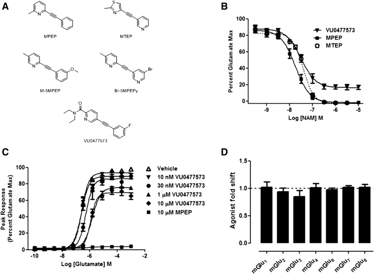
Our group has previously reported the discovery of mGlu5 NAMs that show weak negative cooperativity, including both Br-MPEPy and M-5-methyl-2-phenylethynyl-pyridine (5MPEP) (Rodriguez et al., 2005). In addition, compounds with a similar structure, including 5MPEP and ML353, act as an NALs at mGlu5 (Rodriguez et al., 2005; Gregory and Conn, 2015). We searched our available mGlu5 allosteric antagonists to identify novel compounds with weak negative cooperativity. In screening compounds within the picolinamide acetylene class series (Stauffer, 2011; Bridges et al., 2013; Turlington et al., 2013; Gregory et al., 2015), we noted that the mGlu5 NAM VU0477573 (C18,H17,FN2O; molecular weight = 296.34) showed limited negative cooperativity in inhibiting the glutamate EC80-induced intracellular calcium release in HEK293A cells stably expressing rat mGlu5 (HEK293A-rat-mGlu5-low). The structure of VU0477573 is shown in comparison with the prototypical biaryl acetylene scaffold of the mGlu5 NAMs MPEP and MTEP (Fig. 1A) (Gasparini et al., 1999, 2001; Cosford et al., 2003a,b).
Fig. 1.
Discovery of the partial mGlu5 NAM VU0477573. (A) Chemical structures of mGlu5 negative allosteric modulators: MPEP, MTEP, 5-MPEP, Br-MPEPy, and VU0477573. (B) VU0477573 (black inverted triangles) shows dose-dependent partial NAM activity in glutamate-stimulated intracellular calcium release in HEK293A-rat-mGlu5-low cells in comparison with MPEP (black squares) and MTEP (open circles). (C) Increasing concentrations of VU0477573 (10 nM inverted black triangles, 30 nM black circles, 1 μM black triangles, 10 μM black diamonds) induce a rightward shift in the potency and decreased maximal response to glutamate-stimulated intracellular calcium release in HEK293A-rat-mGlu5-low cells compared with MPEP (10 μM black squares) and vehicle (open triangles). Data are plotted as a percentage of the maximal response to glutamate and error shown as S.E.M. with n = 3 experiments, points performed with two to four replicates. (D) VU0477573 is selective for the mGlu5 receptor subtype. HEK293 cells stably expressing each mGlu subtypes were assessed for either intracellular calcium or thallium flux and the leftward or rightward fold shift of the agonist concentration-response curve was calculated. Data represent the mean ± S.D. of two individual experiments with four to six replicates each.
VU0477573 shows approximately 80% inhibition of the response to an EC80 concentration of glutamate in vitro in inducing intracellular calcium mobilization, whereas MPEP and MTEP completely inhibit the response to an EC80 glutamate concentration (Fig. 1B; Table 1). Therefore, the percent glutamate maximal response is reduced to approximately 20% by VU0477573. The high potency of the compound (32 nM) is similar to that of both MPEP (13 nM) and MTEP (52 nM). To test the mode of action of VU0477573, we examined the effect of a fixed concentration of compound in HEK293A-rat-mGlu5-low cells in the presence of increasing concentrations of glutamate. In the presence of increasing concentrations of VU0477573, the glutamate concentration-response curve shifts to the right along with a decrease in the maximal effect of glutamate that reaches a limit, consistent with a noncompetitive allosteric antagonist (Fig. 1C). Even at a concentration of 10 μM, a concentration approximately 300-fold over the in vitro IC50, VU0477573 did not result in full blockade of the glutamate response compared with treatment with 10 μM MPEP.
TABLE 1.
VU0477573 in vitro activity on rat mGlu5 in functional assays and radioligand binding
| Compound | Ca assay % Glut Max | Ca assay pIC50 | Ca assay IC50 | Binding pKi | Binding Ki |
|---|---|---|---|---|---|
| VU0477573a | ±S.E.M. | ±S.E.M. | nM | ±S.E.M. | nM |
| 16.43 ± 6.20 | 7.49 ± 0.16 | 32 | 7.85 ± 0.06 | 14 | |
| MPEP | −1.56 ± 0.89 | 7.87 ± 0.23 | 13 | 8.19 ± 0.03 | 6 |
| MTEP | −3.11 ± 1.26 | 7.28 ± 0.15 | 52 | 7.65 ± 0.03 | 22 |
The operational model of allosterism parameters for negative allosteric modulation of glutamate-mediated intracellular calcium mobilization in HEK293A-rat-mGlu5-low cells by VU0477573 are logτA = 0.57 ± 0.13, pKB = 7.73 ± 0.10, logβ = -0.44 ± 0.10.
The percent inhibition of a weakly cooperative NAM depends on the concentration of orthosteric agonist used in the assay. In the fold-shift assay (Fig. 1C), 10 μM VU0477573 inhibits an approximate EC90–EC95 concentration of glutamate (1 µM) by approximately 50% and completely inhibits the response to an EC50-60 concentration of glutamate (0.3 µM) (Fig. 1C). In the CRC assay, which uses an EC80 value of glutamate, we see an 80% inhibition by VU0477573 of the EC80 response (Fig. 1B). Therefore, the percent glutamate maximal response is reduced to approximately 20% by VU0477573. In the NAM fold-shift assay, an EC80 of glutamate would be between 0.3 and 1 µM. Therefore, good correlation has been observed between the NAM CRC (Fig. 1B) and fold-shift assays (Fig. 1C) for the partial inhibition by VU0477573 of glutamate-mediated intracellular calcium release. The failure of VU0477573 to fully block the glutamate response is consistent with weak, saturable negative cooperativity, or “partial NAM” activity, for mGlu5 in this assay.
To be useful as an in vivo tool for mGlu5 behavioral assays, compounds need to show mGlu5 subtype selectivity among the metabotropic glutamate receptors. We assessed the selectivity profile of VU0477573 using stable cell lines individually expressing the other metabotropic glutamate receptor subtypes. We examined the effect of 10 μM of VU0477573 against an agonist concentration-response curve. Individual cell lines were assessed for either glutamate—or, for mGlu7, l-AP4—mediated intracellular calcium release (mGlu1) or thallium flux (mGlu2, 3, 4, 6, 7, 8), as indicated. We observed that VU0477573 shows no activity against the other mGlu receptor subtypes, indicating that it is functionally selective for mGlu5 (Fig. 1D).
VU0477573 Binds to the MPEP Site.
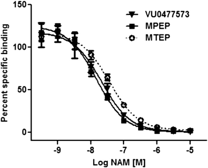
Most mGlu5 allosteric modulators discovered to date bind to the MPEP site of mGlu5. Evidence that allosteric modulators are able to bind to allosteric sites distinct from the MPEP site is increasing. A small subset of mGlu5 PAMs, including CPPHA (O’Brien et al., 2004; Zhao et al., 2007; Chen et al., 2008), VU0357121 (Hammond et al., 2010), VU0028316 (Rodriguez et al., 2010), and NCFP (Zhao et al., 2007; Noetzel et al., 2013), interact noncompetitively with the MPEP site. In addition, the mGlu5 partial NAM VU0029251 reduced maximal [3H]-methoxyPEPy binding by only approximately 50% (Rodriguez et al., 2010). To determine whether VU0477573 binds to the prototypical MPEP allosteric binding site, we performed inhibition binding studies. Using the allosteric antagonist [3H]-methoxyPEPy, known to bind to the MPEP site, we demonstrate that VU0477573 fully displaces the radioligand, consistent with a competitive interaction with the MPEP site (Fig. 2A; Table 1).
Fig. 2.
VU0477573 binds to the MPEP site VU0477573 fully competes with [3H]-methoxyPEPy binding to the MPEP site. Membranes from HEK293A-rat-mGlu5-low cells were incubated with increasing concentrations of VU0477573 (black inverted triangles), MPEP (black squares), or MTEP (open circles) in the presence of 3 nM of [3H]methoxyPEPy. Data represent the mean ± S.E.M. of three individual experiments performed in duplicate. Data are plotted as the percentage of the total [3H]methoxyPEPy binding.
VU0477573 Shows Partial NAM Activity at mGlu5 in Measures of IP Accumulation.
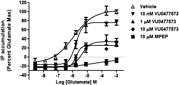
To assess the negative allosteric activity of VU0477573 in an endpoint assay, where the duration of incubation with agonist allows the agonist to come to equilibrium, we examined glutamate-induced IP accumulation in the HEK293A-rat-mGlu5-low cells. Similar to intracellular calcium release, increasing concentrations of VU0477573 caused the glutamate CRC to shift to the right along with a decrease in the maximal effect of glutamate. We again observed that this effect reached a limit, consistent with VU0477573 acting as a noncompetitive allosteric antagonist (Fig. 3). Even at a maximal concentration of 10 μM, VU0477573 did not result in full blockade of the glutamate-induced IP accumulation compared with treatment with 10 μM MPEP. This assay provides additional supportive data that VU0477573 does not fully block the maximal glutamate response in multiple in vitro assays of receptor activity, consistent with weak negative cooperativity, or “partial NAM” activity, for mGlu5.
Fig. 3.
VU0477573 shows partial mGlu5 NAM activity in [3H]IP accumulation. Increasing concentrations of VU0477473 (10 nM black inverted triangles, 1 μM black triangles, 10 μM black diamonds) show a dose-dependent rightward shift in the potency and decreased maximal response to glutamate in HEK293A-rat-mGlu5-low cells as related to MPEP (10 μM black squares) and vehicle (clear triangles). Data are plotted as a percentage of the maximal response to glutamate and error shown as S.D. with n = 3 experiments, with data points performed in duplicate.
VU0477573 is an mGlu5 Partial NAM in Rat Cortical Astrocytes.
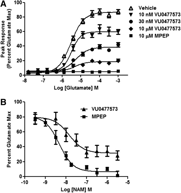
To verify that the partial negative allosteric activity observed in HEK293A-rat-mGlu5-low cells is also observed in a native system, we evaluated the activity of VU0477573 in rat cortical astrocytes. Cortical astrocytes endogenously express mGlu5 without the expression of other mGlu subtypes and serve as an excellent in vitro native experimental system (Peavy et al., 2001, 2002). In addition, the astrocyte level of mGlu5 expression is similar to that of HEK293A-rat-mGlu5-low cells (Noetzel et al., 2012). We first examined the effect of a fixed concentration of VU0477573 in rat cortical astrocytes (Lonza) in the presence of increasing concentrations of glutamate. The compound shows a rightward shift of the glutamate concentration response curves and a decrease in the maximal effect of glutamate, consistent with a noncompetitive allosteric antagonist (Fig. 4A). As in HEK293A-rat-mGlu5-low cells, the 10-μM maximal concentration of VU0477573 did not fully block the glutamate response compared with 10 μM MPEP, consistent with weak negative cooperativity. The compound also shows concentration-dependent, but saturable, inhibition of intracellular calcium release in the presence of an EC80 concentration of glutamate. VU0477573 inhibits the intracellular calcium release by approximately 70% of the glutamate maximal response in rat cortical astrocytes compared with MPEP as a control (Fig. 4B). Therefore, the percent glutamate maximal response is reduced to approximately 30% by VU0477573. The pIC50 for VU0477573 is 7.90 ± 0.18 (13 nM) and for MPEP is 8.26 ± 0.33 (5.5 nM) in astrocytes. The percent glutamate maximal response is 31.5 ± 4.7 for VU0477573 and 6.5 ± 2.9 for MPEP. Comparing the activity of VU0477573 in the NAM CRC assay (Fig. 4B), which uses an EC80 value of glutamate, and the fold-shift assay (Fig. 4A). The maximal effect of 10 μM VU0477573 in the fold-shift assay (Fig. 4A) at 50 μM glutamate is approximately 80% and at 1 μM glutamate is 100% inhibition (Fig. 4A). The EC80 of glutamate for the fold-shift assay is approximately 25 μM (Fig. 4A), and 10 μM VU0477573 demonstrated approximately 80% inhibition (Fig. 4A). This value correlates well with the maximal inhibition observed in the NAM CRC assay (Fig. 4B) in rat cortical astrocytes. Again, consistent with the results observed in HEK293A-rat-mGlu5-low cells, VU0477573 also shows weak negative cooperativity, or partial NAM activity, for mGlu5 in this native in vitro system.
Fig. 4.
VU0477573 shows partial mGlu5 NAM activity in rat cortical astrocytes. (A) Increasing concentrations of VU0477473 (10 nM inverted black triangles, 30 nM black circles, 10 μM black diamonds) show a dose-dependent rightward shift in the potency and decreased maximal response to glutamate in rat cortical astrocytes as related to MPEP (10 μM black squares) and vehicle (clear triangles). Data are plotted as a percentage of the maximal response to glutamate and error shown as S.E.M. with n = 3 or 4 experiments, with two to four replicates per experiment. (B) VU0477573 (triangles) shows dose-dependent partial NAM activity in agonist-stimulated intracellular calcium release in rat cortical astrocytes compared with MPEP (squares). Data are plotted as a percentage of the maximal response to glutamate and error shown as S.E.M. with n = 6 experiments, with two to four replicates per experiment.
VU0477573 Shows Partial Blockade of DHPG-Induced Inhibition of Excitatory Transmission at SC-CA1 Synapses.
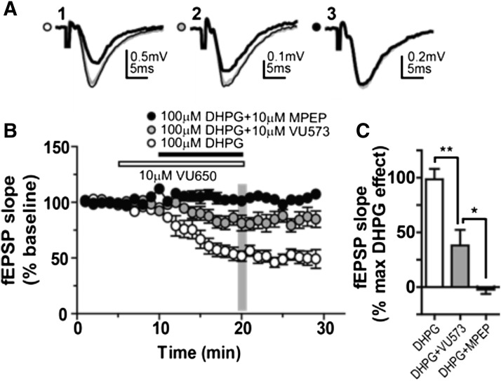
To determine whether VU0477573 has partial NAM activity at mGlu5 in a native system, we examined DHPG-induced inhibition of synaptic transmission at the Schaffer collateral (SC)–CA1 synapse in mouse hippocampal brain slice preparation. Since both mGlu1 and mGlu5 have been shown to play a role in modulation of glutamatergic transmission at SC-CA1 synapses (Huber et al., 2001; Mannaioni et al., 2001; Faas et al., 2002), we performed the experiments in the presence of the known mGlu1 NAM VU0469650 (Lovell et al., 2013) to block mGlu1 activity. In the presence of VU0469650 pretreatment, 100μM DHPG inhibited the extracellular field excitatory postsynaptic potential (fEPSP) slope (Fig. 5, A and B). The addition of 10 μM VU0477573 partially blocked this response, whereas the addition of 10 μM MPEP fully blocked the response (Fig. 5, A and B). The effects of VU0477573 and MPEP on DHPG-induced inhibition were statistically significant at the 20-minute time point, with VU0477573 showing partial activity, blocking the DHPG-mediated fEPSP slope by approximately 40% compared with full blockade by MPEP (Fig. 5C). Therefore, as in the assays using mGlu5 expressing cell lines described here, VU0477573 shows partial activity, compared with full blockade induced by MPEP, of DHPG-mediated inhibition of glutamatergic transmission at the SC-CA1 synapse in mice.
Fig. 5.
Effects of VU0477573 and MPEP on DHPG-induced inhibition of glutamatergic transmission at SC-CA1 synapses. (A) Representative traces of fEPSPs in control (thin black traces) and after applications of mGlu1 NAM VU0469650 (10 µM, gray traces) followed by combination of VU0469650 and 100 µM DHPG, VU0469650 and 10 µM VU0477573, and VU0469650 and 10 µM MPEP (thick black traces in A1, A2, and A3, respectively). (B) Average time courses of effects of VU0469650 followed by combination of VU0469650 with DHPG (n = 6), VU0477573 (n = 9) or MPEP (n = 8) on the slope of fEPSPs at SC-CA1 synapses. The gray vertical bar indicates the time point at which the data were taken to make statistical comparisons. (C) Summary bar graph for the data taken from the time point indicated by the gray vertical bar in (B) and presented as the percentage of maximal DHPG effect, demonstrating that VU0477573 partially blocked the DHPG effect to 38.1 ± 14.2% of maximum DHPG response, whereas MPEP completely blocked the DHPG effect to -2.3 ± 3.8% of maximum DHPG response. One-way analysis of variance revealed significant differences between groups [F(2,20) = 19.39, P < 0.0001]. The post-hoc Bonferroni’s test showed significant differences between DHPG+VU0469650 and DHPG+VU0469650+VU0477573 groups (**P < 0.01) and between DHPG+VU0469650+VU0477573 and DHPG+VU0469650+MPEP groups (*P < 0.05).
VU0477573 Fully Inhibits Glutamate-Induced Extracellular Signal-Related Kinase 1/2 Phosphorylation.
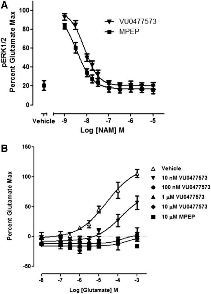
Allosteric ligands have the capacity to show differential efficacy in downstream signaling pathways. This property has several labels, including functional selectivity, biased agonism, or stimulus trafficking (Conn et al., 2009; Kenakin, 2011). mGlu5 allosteric ligands are known to show functional bias, with varying levels of efficacy in different signaling pathways (Zhang et al., 2005). Numerous examples exist showing functional selectivity among allosteric modulators for other GPCRs (Maj et al., 2003; Sachpatzidis et al., 2003; Wei et al., 2003; Mathiesen et al., 2005; Sheffler and Conn, 2008; Kenakin and Miller, 2010; Niswender et al., 2010; Digby et al., 2012; Noetzel et al., 2013). We examined the effect of VU0477573 on glutamate-stimulated extracellular signal-related kinase 1 and 2 phosphorylation (phosphoERK1/2) in HEK293A-rat-mGlu5-low cells using the ERK Surefire Assay Kit. VU0477573 induces full inhibition of glutamate-mediated phosphoERK1/2 phosphorylation, similar to MPEP (Fig. 6A), returning the levels of phosphoERK1/2 to basal levels comparable to vehicle-treated cells. The pIC50 is 8.13 ± 0.70 (7.41 nM) for VU0477573 compared with 8.59 ± 0.31 (2.57 nM) for MPEP. In addition, VU0477573 induces a rightward shift of the glutamate CRC and full inhibition of the maximal effect of glutamate, consistent with a full noncompetitive allosteric antagonist (Fig. 6B).
Fig. 6.
VU0477573 fully inhibits glutamate-mediated ERK1/2 phosphorylation. (A) VU0477573 (inverted triangles) shows full dose-dependent inhibition of glutamate-mediated ERK1/2 phosphorylation to basal levels comparable to vehicle (left black square) and MPEP (squares) in HEK293A-rat-mGlu5-low cells. The concentration response curve represents the mean ± S.E.M from four independent experiments with four replicates per condition. (B) Increasing concentrations of VU0477573 (10 nM black inverted triangles, 100 nM black circles, 1 μM black triangles, 10 μM black diamonds) induce a rightward shift in the potency and decreased maximal response to glutamate-stimulated intracellular calcium release in HEK293A-rat-mGlu5-low cells in glutamate-mediated ERK1/2 phosphorylation compared with MPEP (black squares) and vehicle (clear triangles). Data are plotted as a percentage of the maximal response to glutamate and error shown as S.E.M. with n = 5 experiments, with three to six replicates per experiment.
These data may suggest that VU0477573 has differential cooperativity with glutamate, depending on the measure of receptor function. As reported previously, however, mGlu5 is coupled more weakly to phosphoERK1/2 responses in HEK293A-rat-mGlu5-low cells relative to calcium mobilization, such that some NAMs with lower cooperativity in calcium assays fully inhibit glutamate-mediated phosphoERK1/2 (Gregory et al., 2012). To compare these responses, we used the operational model of allosterism (Leach et al., 2007) to estimate VU0477573 affinity (pKB) and cooperativity (logβ) values (Table 1) from the shift in glutamate concentration-response curves for intracellular calcium mobilization (Fig. 1B). In this analysis, we assumed that affinity modulation was neutral and the pKA was held constant as validated previously (Gregory et al., 2012). We then used the values obtained from the operational analysis of the Ca2+ mobilization data for VU0477573 to simulate the phosphoERK1/2 data (Supplemental Fig. 1). The logτA was constrained at −0.36, according to previously published results reflecting the decreased coupling efficiency of glutamate for phosphoERK1/2 compared with calcium mobilization in this cell line (Gregory et al., 2012). If we assume that VU0477573 has the same apparent affinity and cooperativity with glutamate between Ca2+ assays and phosphoERK1/2, then the simulation predicts that the maximal glutamate response for phosphoERK1/2 will be fully inhibited (Supplemental Fig. 1). In support of this assumption, simulation of the VU0477573 EC80 inhibition curves in both Ca2+ and phosphoERK1/2 assays predicts that VU0477573 will be more potent in phosphoERK1/2 than Ca2+, as we observed. Therefore, the full blockade of glutamate observed in phosphoERK1/2 is most likely due to the low stimulus response coupling of the system rather than biased cooperativity of VU0477573 between these two different endpoints. Fitting the fold-shift data (Fig. 6B, expressed as -fold over basal) to the operational model of allosterism, the apparent pKB is 6.88 ± 0.10 (with the constraints of logτB = −100, logKA = −6.155, log α = 0, log β = −100, Em = 9.1 for FBS maximal response and basal = 1.0)
VU0477573 Does Not Show Inverse Agonist Activity in a PI Hydrolysis Assay.
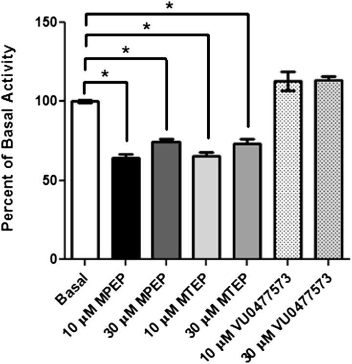
Ligands that bind to GPCRs have the potential to show a range of modes of action, including full agonism, antagonism, full inverse agonism, and neutral interactions that do not show functional effects. The mGlu5 NAMs MPEP and MTEP act as inverse agonists, decreasing the basal level of inositol phosphate accumulation (Gasparini et al., 1999; Roppe et al., 2004; Lea and Faden, 2006). Fenobam, the atypical anxiolytic and mGlu5 NAM, also exhibits inverse agonist properties (Porter et al., 2005). The inverse agonist activity seen with mGlu5 allosteric ligands such as MPEP has been proposed to correlate with the adverse side effects associated with mGlu5 NAMs (Gasparini et al., 1999; Roppe et al., 2004; Porter et al., 2005; Lea and Faden, 2006; Rodriguez et al., 2010). Examination of mGlu5 NAM candidates for inverse agonist activity may prove important as these agents are further developed and characterized. When expressed in heterologous systems, ligand-independent signal transduction is correlated with an increased level of heterologous GPCR expression (Tiberi and Caron, 1994). To assess inverse agonist activity of VU0477573, we used an [3H]IP accumulation assay and HEK293A cells stably expressing an HA-tagged N-terminally truncated rat mGlu5 (Noetzel et al., 2013). This receptor lacks the extracellular N-terminal orthosteric agonist binding domain and is unresponsive to glutamate stimulation. Therefore, the accumulated [3H]IP in this assay using this cell line represents constitutive mGlu5 receptor activity and is not complicated by the presence of glutamate. These studies reveal that VU0477573 (10 μM and 30 μM) does not show inverse agonist activity compared with the prototypical mGlu5 NAMs MPEP and MTEP at 10 μM and 30 μM concentrations (Fig. 7).
Fig. 7.
VU0477573 does not show inverse agonist activity in [3H]inositol phosphate accumulation. The basal level of [3H]IP accumulation was determined as a measure of inverse agonist activity in HEK293A cells stably expressing HA-tagged N-terminally truncated mGlu5. The bar graph represents the mean ± S.E.M from three to five independent experiments performed with three to six replicates per treatment, expressed as a percentage of the basal [3H]IP accumulation. *P < 0.0001 versus vehicle (one-way analysis of variance followed by Bonferroni’s multiple comparison test).
VU0477573 Demonstrates Excellent Pharmacokinetic Properties.
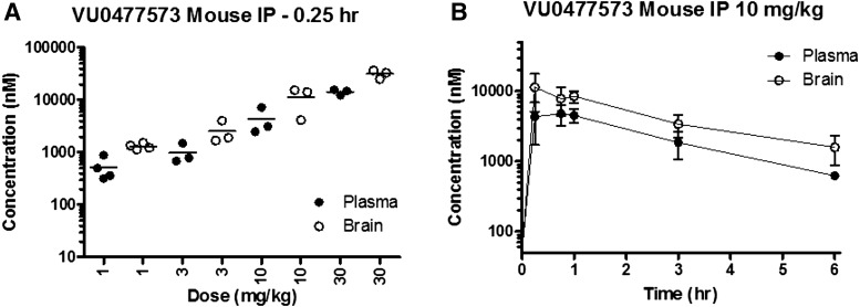
To determine whether VU0477573 achieved the brain exposure necessary for in vivo behavioral assays, we first performed a dose-response exposure study in mice. VU0477573 was well solubilized in the 10% Tween80/90% water vehicle used for these studies. The clogP value for the compound is 3.35, which was calculated using ChemBioDraw Ultra v. 14.0 (Cambridge Software Corporation, Cambridge, UK). We performed in vivo pharmacokinetic studies of VU0477573 in male CD-1 mice after i.p. administration of 1, 3, 10, and 30 mg/kg. We obtained blood and brain samples at both 15- and 45-minute time points (Fig. 6A; Table 2). VU0477573 showed excellent brain penetrance, with a good correlation between the unbound brain concentration (43.7 nM at a 3mg/kg dose at 15 minutes postdosing) and efficacy in the intracellular calcium release assay (IC50 = 32 nM). At the 15-minute exposure time point, VU0477573 showed an approximately linear increase in total brain and plasma exposure with increasing dose in CD-1 mice (Fig. 8A). The unbound brain-to-plasma concentration ratio (Kp uu) is 0.77 for the 3 mg/kg dose of compound at 15 minutes, demonstrating excellent brain exposure of VU0477573 in mice. We next performed a time-course study at the 10mg/kg dose. The Tmax at the 10 mg/kg dose in the time course is 15 minutes (Fig. 8B). The compound shows sufficient brain exposure over the hour needed to run behavioral assays. The suitable brain exposure of the compound prompted us to progress VU0477573 into behavioral studies in vivo.
TABLE 2.
Mouse pharmacokinetic results for VU0477573
| Dose | Time | Unbound Plasma | Unbound Brain |
|---|---|---|---|
| mg/kg1 | h | nM | nM |
| 1 | 0.25 | 36.4 | 27.7 |
| 0.75 | 16.4 | 20.9 | |
| 3 | 0.25 | 69.7 | 53.7 |
| 0.75 | 82.0 | 43.7 | |
| 10 | 0.25 | 302 | 239 |
| 0.75 | 335 | 164 | |
| 30 | 0.25 | 1009 | 670 |
| 0.75 | 783 | 569 |
Fig. 8.
VU0477573 has an excellent pharmacokinetic profile in CD-1 mice. Pharmacokinetic studies in CD-1 male mice show (A) VU0477573 is brain penetrant at 15 minutes postdosing with the total brain level increasing in an approximately linear fashion with increasing dose. (B) Time exposure data at 10 mg/kg demonstrates excellent brain exposure over time with a Tmax of 0.25 hours. Data are mean ± S.E.M. with three to four animals per group.
VU0477573 Achieves Full Occupancy of mGlu5 In Vivo.
Based on its excellent brain exposure, we would expect VU0477573 to achieve high occupancy of mGlu5 in the CNS after systemic dosing. Before using this compound for in vivo behavioral studies, we performed studies to definitively confirm determine whether the compound achieved full occupancy of the receptor in vivo. Using CD-1 mice, we assessed the occupancy of the compound at mGlu5 using displacement of the radioligand [3H]-methoxyPEPy (Fig. 9A). Increasing doses of VU0477573 resulted in full occupancy of the receptor with an RO50 of 2.6 ± 1.9 mg/kg, corresponding to 50% receptor occupancy. For this assay, we used the cerebellum as a reference region with low mGlu5 expression levels to confirm low radioactive counts, similar to the method detailed in Anderson et al. (2003) (Supplemental Fig. 2).
Fig. 9.
VU0577573 displays efficacy in marble burying and achieves full receptor in vivo occupancy. (A) Systemic administration of VU0477573 (1–30 mg/kg i.p.) dose-dependently displaces hippocampal [3H]-methoxyPEPy binding in CD-1 mice. Data are mean ± S.D. of 6–12 animals per group. (B) Systemic administration of VU0477573 (1–30 mg/kg i.p.) dose-dependently inhibits marble burying in CD-1 mice, with a control MTEP dose of 15 mg/kg. Data are mean ± S.E.M. of five to eight animals per group. *P < 0.05 versus vehicle (one-way analysis of variance followed by Dunnett’s test). Inhibition of marble burying correlates with increased brain exposure. Bars denote marbles buried. Open circles correspond to unbound brain exposure.
VU0477573 Displays Dose-Dependent Efficacy in a Marble Burying Behavioral Model of Anxiety.
To determine the potential efficacy of VU0477573, we performed a marble-burying assay in CD-1 mice, which is a standard behavioral model of novelty-induced anxiety (Spooren et al., 2000). Mice are known to bury foreign objects in deep bedding (Deacon, 2006), and the anxiolytic benzodiazepine inhibits burying behavior (Broekkamp et al., 1986; Njung'e and Handley, 1991). The prototypical mGlu5 NAM MPEP as well as fenobam are also effective in this behavioral assay (Spooren et al., 2000; Nicolas et al., 2006). VU0477573 dose-dependently reduced the number of marbles buried as compared with vehicle (Fig. 9B). The mGlu5 NAM MTEP at 15 mg/kg was included as a positive control. Whereas 1 mg/kg of VU0477573 did not result in a significant decrease in the number of marbles buried, the 3, 10, and 30 mg/kg doses did result in a statistically significant reduction in total marbles buried. Compared with the potency value (IC50) of 32 nM in the calcium release assay in HEK293A-rat-mGlu5-low cells, the efficacy in the marble-burying assay observed at 3 mg/kg corresponds well to the unbound brain concentration of 43.7 nM. The 1 mg/kg dose provides unbound brain concentrations of approximately 24 nM, which is lower than the in vitro IC50 of the compound and did not exhibit efficacy in the marble-burying assay. Therefore, there is good correlation between the unbound brain concentration, the efficacy seen in the marble burying assay, and the observed potency of the compound in the intracellular calcium release assay (Fig. 9B). Also, these data are in close agreement with the doses of RO50 value, corresponding to 50% receptor occupancy, determined in the receptor occupancy studies (Fig. 9A). This behavioral assay demonstrates that VU0477573 shows efficacy in a standard rodent model of anxiety. Therefore, compounds with weak negative cooperativity, or partial NAMs, of mGlu5 may show potential as therapeutics in anxiety disorders.
Discussion
The discovery of allosteric modulators of metabotropic glutamate receptors has led to an abundance of research to identify therapeutic candidates for CNS disorders. CNS indications for mGlu5 NAMs include fragile X syndrome, l-dopa induced dyskinesia in Parkinson disease, Alzheimer disease, addiction, and anxiety. Unfortunately, mGlu5 NAMs have not been successful thus far as clinical therapeutics, in part because of the induction of side effects, including psychotomimetic activity (Pecknold et al., 1982; Porter et al., 2005; Rodriguez et al., 2005; Abou Farha et al., 2014) or the lack of efficacy in the treatment of l-DOPA–induced dyskinesia (Petrov et al., 2014) and fragile X syndrome [FRAXA mavoglurant posting (Novartis; http://www.fraxa.org/novartis-discontinues-development-mavoglurant-afq056-fragile-x-syndrome/. in), FRAXA basimglurant posting (Roche; basimglurant, FRAXA, http://www.fraxa.org/roche-reports-clinical-trial-negative-results/, in.)] and the doses tested. Recent strategies aimed at avoiding the side effect liabilities of mGlu5 NAMs include the use of new chemical scaffolds different from MPEP, identifying compounds with partial NAM activity, and assessing inverse agonist activity as a potential predictor of side effect liability. We present here the discovery and characterization of VU0477573 a high affinity mGlu5 selective partial NAM (for glutamate-mediated calcium mobilization) with no inverse activity and pharmacokinetic properties ideal for in vivo studies.
The mGlu5 NAMs have been associated with adverse side effects in both preclinical species and humans. One adverse event observed in patients is the induction of psychomimetic-like behaviors. Previous human studies have demonstrated that noncompetitive, use-dependent N-methyl-d-aspartate subtype of glutamate receptors (NMDAR) antagonists, such as phencyclidine (PCP) and ketamine, exacerbate all three symptom clusters observed in individuals with schizophrenia and are psychotomimetic in healthy individuals (Krystal et al., 1994; Adler et al., 1998; Domino and Luby, 2012). mGlu5 is a closely associated signaling partner of NMDAR. Therefore, complete inhibition of mGlu5 may mimic the effects of NMDAR blockade in vivo. Early clinical trials using fenobam reported effects that dissociative side effects (Pecknold et al., 1982) and serious psychotomimetic side effects were recently reported for a newer class of mGlu5 NAM (Swedberg and Raboisson, 2014). Unfortunately, these adverse effects were not predicted in preclinical studies; however, partial NAMs may be useful in treatment regimens where one desires to maintain some measure of receptor activity while inhibiting the ability of the native orthosteric agonist to induce excessive receptor activation. The discovery of VU0029251 as a potent systemically active partial NAM that achieves high CNS occupancy after systemic dosing provides an exciting new tool for fully evaluating the range of effects of mGlu5 partial NAMs in animal models. Also, the finding that VU0477573 has robust efficacy in at least one rodent model of anxiolytic activity suggests that complete blockade of mGlu5 and mGlu5-mediated inverse agonist activity are not required for efficacy in this in vivo measure. An important finding is that VU0477573 has ∼10-fold higher affinity for mGlu5 than the previously reported partial NAMs, -5MPEP and Br-MPEPy (Rodriguez et al., 2005) and greater than 30-fold higher affinity than VU0029251 (Rodriguez et al., 2010). Moreover, VU0477573 has acceptable pharmacokinetic properties necessary to evaluate the effect of partial NAMs in vivo.
For the most part, discovery programs for mGlu5 allosteric modulators continue to rely on a single measure of receptor activation (i.e., glutamate-mediated intracellular calcium mobilization) to classify allosteric modulator pharmacology. It has become increasingly apparent that allosteric ligands can exhibit biased modulation of the orthosteric agonist (Zhang et al., 2005; Gregory et al., 2012; Noetzel et al., 2013; Rook et al., 2015). M-5MPEP has been suggested to have differential negative cooperativity, partially inhibiting agonist-stimulated [3H]IP accumulation and completely blocking agonist-stimulated Ca2+ oscillations (Bradley et al., 2011). VU0477573 only partially inhibited agonist-mediated intracellular calcium release and fully inhibited ERK1/2 phosphorylation. These phenotypic differences in the pharmacology of VU0477573 on the ERK1/2 phosphorylation versus intracellular calcium release pathways raise the possibility of identifying partial NAMs with functional bias. This feature may be leveraged in future development efforts to achieve activity in the desired downstream pathway from mGlu5. If specific signaling pathways are linked to clinical applications, ligands that show functional bias may allow for the development of tailored therapies; however, as we demonstrated, these phenotypic differences can also arise from differential system bias. In this case, VU0477573 most likely causes complete blockade of glutamate-induced ERK1/2 phosphorylation because mGlu5 is weakly coupled to phosphoERK1/2 relative to calcium mobilization. Similar conclusions were drawn regarding the phenotypic differences for the partial NAMs VU0366248 and M-5MPEP (between glutamate stimulated Ca2+ mobilization compared with phosphoERK1/2) (Gregory et al., 2012). Nevertheless, VU0366249, which has lower cooperativity with glutamate in the Ca2+ assay than VU0366248, M-5MPEP, and VU0477573, retains its partial NAM phenotype in the phosphoERK1/2 assay (Gregory et al., 2012). These data highlight the importance of considering not only ligand pharmacology but also the signal transduction pathways mediating receptor effects when classifying ligands.
The inverse agonist activity seen with mGlu5 allosteric ligands such as MPEP (Gasparini et al., 1999; Roppe et al., 2004; Lea and Faden, 2006) has been proposed to correlate with the adverse side effects observed for the mGlu5 NAMs (Rodriguez et al., 2010). It remains to be seen whether the lack of inverse agonism for VU0477573 is associated with decreased adverse effect liability. Additional complementary screening assays predictive of adverse side effects are needed. Comparison of the available mGlu5 full and partial NAMs in assays indicative of psychotomimetic-like activity, including disruption of prepulse inhibition of the acoustic startle response and potentiation of PCP-induced hyperlocomotion, may elucidate the predictive value of inverse agonist activity on potential psychotomimetic side effects (Spooren et al., 2000). Importantly, VU0477573 has anxiolytic efficacy at doses that correlate well with receptor occupancy, suggesting that inverse agonist activity, as detected using PI hydrolysis experiments, is not required for anxiolytic efficacy. As more mGlu5 partial NAMs are identified, further characterization will shed light on their potential utility as therapeutic agents and the relative contribution of affinity, cooperativity, and inverse agonist activity. The ideal scenario to assess the potential utility of mGlu5 partial NAMs would be identification of a set of compounds that show similar affinities and pharmacokinetic properties but with varying degrees of cooperativity with glutamate at mGlu5 from a small (i.e., 25%) to high (i.e., 80% inhibition) extent. With this putative group of compounds showing a range of mGlu5 partial NAM activity, it would then be possible to assess the degree of cooperativity needed to show efficacy while not eliciting side effects. One could examine the desired balance between the activity needed for efficacy and that associated with adverse effects in preclinical models.
In summary, the discovery of the mGlu5 partial NAM VU0477573, which shows efficacy in the anxiolytic behavioral model of marble burying and achieves 100% in vivo receptor occupancy, provides a promising step in the development of mGlu5 allosteric agents. The identification of VU0477573 is proof-of-concept that mGlu5 NAMs can have limited cooperativity and can achieve full receptor occupancy in vivo and demonstrate efficacy in a preclinical model of anxiety. Ongoing studies will determine the efficacy of mGlu5 partial NAMs in additional behavioral models of psychosis, addiction, and depression.
Supplementary Material
Acknowledgments
Experiments and data analysis were performed in part through the use of the Murine Neurobehavior Core Laboratory at the Vanderbilt University Medical Center. The HA-tagged N-terminally truncated rat mGlu5 construct was generously provided by the Jean Phillippe Pin Laboratory.
Abbreviations
- 5MPEP
5-methyl-2-phenylethynyl-pyridine
- ACSF
artificial cerebrospinal fluid
- AGM
assay growth media
- Br-5MPEPy
3-(6-methylpyridin-2-ylethynyl)-5-bromopyridine
- CNS
central nervous system
- CRC
concentration-response curve
- DMEM
Dulbecco’s modified Eagle’s medium
- DMSO
dimethylsulfoxide
- ERK
receptor-mediated extracellular signal-regulated kinase
- FBS
fetal bovine serum
- FDSS
Functional Drug Screening System
- fEPSP
field excitatory postsynaptic potentials
- GIRK
G-protein inwardly rectifying potassium
- GPCR
G protein–coupled receptor
- HBSS
Hanks’ balanced saline solution
- HEK293
human embryonic kidney cell line
- IP
[3H]inositol phosphate
- l-AP4
(l-(+)-2-amino-4-phosphaonobutyric acid)
- M-5MPEP
2-(3-methoxyphenylethynyl)-5-methylpyridine
- mGlu
metabotropic glutamate receptor
- methoxy-PEPy
3-methoxy-5-(2-pyridinylethynyl)pyridine
- MPEP
2-methyl-6-(phenylethyl)pyridine
- MTEP
3-[(2-methyl-1,3-thiazol-4-yl)ethynyl]pyridine
- NAL
neutral allosteric modulator
- NAM
negative allosteric modulator
- PAM
positive allosteric modulator
- PI
phosphotidylinositol
- SF
serum/glutamate-free medium
- VU0477573
N,N-diethyl-5-((3-fluorophenyl)ethynyl)picolinamide
Authorship Contributions
Participated in research design: Nickols, Gregory, Daniels, Emmitte, Bubser, Xiang, Niswender, Jones, Conn, Lindsley.
Conducted experiments: Nickols, Yuh, Bates, Morrison, Peng, Bubser, Jones, Thompson, Nedelcovych, Lv.
Performed data analysis: Nickols, Bubser, Thompson, Morrison, Lv, Xiang.
Wrote or contributed to the writing of the manuscript: Nickols, Yuh, Gregory, Xiang, Niswender, Conn.
Footnotes
Funding for this research was provided by the National Institutes of Health [Grants R01MH062646, R37NS031373, U19MH097056, R01DA023947]. H.H.N. is supported by startup funds from the Vanderbilt Department of Pathology, Microbiology, and Immunology. K.J.G. is supported by a National Health and Medical Research Council (Australia) C. J. Martin postdoctoral training fellowship. All animal studies were performed using Institutional Animal Care and Use Committee–approved protocols.
 This article has supplemental material available at jpet.aspetjournals.org.
References
- Abe T, Sugihara H, Nawa H, Shigemoto R, Mizuno N, Nakanishi S. (1992) Molecular characterization of a novel metabotropic glutamate receptor mGluR5 coupled to inositol phosphate/Ca2+ signal transduction. J Biol Chem 267:13361–13368. [PubMed] [Google Scholar]
- Abou Farha K, Bruggeman R, Baljé-Volkers C. (2014) Metabotropic glutamate receptor 5 negative modulation in phase I clinical trial: potential impact of circadian rhythm on the neuropsychiatric adverse reactions-do hallucinations matter? ISRN Psychiatry 2014:1–8. [DOI] [PMC free article] [PubMed] [Google Scholar]
- Achat-Mendes C, Platt DM, Spealman RD. (2012) Antagonism of metabotropic glutamate 1 receptors attenuates behavioral effects of cocaine and methamphetamine in squirrel monkeys. J Pharmacol Exp Ther 343:214–224. [DOI] [PMC free article] [PubMed] [Google Scholar]
- Adler CM, Goldberg TE, Malhotra AK, Pickar D, Breier A. (1998) Effects of ketamine on thought disorder, working memory, and semantic memory in healthy volunteers. Biol Psychiatry 43:811–816. [DOI] [PubMed] [Google Scholar]
- Amato RJ, Felts AS, Rodriguez AL, Venable DF, Morrison RD, Byers FW, Daniels JS, Niswender CM, Conn PJ, Lindsley CW, et al. (2013) Substituted 1-Phenyl-3-(pyridin-2-yl)urea negative allosteric modulators of mGlu5: discovery of a new tool compound VU0463841 with activity in rat models of cocaine addiction. ACS Chem Neurosci 4:1217–1228. [DOI] [PMC free article] [PubMed] [Google Scholar]
- Anderson JJ, Bradbury MJ, Giracello DR, Chapman DF, Holtz G, Roppe J, King C, Cosford ND, Varney MA. (2003) In vivo receptor occupancy of mGlu5 receptor antagonists using the novel radioligand [3H]3-methoxy-5-(pyridin-2-ylethynyl)pyridine). Eur J Pharmacol 473:35–40. [DOI] [PubMed] [Google Scholar]
- Bao WL, Williams AJ, Faden AI, Tortella FC. (2001) Selective mGluR5 receptor antagonist or agonist provides neuroprotection in a rat model of focal cerebral ischemia. Brain Res 922:173–179. [DOI] [PubMed] [Google Scholar]
- Bear MF, Huber KM, Warren ST. (2004) The mGluR theory of fragile X mental retardation. Trends Neurosci 27:370–377. [DOI] [PubMed] [Google Scholar]
- Bird MK, Lawrence AJ. (2009) Group I metabotropic glutamate receptors: involvement in drug-seeking and drug-induced plasticity. Curr Mol Pharmacol 2:83–94. [DOI] [PubMed] [Google Scholar]
- Bradley SJ, Langmead CJ, Watson JM, Challiss RA. (2011) Quantitative analysis reveals multiple mechanisms of allosteric modulation of the mGlu5 receptor in rat astroglia. Mol Pharmacol 79:874–885. [DOI] [PMC free article] [PubMed] [Google Scholar]
- Bridges TM, Rook JM, Noetzel MJ, Morrison RD, Zhou Y, Gogliotti RD, Vinson PN, Xiang Z, Jones CK, Niswender CM, et al. (2013) Biotransformation of a novel positive allosteric modulator of metabotropic glutamate receptor subtype 5 contributes to seizure-like adverse events in rats involving a receptor agonism-dependent mechanism. Drug Metab Dispos 41:1703–1714. [DOI] [PMC free article] [PubMed] [Google Scholar]
- Broekkamp CL, Rijk HW, Joly-Gelouin D, Lloyd KL. (1986) Major tranquillizers can be distinguished from minor tranquillizers on the basis of effects on marble burying and swim-induced grooming in mice. Eur J Pharmacol 126:223–229. [DOI] [PubMed] [Google Scholar]
- Bruno V, Ksiazek I, Battaglia G, Lukic S, Leonhardt T, Sauer D, Gasparini F, Kuhn R, Nicoletti F, Flor PJ. (2000) Selective blockade of metabotropic glutamate receptor subtype 5 is neuroprotective. Neuropharmacology 39:2223–2230. [DOI] [PubMed] [Google Scholar]
- Campbell UC, Lalwani K, Hernandez L, Kinney GG, Conn PJ, Bristow LJ. (2004) The mGluR5 antagonist 2-methyl-6-(phenylethynyl)-pyridine (MPEP) potentiates PCP-induced cognitive deficits in rats. Psychopharmacology (Berl) 175:310–318. [DOI] [PubMed] [Google Scholar]
- Chaki S, Ago Y, Palucha-Paniewiera A, Matrisciano F, Pilc A. (2013) mGlu2/3 and mGlu5 receptors: potential targets for novel antidepressants. Neuropharmacology 66:40–52. [DOI] [PubMed] [Google Scholar]
- Chen Y, Goudet C, Pin JP, Conn PJ. (2008) N-4-Chloro-2-[(1,3-dioxo-1,3-dihydro-2H-isoindol-2-yl)methyl]phenyl-2-hydroxybenzamide (CPPHA) acts through a novel site as a positive allosteric modulator of group 1 metabotropic glutamate receptors. Mol Pharmacol 73:909–918. [DOI] [PubMed] [Google Scholar]
- Christopoulos A. (2014) Advances in G protein-coupled receptor allostery: from function to structure. Mol Pharmacol 86:463–478. [DOI] [PubMed] [Google Scholar]
- Christopoulos A, Kenakin T. (2002) G protein-coupled receptor allosterism and complexing. Pharmacol Rev 54:323–374. [DOI] [PubMed] [Google Scholar]
- Conn PJ, Christopoulos A, Lindsley CW. (2009) Allosteric modulators of GPCRs: a novel approach for the treatment of CNS disorders. Nat Rev Drug Discov 8:41–54. [DOI] [PMC free article] [PubMed] [Google Scholar]
- Conn PJ, Pin JP. (1997) Pharmacology and functions of metabotropic glutamate receptors. Annu Rev Pharmacol Toxicol 37:205–237. [DOI] [PubMed] [Google Scholar]
- Cosford ND, Roppe J, Tehrani L, Schweiger EJ, Seiders TJ, Chaudary A, Rao S, Varney MA. (2003a) [3H]-methoxymethyl-MTEP and [3H]-methoxy-PEPy: potent and selective radioligands for the metabotropic glutamate subtype 5 (mGlu5) receptor. Bioorg Med Chem Lett 13:351–354. [DOI] [PubMed] [Google Scholar]
- Cosford ND, Tehrani L, Roppe J, Schweiger E, Smith ND, Anderson J, Bristow L, Brodkin J, Jiang X, McDonald I, et al. (2003b) 3-[(2-Methyl-1,3-thiazol-4-yl)ethynyl]-pyridine: a potent and highly selective metabotropic glutamate subtype 5 receptor antagonist with anxiolytic activity. J Med Chem 46:204–206. [DOI] [PubMed] [Google Scholar]
- Deacon RM. (2006) Digging and marble burying in mice: simple methods for in vivo identification of biological impacts. Nat Protoc 1:122–124. [DOI] [PubMed] [Google Scholar]
- Dekundy A, Pietraszek M, Schaefer D, Cenci MA, Danysz W. (2006) Effects of group I metabotropic glutamate receptors blockade in experimental models of Parkinson’s disease. Brain Res Bull 69:318–326. [DOI] [PubMed] [Google Scholar]
- Digby GJ, Noetzel MJ, Bubser M, Utley TJ, Walker AG, Byun NE, Lebois EP, Xiang Z, Sheffler DJ, Cho HP, et al. (2012) Novel allosteric agonists of M1 muscarinic acetylcholine receptors induce brain region-specific responses that correspond with behavioral effects in animal models. J Neurosci 32:8532–8544. [DOI] [PMC free article] [PubMed] [Google Scholar]
- Domino EF, Luby ED. (2012) Phencyclidine/schizophrenia: one view toward the past, the other to the future. Schizophr Bull 38:914–919. [DOI] [PMC free article] [PubMed] [Google Scholar]
- Faas GC, Adwanikar H, Gereau RW, IV, Saggau P. (2002) Modulation of presynaptic calcium transients by metabotropic glutamate receptor activation: a differential role in acute depression of synaptic transmission and long-term depression. J Neurosci 22:6885–6890. [DOI] [PMC free article] [PubMed] [Google Scholar]
- Felts AS, Lindsley SR, Lamb JP, Rodriguez AL, Menon UN, Jadhav S, Jones CK, Conn PJ, Lindsley CW, Emmitte KA. (2010) 3-Cyano-5-fluoro-N-arylbenzamides as negative allosteric modulators of mGlu(5): Identification of easily prepared tool compounds with CNS exposure in rats. Bioorg Med Chem Lett 20:4390–4394. [DOI] [PMC free article] [PubMed] [Google Scholar]
- Felts AS, Rodriguez AL, Morrison RD, Venable DF, Manka JT, Bates BS, Blobaum AL, Byers FW, Daniels JS, Niswender CM, et al. (2013) Discovery of VU0409106: A negative allosteric modulator of mGlu5 with activity in a mouse model of anxiety. Bioorg Med Chem Lett 23:5779–5785. [DOI] [PMC free article] [PubMed] [Google Scholar]
- Ferraguti F, Shigemoto R. (2006) Metabotropic glutamate receptors. Cell Tissue Res 326:483–504. [DOI] [PubMed] [Google Scholar]
- Frisby CL, Mattsson JP, Jensen JM, Lehmann A, Dent J, Blackshaw LA. (2005) Inhibition of transient lower esophageal sphincter relaxation and gastroesophageal reflux by metabotropic glutamate receptor ligands. Gastroenterology 129:995–1004. [DOI] [PubMed] [Google Scholar]
- Gasparini F, Bilbe G, Gomez-Mancilla B, Spooren W. (2008) mGluR5 antagonists: discovery, characterization and drug development. Curr Opin Drug Discov Devel 11:655–665. [PubMed] [Google Scholar]
- Gasparini F, Floersheim P, Flor PJ, Heinrich M, Inderbitzin W, Ott D, Pagano A, Stierlin C, Stoehr N, Vranesic I, et al. (2001) Discovery and characterization of non-competitive antagonists of group I metabotropic glutamate receptors. Farmaco 56:95–99. [DOI] [PubMed] [Google Scholar]
- Gasparini F, Kuhn R, Pin JP. (2002) Allosteric modulators of group I metabotropic glutamate receptors: novel subtype-selective ligands and therapeutic perspectives. Curr Opin Pharmacol 2:43–49. [DOI] [PubMed] [Google Scholar]
- Gasparini F, Lingenhöhl K, Stoehr N, Flor PJ, Heinrich M, Vranesic I, Biollaz M, Allgeier H, Heckendorn R, Urwyler S, et al. (1999) 2-Methyl-6-(phenylethynyl)-pyridine (MPEP), a potent, selective and systemically active mGlu5 receptor antagonist. Neuropharmacology 38:1493–1503. [DOI] [PubMed] [Google Scholar]
- Goudet C, Gaven F, Kniazeff J, Vol C, Liu J, Cohen-Gonsaud M, Acher F, Prézeau L, Pin JP. (2004) Heptahelical domain of metabotropic glutamate receptor 5 behaves like rhodopsin-like receptors. Proc Natl Acad Sci USA 101:378–383. [DOI] [PMC free article] [PubMed] [Google Scholar]
- Gregory KJ, Conn PJ. (2015) Molecular Insights into Metabotropic Glutamate Receptor Allosteric Modulation. Mol Pharmacol 88:188–202. [DOI] [PMC free article] [PubMed] [Google Scholar]
- Gregory KJ, Noetzel MJ, Rook JM, Vinson PN, Stauffer SR, Rodriguez AL, Emmitte KA, Zhou Y, Chun AC, Felts AS, et al. (2012) Investigating metabotropic glutamate receptor 5 allosteric modulator cooperativity, affinity, and agonism: enriching structure-function studies and structure-activity relationships. Mol Pharmacol 82:860–875. [DOI] [PMC free article] [PubMed] [Google Scholar]
- Gregory KJMC, Turlington M, Morrison R, Vinson P, Daniels JS, Jones C, Niswender CM, Conn PJ, Lindsley SW, and Stauffer SR (2015) Identification of a high affinity MPEP-site silent allosteric modulator (SAM) for the metabotropic glutamate subtype 5 receptor (mGlu5), in Probe Reports from the NIH Molecular Libraries Program pp 1–6, National Center for Biotechnology Information, Bethesda, MD. [PubMed] [Google Scholar]
- Hammond AS, Rodriguez AL, Townsend SD, Niswender CM, Gregory KJ, Lindsley CW, Conn PJ. (2010) Discovery of a novel chemical class of mGlu(5) allosteric ligands with distinct modes of pharmacology. ACS Chem Neurosci 1:702–716. [DOI] [PMC free article] [PubMed] [Google Scholar]
- Huber KM, Roder JC, Bear MF. (2001) Chemical induction of mGluR5- and protein synthesis--dependent long-term depression in hippocampal area CA1. J Neurophysiol 86:321–325. [DOI] [PubMed] [Google Scholar]
- Hughes ZA, Neal SJ, Smith DL, Sukoff Rizzo SJ, Pulicicchio CM, Lotarski S, Lu S, Dwyer JM, Brennan J, Olsen M, et al. (2013) Negative allosteric modulation of metabotropic glutamate receptor 5 results in broad spectrum activity relevant to treatment resistant depression. Neuropharmacology 66:202–214. [DOI] [PubMed] [Google Scholar]
- Jaeschke GJS, Lindemann L, Ricci A, Rueher D, Stadler H, Viere E (2012) inventors, Hoffmann-La Roche Inc., assignee. Phenyl or pyridinyl-ethynyl derivatives. U.S. patent 8772300 B2. 2014 Jul 8. [Google Scholar]
- Jensen J, Lehmann A, Uvebrant A, Carlsson A, Jerndal G, Nilsson K, Frisby C, Blackshaw LA, Mattsson JP. (2005) Transient lower esophageal sphincter relaxations in dogs are inhibited by a metabotropic glutamate receptor 5 antagonist. Eur J Pharmacol 519:154–157. [DOI] [PubMed] [Google Scholar]
- Julio-Pieper M, Flor PJ, Dinan TG, Cryan JF. (2011) Exciting times beyond the brain: metabotropic glutamate receptors in peripheral and non-neural tissues. Pharmacol Rev 63:35–58. [DOI] [PubMed] [Google Scholar]
- Kenakin T. (2011) Functional selectivity and biased receptor signaling. J Pharmacol Exp Ther 336:296–302. [DOI] [PubMed] [Google Scholar]
- Kenakin T, Miller LJ. (2010) Seven transmembrane receptors as shapeshifting proteins: the impact of allosteric modulation and functional selectivity on new drug discovery. Pharmacol Rev 62:265–304. [DOI] [PMC free article] [PubMed] [Google Scholar]
- Krystal JH, Karper LP, Seibyl JP, Freeman GK, Delaney R, Bremner JD, Heninger GR, Bowers MB, Jr, Charney DS. (1994) Subanesthetic effects of the noncompetitive NMDA antagonist, ketamine, in humans. Psychotomimetic, perceptual, cognitive, and neuroendocrine responses. Arch Gen Psychiatry 51:199–214. [DOI] [PubMed] [Google Scholar]
- Lea PM, IV, Faden AI. (2006) Metabotropic glutamate receptor subtype 5 antagonists MPEP and MTEP. CNS Drug Rev 12:149–166. [DOI] [PMC free article] [PubMed] [Google Scholar]
- Leach K, Sexton PM, Christopoulos A. (2007) Allosteric GPCR modulators: taking advantage of permissive receptor pharmacology. Trends Pharmacol Sci 28:382–389. [DOI] [PubMed] [Google Scholar]
- Levandis G, Bazzini E, Armentero MT, Nappi G, Blandini F. (2008) Systemic administration of an mGluR5 antagonist, but not unilateral subthalamic lesion, counteracts l-DOPA-induced dyskinesias in a rodent model of Parkinson’s disease. Neurobiol Dis 29:161–168. [DOI] [PubMed] [Google Scholar]
- Li X, Need AB, Baez M, Witkin JM. (2006) Metabotropic glutamate 5 receptor antagonism is associated with antidepressant-like effects in mice. J Pharmacol Exp Ther 319:254–259. [DOI] [PubMed] [Google Scholar]
- Lindsley CW, Bates BS, Menon UN, Jadhav SB, Kane AS, Jones CK, Rodriguez AL, Conn PJ, Olsen CM, Winder DG, et al. (2011) (3-Cyano-5-fluorophenyl)biaryl negative allosteric modulators of mGlu(5): Discovery of a new tool compound with activity in the OSS mouse model of addiction. ACS Chem Neurosci 2:471–482. [DOI] [PMC free article] [PubMed] [Google Scholar]
- Lovell KM, Felts AS, Rodriguez AL, Venable DF, Cho HP, Morrison RD, Byers FW, Daniels JS, Niswender CM, Conn PJ, et al. (2013) N-Acyl-N'-arylpiperazines as negative allosteric modulators of mGlu1: identification of VU0469650, a potent and selective tool compound with CNS exposure in rats. Bioorg Med Chem Lett 23:3713–3718. [DOI] [PMC free article] [PubMed] [Google Scholar]
- Maj M, Bruno V, Dragic Z, Yamamoto R, Battaglia G, Inderbitzin W, Stoehr N, Stein T, Gasparini F, Vranesic I, et al. (2003) (-)-PHCCC, a positive allosteric modulator of mGluR4: characterization, mechanism of action, and neuroprotection. Neuropharmacology 45:895–906. [DOI] [PubMed] [Google Scholar]
- Mannaioni G, Marino MJ, Valenti O, Traynelis SF, Conn PJ. (2001) Metabotropic glutamate receptors 1 and 5 differentially regulate CA1 pyramidal cell function. J Neurosci 21:5925–5934. [DOI] [PMC free article] [PubMed] [Google Scholar]
- Mao L, Yang L, Tang Q, Samdani S, Zhang G, Wang JQ. (2005) The scaffold protein Homer1b/c links metabotropic glutamate receptor 5 to extracellular signal-regulated protein kinase cascades in neurons. J Neurosci 25:2741–2752. [DOI] [PMC free article] [PubMed] [Google Scholar]
- Marino MJ, Awad H, Poisik O, Wittmann M, Conn PJ. (2002) Localization and physiological roles of metabotropic glutamate receptors in the direct and indirect pathways of the basal ganglia. Amino Acids 23:185–191. [DOI] [PubMed] [Google Scholar]
- Marino MJ, Conn PJ. (2002) Modulation of the basal ganglia by metabotropic glutamate receptors: potential for novel therapeutics. Curr Drug Targets CNS Neurol Disord 1:239–250. [DOI] [PubMed] [Google Scholar]
- Mathiesen JM, Ulven T, Martini L, Gerlach LO, Heinemann A, Kostenis E. (2005) Identification of indole derivatives exclusively interfering with a G protein-independent signaling pathway of the prostaglandin D2 receptor CRTH2. Mol Pharmacol 68:393–402. [DOI] [PubMed] [Google Scholar]
- Mcgeehan AJ, Janak PH, Olive MF. (2004) Effect of the mGluR5 antagonist 6-methyl-2-(phenylethynyl)pyridine (MPEP) on the acute locomotor stimulant properties of cocaine, D-amphetamine, and the dopamine reuptake inhibitor GBR12909 in mice. Psychopharmacology (Berl) 174:266–273. [DOI] [PubMed] [Google Scholar]
- Mela F, Marti M, Dekundy A, Danysz W, Morari M, Cenci MA. (2007) Antagonism of metabotropic glutamate receptor type 5 attenuates l-DOPA-induced dyskinesia and its molecular and neurochemical correlates in a rat model of Parkinson’s disease. J Neurochem 101:483–497. [DOI] [PubMed] [Google Scholar]
- Morin N, Grégoire L, Gomez-Mancilla B, Gasparini F, Di Paolo T. (2010) Effect of the metabotropic glutamate receptor type 5 antagonists MPEP and MTEP in parkinsonian monkeys. Neuropharmacology 58:981–986. [DOI] [PubMed] [Google Scholar]
- Morin N, Grégoire L, Morissette M, Desrayaud S, Gomez-Mancilla B, Gasparini F, Di Paolo T. (2013) MPEP, an mGlu5 receptor antagonist, reduces the development of L-DOPA-induced motor complications in de novo parkinsonian monkeys: biochemical correlates. Neuropharmacology 66:355–364. [DOI] [PubMed] [Google Scholar]
- Nickols HH, Conn PJ. (2014) Development of allosteric modulators of GPCRs for treatment of CNS disorders. Neurobiol Dis 61:55–71. [DOI] [PMC free article] [PubMed] [Google Scholar]
- Nicolas LB, Kolb Y, Prinssen EP. (2006) A combined marble burying-locomotor activity test in mice: a practical screening test with sensitivity to different classes of anxiolytics and antidepressants. Eur J Pharmacol 547:106–115. [DOI] [PubMed] [Google Scholar]
- Niswender CM, Conn PJ. (2010) Metabotropic glutamate receptors: physiology, pharmacology, and disease. Annu Rev Pharmacol Toxicol 50:295–322. [DOI] [PMC free article] [PubMed] [Google Scholar]
- Niswender CM, Johnson KA, Luo Q, Ayala JE, Kim C, Conn PJ, Weaver CD. (2008) A novel assay of Gi/o-linked G protein-coupled receptor coupling to potassium channels provides new insights into the pharmacology of the group III metabotropic glutamate receptors. Mol Pharmacol 73:1213–1224. [DOI] [PubMed] [Google Scholar]
- Niswender CM, Johnson KA, Miller NR, Ayala JE, Luo Q, Williams R, Saleh S, Orton D, Weaver CD, Conn PJ. (2010) Context-dependent pharmacology exhibited by negative allosteric modulators of metabotropic glutamate receptor 7. Mol Pharmacol 77:459–468. [DOI] [PMC free article] [PubMed] [Google Scholar]
- Njung’e K, Handley SL. (1991) Effects of 5-HT uptake inhibitors, agonists and antagonists on the burying of harmless objects by mice; a putative test for anxiolytic agents. Br J Pharmacol 104:105–112. [DOI] [PMC free article] [PubMed] [Google Scholar]
- Noetzel MJ, Gregory KJ, Vinson PN, Manka JT, Stauffer SR, Lindsley CW, Niswender CM, Xiang Z, Conn PJ. (2013) A novel metabotropic glutamate receptor 5 positive allosteric modulator acts at a unique site and confers stimulus bias to mGlu5 signaling. Mol Pharmacol 83:835–847. [DOI] [PMC free article] [PubMed] [Google Scholar]
- Noetzel MJ, Rook JM, Vinson PN, Cho HP, Days E, Zhou Y, Rodriguez AL, Lavreysen H, Stauffer SR, Niswender CM, et al. (2012) Functional impact of allosteric agonist activity of selective positive allosteric modulators of metabotropic glutamate receptor subtype 5 in regulating central nervous system function. Mol Pharmacol 81:120–133. [DOI] [PMC free article] [PubMed] [Google Scholar]
- O’Brien JA, Lemaire W, Chen TB, Chang RS, Jacobson MA, Ha SN, Lindsley CW, Schaffhauser HJ, Sur C, Pettibone DJ, et al. (2003) A family of highly selective allosteric modulators of the metabotropic glutamate receptor subtype 5. Mol Pharmacol 64:731–740. [DOI] [PubMed] [Google Scholar]
- O’Brien JA, Lemaire W, Wittmann M, Jacobson MA, Ha SN, Wisnoski DD, Lindsley CW, Schaffhauser HJ, Rowe B, Sur C, et al. (2004) A novel selective allosteric modulator potentiates the activity of native metabotropic glutamate receptor subtype 5 in rat forebrain. J Pharmacol Exp Ther 309:568–577. [DOI] [PubMed] [Google Scholar]
- Peavy RD, Chang MS, Sanders-Bush E, Conn PJ. (2001) Metabotropic glutamate receptor 5-induced phosphorylation of extracellular signal-regulated kinase in astrocytes depends on transactivation of the epidermal growth factor receptor. J Neurosci 21:9619–9628. [DOI] [PMC free article] [PubMed] [Google Scholar]
- Peavy RD, Sorensen SD, Conn PJ. (2002) Differential regulation of metabotropic glutamate receptor 5-mediated phosphoinositide hydrolysis and extracellular signal-regulated kinase responses by protein kinase C in cultured astrocytes. J Neurochem 83:110–118. [DOI] [PubMed] [Google Scholar]
- Pecknold JC, McClure DJ, Appeltauer L, Wrzesinski L, Allan T. (1982) Treatment of anxiety using fenobam (a nonbenzodiazepine) in a double-blind standard (diazepam) placebo-controlled study. J Clin Psychopharmacol 2:129–133. [PubMed] [Google Scholar]
- Petrov D, Pedros I, de Lemos ML, Pallàs M, Canudas AM, Lazarowski A, Beas-Zarate C, Auladell C, Folch J, Camins A. (2014) Mavoglurant as a treatment for Parkinson’s disease. Expert Opin Investig Drugs 23:1165–1179. [DOI] [PubMed] [Google Scholar]
- Pietraszek M, Rogóz Z, Wolfarth S, Ossowska K. (2004) Opposite influence of MPEP, an mGluR5 antagonist, on the locomotor hyperactivity induced by PCP and amphetamine. J Physiol Pharmacol 55:587–593. [PubMed] [Google Scholar]
- Pilc A, Wierońska JM, Skolnick P. (2013) Glutamate-based antidepressants: preclinical psychopharmacology. Biol Psychiatry 73:1125–1132. [DOI] [PubMed] [Google Scholar]
- Porter RH, Jaeschke G, Spooren W, Ballard TM, Büttelmann B, Kolczewski S, Peters JU, Prinssen E, Wichmann J, Vieira E, et al. (2005) Fenobam: a clinically validated nonbenzodiazepine anxiolytic is a potent, selective, and noncompetitive mGlu5 receptor antagonist with inverse agonist activity. J Pharmacol Exp Ther 315:711–721. [DOI] [PubMed] [Google Scholar]
- Rodriguez AL, Grier MD, Jones CK, Herman EJ, Kane AS, Smith RL, Williams R, Zhou Y, Marlo JE, Days EL, et al. (2010) Discovery of novel allosteric modulators of metabotropic glutamate receptor subtype 5 reveals chemical and functional diversity and in vivo activity in rat behavioral models of anxiolytic and antipsychotic activity. Mol Pharmacol 78:1105–1123. [DOI] [PMC free article] [PubMed] [Google Scholar]
- Rodriguez AL, Nong Y, Sekaran NK, Alagille D, Tamagnan GD, Conn PJ. (2005) A close structural analog of 2-methyl-6-(phenylethynyl)-pyridine acts as a neutral allosteric site ligand on metabotropic glutamate receptor subtype 5 and blocks the effects of multiple allosteric modulators. Mol Pharmacol 68:1793–1802. [DOI] [PubMed] [Google Scholar]
- Rook JM, Tantawy MN, Ansari MS, Felts AS, Stauffer SR, Emmitte KA, Kessler RM, Niswender CM, Daniels JS, Jones CK, et al. (2015) Relationship between in vivo receptor occupancy and efficacy of metabotropic glutamate receptor subtype 5 allosteric modulators with different in vitro binding profiles. Neuropsychopharmacology 40:755–765. [DOI] [PMC free article] [PubMed] [Google Scholar]
- Roppe JR, Wang B, Huang D, Tehrani L, Kamenecka T, Schweiger EJ, Anderson JJ, Brodkin J, Jiang X, Cramer M, et al. (2004) 5-[(2-Methyl-1,3-thiazol-4-yl)ethynyl]-2,3′-bipyridine: a highly potent, orally active metabotropic glutamate subtype 5 (mGlu5) receptor antagonist with anxiolytic activity. Bioorg Med Chem Lett 14:3993–3996. [DOI] [PubMed] [Google Scholar]
- Rylander D, Iderberg H, Li Q, Dekundy A, Zhang J, Li H, Baishen R, Danysz W, Bezard E, Cenci MA. (2010) A mGluR5 antagonist under clinical development improves L-DOPA-induced dyskinesia in parkinsonian rats and monkeys. Neurobiol Dis 39:352–361. [DOI] [PubMed] [Google Scholar]
- Rylander D, Recchia A, Mela F, Dekundy A, Danysz W, Cenci MA. (2009) Pharmacological modulation of glutamate transmission in a rat model of L-DOPA-induced dyskinesia: effects on motor behavior and striatal nuclear signaling. J Pharmacol Exp Ther 330:227–235. [DOI] [PMC free article] [PubMed] [Google Scholar]
- Sachpatzidis A, Benton BK, Manfredi JP, Wang H, Hamilton A, Dohlman HG, Lolis E. (2003) Identification of allosteric peptide agonists of CXCR4. J Biol Chem 278:896–907. [DOI] [PubMed] [Google Scholar]
- Sams AG, Mikkelsen GK, Brodbeck RM, Pu X, Ritzén A. (2011) Efficacy switching SAR of mGluR5 allosteric modulators: highly potent positive and negative modulators from one chemotype. Bioorg Med Chem Lett 21:3407–3410. [DOI] [PubMed] [Google Scholar]
- Sheffler DJ, Conn PJ. (2008) Allosteric potentiators of metabotropic glutamate receptor subtype 1a differentially modulate independent signaling pathways in baby hamster kidney cells. Neuropharmacology 55:419–427. [DOI] [PMC free article] [PubMed] [Google Scholar]
- Spooren WP, Vassout A, Neijt HC, Kuhn R, Gasparini F, Roux S, Porsolt RD, Gentsch C. (2000) Anxiolytic-like effects of the prototypical metabotropic glutamate receptor 5 antagonist 2-methyl-6-(phenylethynyl)pyridine in rodents. J Pharmacol Exp Ther 295:1267–1275. [PubMed] [Google Scholar]
- Stauffer SR. (2011) Progress toward positive allosteric modulators of the metabotropic glutamate receptor subtype 5 (mGluR5). ACS Chem Neurosci 2:450–470. [DOI] [PMC free article] [PubMed] [Google Scholar]
- Swanson CJ, Bures M, Johnson MP, Linden AM, Monn JA, Schoepp DD. (2005) Metabotropic glutamate receptors as novel targets for anxiety and stress disorders. Nat Rev Drug Discov 4:131–144. [DOI] [PubMed] [Google Scholar]
- Swedberg MD, Raboisson P. (2014) AZD9272 and AZD2066: selective and highly central nervous system penetrant mGluR5 antagonists characterized by their discriminative effects. J Pharmacol Exp Ther 350:212–222. [DOI] [PubMed] [Google Scholar]
- Tiberi M, Caron MG. (1994) High agonist-independent activity is a distinguishing feature of the dopamine D1B receptor subtype. J Biol Chem 269:27925–27931. [PubMed] [Google Scholar]
- Turlington M, Noetzel MJ, Chun A, Zhou Y, Gogliotti RD, Nguyen ED, Gregory KJ, Vinson PN, Rook JM, Gogi KK, et al. (2013) Exploration of allosteric agonism structure-activity relationships within an acetylene series of metabotropic glutamate receptor 5 (mGlu5) positive allosteric modulators (PAMs): discovery of 5-((3-fluorophenyl)ethynyl)-N-(3-methyloxetan-3-yl)picolinamide (ML254). J Med Chem 56:7976–7996. [DOI] [PMC free article] [PubMed] [Google Scholar]
- Wei H, Ahn S, Shenoy SK, Karnik SS, Hunyady L, Luttrell LM, Lefkowitz RJ. (2003) Independent beta-arrestin 2 and G protein-mediated pathways for angiotensin II activation of extracellular signal-regulated kinases 1 and 2. Proc Natl Acad Sci USA 100:10782–10787. [DOI] [PMC free article] [PubMed] [Google Scholar]
- Zerbib F, Bruley des Varannes S, Roman S, Tutuian R, Galmiche JP, Mion F, Tack J, Malfertheiner P, Keywood C. (2011) Randomised clinical trial: effects of monotherapy with ADX10059, a mGluR5 inhibitor, on symptoms and reflux events in patients with gastro-oesophageal reflux disease. Aliment Pharmacol Ther 33:911–921. [DOI] [PubMed] [Google Scholar]
- Zhang Y, Rodriguez AL, Conn PJ. (2005) Allosteric potentiators of metabotropic glutamate receptor subtype 5 have differential effects on different signaling pathways in cortical astrocytes. J Pharmacol Exp Ther 315:1212–1219. [DOI] [PubMed] [Google Scholar]
- Zhao Z, Wisnoski DD, O’Brien JA, Lemaire W, Williams DL, Jr, Jacobson MA, Wittman M, Ha SN, Schaffhauser H, Sur C, et al. (2007) Challenges in the development of mGluR5 positive allosteric modulators: the discovery of CPPHA. Bioorg Med Chem Lett 17:1386–1391. [DOI] [PubMed] [Google Scholar]
Associated Data
This section collects any data citations, data availability statements, or supplementary materials included in this article.


