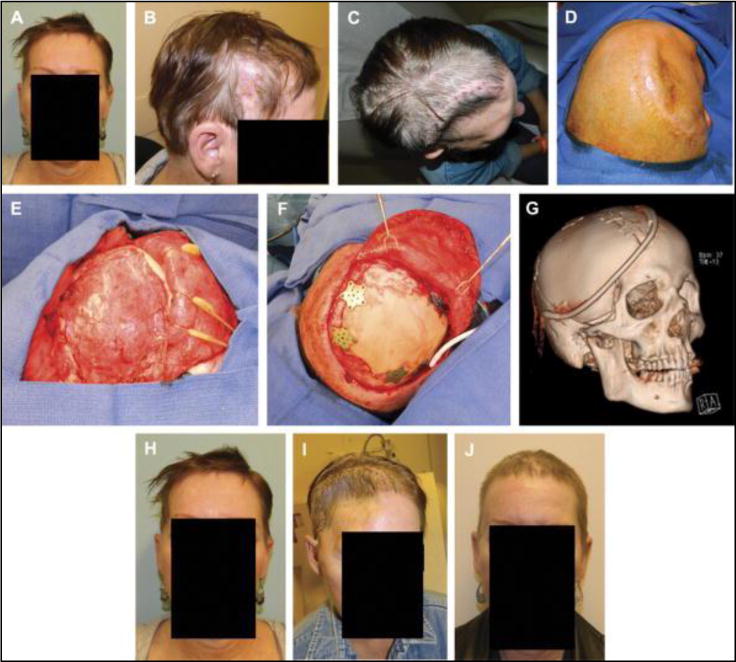
FIGURE 2.

Frontal view (A) and right profile view (B) of the original presentation with bone flap osteomyelitis. Aerial view (C) of the patient two weeks after removal of infected bone flap. Cephalad (D) on-table view before secondary cranial reconstruction 4 months after the bone flap removal. Photograph of vascularized pericranial-onlay dissection covering the dura (E) before insetting of the custom cranial implant with plates and screws (F). Postoperative 3-dimensional computed tomographic scan demonstrating the relative size of the implant, drain placement, and large amount of accompanying soft tissue reconstruction required with temporalis reinsertion (G). Frontal view photographs at the time of bone flap infection (H), at 3 months after bone flap removal (I), and at 6 months after reconstruction demonstrating acceptable contour and temporal symmetry (J).
