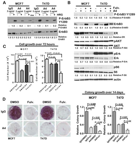
Figure 1. Fulvestrant-mediated ErbB3 up-regulation is neutralized by the ErbB3 antibody A4.
A. Western analysis of whole cell lysates collected from serum-starved MCF7 and T47D cells treated 24 h with antibody A4 (2–5 μg/ml) or non-specific human IgG, and for the final 10 min of culture with recombinant HRGβ1 (2 pg/ml).
B. Western analysis of whole cell lysates cultured in 10% serum and treated 24 h with fulvestrant (1μM) or antibody A4 (2 μg/ml).
C. Cells cultured for 3 days in 10% serum in the presence or absence of fulvestrant (1μM) or antibody A4 (2 μg/ml) were collected by trypsinization and counted. Average cell number (± S.D.) is shown. N = 5, each counted in duplicate. Student’s T-test.
D. Left panel: Representative images of crystal violet-stained cells after 14 d culture in 10% serum in the presence or absence of fulvestrant (1μM) and antibody A4 (2 μg/ml). Right panel: Crystal violet fluorescence was measured on the Odyssey scanner, and used as a relative measure of total cell number, setting the average fluorescence value for cells cultured in IgG/DMSO equal to 1. Average ± S.D. is shown. N = 3, each assessed in triplicate. Student’s T-test.

