Abstract
Pulmonary infection with Burkholderia cepacia complex in cystic fibrosis (CF) patients is associated with more-rapid lung function decline and earlier death than in CF patients without this infection. In this study, we used confocal microscopy to visualize the effects of various concentrations of tobramycin, achievable with systemic and aerosolized drug administration, on mature B. cepacia complex biofilms, both in the presence and absence of CF sputum. After 24 h of growth, biofilm thickness was significantly reduced by exposure to 2,000 μg/ml of tobramycin for Burkholderia cepacia, Burkholderia multivorans, and Burkholderia vietnamiensis; 200 μg/ml of tobramycin was sufficient to reduce the thickness of Burkholderia dolosa biofilm. With a more mature 48-h biofilm, significant reductions in thickness were seen with tobramycin at concentrations of ≥100 μg/ml for all Burkholderia species. In addition, an increased ratio of dead to live cells was observed in comparison to control with tobramycin concentrations of ≥200 μg/ml for B. cepacia and B. dolosa (24 h) and ≥100 μg/ml for Burkholderia cenocepacia and B. dolosa (48 h). Although sputum significantly increased biofilm thickness, tobramycin concentrations of 1,000 μg/ml were still able to significantly reduce biofilm thickness of all B. cepacia complex species with the exception of B. vietnamiensis. In the presence of sputum, 1,000 μg/ml of tobramycin significantly increased the dead-to-live ratio only for B. multivorans compared to control. In summary, although killing is attenuated, high-dose tobramycin can effectively decrease the thickness of B. cepacia complex biofilms, even in the presence of sputum, suggesting a possible role as a suppressive therapy in CF.
INTRODUCTION
Burkholderia cepacia complex is a group of closely related Gram-negative bacterial species that infect approximately 3% of cystic fibrosis (CF) patients in the United States and 5% of Canadian CF patients (1, 2). Pulmonary infection with B. cepacia complex species has been associated with increased lung function decline and higher mortality rates in individuals with CF than in those who do not have B. cepacia complex infection (3, 4). Despite these poor outcomes, there are currently no effective chronic suppressive antimicrobial therapies for B. cepacia complex infection in individuals with CF. Treatment of B. cepacia complex is difficult due to these organisms' intrinsic possession of mechanisms of antimicrobial resistance to multiple classes of antibiotics, including aminoglycosides, β-lactams, and fluoroquinolones. These mechanisms include efflux pumps, chromosomally encoded β-lactamases, and decreased outer membrane permeability (5). In addition, B. cepacia complex species are known to grow in the CF lung as a biofilm or in clusters of bacteria (6), which can act as a significant barrier to the effective delivery of drug intracellularly.
We have previously shown that high concentrations of tobramycin achievable by newer inhalation devices such as the podhaler (7) can inhibit B. cepacia complex species when grown planktonically or as a biofilm (8). However, these static models of biofilm formation on plastic pegs examine the inhibition of visible growth in a well (9) and provide limited information on the effects of antibiotics on bacterial biofilms within the CF lung. More-dynamic models that examine antimicrobial penetration and bactericidal activity on mature biofilm structures are needed. We are currently conducting a clinical trial of tobramycin inhalation powder in CF patients with B. cepacia complex infection (ClinicalTrials.gov identifier NCT02212587), with the aim of delivering high concentrations of drug that can penetrate the biofilm matrix and kill the bacterial cells within, as has been previously demonstrated in vitro (10). We thus developed a live-cell model that could help visualize the effects of antibiotics both on biofilm thickness and on viability, in order to understand responses to therapy.
The objectives of this study were thus to visualize the effects of various concentrations of tobramycin, achievable with systemic and aerosolized drug administration, on mature B. cepacia complex biofilms, both in the presence and absence of CF sputum, using confocal microscopy. The long-range goal is to identify potential effective therapy for CF individuals with B. cepacia complex pulmonary infections.
MATERIALS AND METHODS
Isolate collection.
B. cepacia complex isolates were collected from the Hospital for Sick Children, Toronto, Canada (Burkholderia cenocepacia HSC 64), St. Michael's Hospital, Toronto, Canada (Burkholderia multivorans SMH 46), the Cystic Fibrosis Foundation Burkholderia cepacia Research Repository at the University of Michigan, Ann Arbor (Burkholderia dolosa AU 2167), and the Canadian Burkholderia cepacia complex Research and Referral Repository at the University of British Columbia, Vancouver, Canada (Burkholderia vietnamiensis VC 9237 and Burkholderia cepacia VC 14106).
Conventional and biofilm susceptibility testing.
The MIC based on planktonic growth and biofilm inhibitory concentration (BIC) of tobramycin for each isolate were determined as previously described (8). In brief, antimicrobial susceptibility testing was performed on isolates grown planktonically by broth microdilution, using methods as per Clinical and Laboratory Standards Institute (CLSI) guidelines (11). Antimicrobial susceptibility testing was performed on isolates grown as a biofilm, using a modified method of the Calgary biofilm technique (9, 12). The antibiotic panels contained tobramycin at concentrations of 0, 10, 100, 200, 400, 800, 1,600, and 3,200 μg/ml. The MIC based on planktonic growth and the BIC of tobramycin for each isolate were determined by visually assessing the turbidity of each well.
Biofilm visualization using confocal microscopy.
Bacterial isolates were retrieved from frozen glycerol stocks and plated onto Columbia blood agar with 5% sheep blood (Thermo Scientific, Mississauga, ON, Canada). Overnight cultures were inoculated with a single colony into 4 ml of lysogeny broth (LB) and grown in 15-ml Falcon tubes for approximately 15 h at 37°C with shaking at 250 rpm. These cultures were then diluted 1/1,000 and grown to an optical density at 600 nm (OD600) of 0.6 before being diluted to an OD600 of 0.1. A 250-μl volume of this 0.1 OD culture was used to seed each chamber of an eight-chambered coverslip slide (LabTek II chamber slide on 1.5 borosilicate; VWR, Mississauga, ON, Canada). Static biofilm was allowed to form in these wells for 24 or 48 h at 37°C without shaking. Following this period, medium was removed and bacterial biofilms were either subjected to various concentrations of drug (tobramycin; Sigma-Aldrich, Oakville, ON, Canada) in a total volume of 250 μl for 24 h or fed with fresh LB medium. After 24 h, biofilms exposed to drug were stained and visualized, while biofilms that were fed were subjected to the same drug treatment as previously described before being stained and visualized.
Biofilm staining was performed using the Filmtracer Live/Dead biofilm viability kit (Life Technologies, Burlington, ON, Canada). Medium was gently removed from the chambers, and 200 μl total of the Live/Dead stain was added to the chambers. After 45 min of incubation, the stain was removed and fresh medium was placed in the wells. These chamber slides were used for confocal imaging.
Confocal images were acquired using a Quorum WaveFX spinning disk confocal system (Quorum Technologies Inc., Guelph, Canada). All images were acquired using a 25× water objective (total magnification, ×250) on a Zeiss AxioVert 200M Microscope. Spectral borealis lasers (green, 491 nm; red, 561 nm) were used for excitation. Emission filter sets of 515/40 and 624/40 were used to visualize the SYTO9 and propidium iodide stains, respectively. Images in the z-stack were obtained at a distance of 0.8 μm to obtain the depth of the biofilm. For every experiment, at least 30 depths of the biofilm were measured for each image captured. A total of three images of three separate views were taken for each slide chamber, and the mean of all images (total, 324 depth measurements) was used to calculate the thickness of the biofilm. Volocity software (PerkinElmer, Guelph, Canada) was used for acquisition and analysis of images.
Biofilm growth with sputum supernatant.
Sputum was collected from 5 CF patients from the Hospital for Sick Children (Research Ethics Board number 1000026462). Following collection, sputum was diluted in 3× phosphate-buffered saline (PBS) (vol/vol) and spun down at 4,400× relative centrifugal force (RCF) for 10 min. Supernatants were then collected and filter sterilized through 0.45-μm syringe filters (Sarstedt, St Leonard, Quebec, Canada). Following this, sputum supernatants were stored at −20°C until use. Biofilms were generated as described above in the presence of 10% (vol/vol) sputum supernatants (final sputum dilution, 3.33%) in LB for 24 h. Following this time period, medium was removed and replaced with fresh medium containing 10% sputum supernatants in the presence or absence of tobramycin. After 24 h, the biofilms underwent staining and imaging as described above.
Statistical analysis.
Comparison of continuous data within groups was done using the Kruskal-Wallis test with a Dunn's multiple comparison posttest. Correlations between measures were calculated using the Spearman correlation coefficient. A P value of <0.05 was considered significant. All analyses were done using GraphPad Prism version 5.04.
RESULTS
Selection of CF B. cepacia complex clinical isolates.
A sample of different species within the B. cepacia complex most commonly isolated from CF patients were selected for study (Table 1). Isolates were chosen with tobramycin MICs and BICs that were representative of the MIC50 and BIC50 for tobramycin for a large collection of CF B. cepacia complex isolates (8).
TABLE 1.
Tobramycin MIC and BIC for Burkholderia cepacia complex CF isolates
| Burkholderia isolate | Tobramycin |
|
|---|---|---|
| MICa (μg/ml) | BICb (μg/ml) | |
| B. cepacia VC 14106 | 10 | 100 |
| B. multivorans SMH 46 | 100 | 100 |
| B. cenocepacia HSC 64 | 100 | 100 |
| B. dolosa AU 2167 | 200 | 400 |
| B. vietnamiensis VC 9237 | 10 | 100 |
MIC is for planktonic cells.
BIC, biofilm inhibitory concentration.
Effects of tobramycin on biofilm thickness.
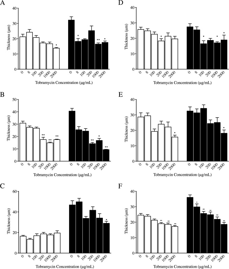
The thickness of biofilm after 24 h of growth was significantly reduced after exposure to 2,000 μg/ml of tobramycin for B. cepacia, B. multivorans, and B. vietnamiensis; significant reductions were also seen at 200 μg/ml and 1,000 μg/ml for B. multivorans and 200 μg/ml alone for B. dolosa (Fig. 1). There was no effect seen for B. cenocepacia.
FIG 1.
Average thickness (in micrometers) of biofilms in the presence of 0, 8, 100, 200, 1,000, and 2,000 μg/ml of tobramycin after 24 h (white bars) and 48 h (black bars) of growth of isolates of Burkholderia cepacia (A), Burkholderia multivorans (B), Burkholderia cenocepacia (C), Burkholderia dolosa (D), and Burkholderia vietnamiensis (E) and all strains combined (F). *, P < 0.05; **, P < 0.001 compared to control (0 μg/ml) using the Kruskal-Wallis test with Dunn's multiple comparison posttest.
Even with a more mature biofilm after 48 h of growth, significant reductions in biofilm thickness were still observed at the higher tobramycin concentrations (100 μg/ml or greater) for all Burkholderia species. Of note, only B. cepacia had significant reductions in biofilm thickness with 8 μg/ml of tobramycin. When the sum of all strains was examined, there was a dose-dependent response in terms of decreased thickness with increasing drug concentration after 24 as well as 48 h (Fig. 1F).
Cidal activity of tobramycin against biofilms.
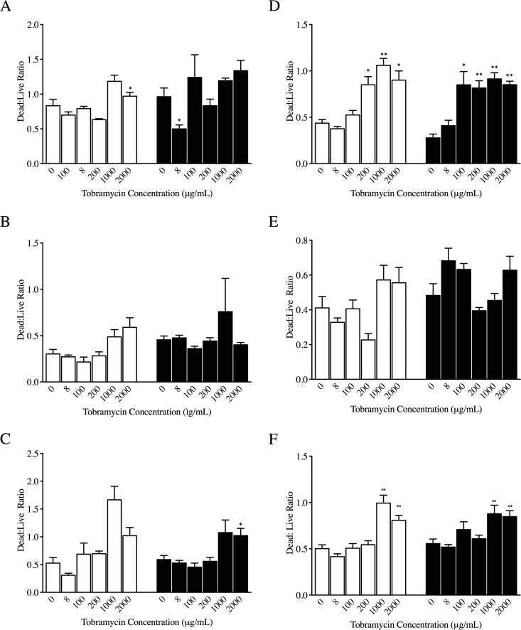
The ability of tobramycin to kill B. cepacia complex within biofilms was then examined. A significantly increased ratio of dead to live cells was observed in comparison to control at tobramycin concentrations of 200 μg/ml or greater after 24 h of growth of B. cepacia and B. dolosa (Fig. 2). After 48 h of growth, there was a higher ratio of dead to live cells compared to control with tobramycin concentrations of 100 μg/ml or greater for B. cenocepacia and B. dolosa. At the lower tobramycin concentration of 8 μg/ml, there was, in fact, a lower dead-to-live ratio, indicating more live cells in a 48-h biofilm of B. cepacia than in the control. When all strains were examined as a group, higher tobramycin concentrations (1,000 and 2,000 μg/ml) were associated with an increased dead-to-live ratio compared to controls (Fig. 2F).
FIG 2.
Dead-to-live ratio of biofilms in the presence of 0, 8, 100, 200, 1,000, and 2,000 μg/ml of tobramycin after 24 h (white bars) and 48 h (black bars) of growth of isolates of Burkholderia cepacia (A), Burkholderia multivorans (B), Burkholderia cenocepacia (C), Burkholderia dolosa (D), and Burkholderia vietnamiensis (E) and all strains combined (F). *, P < 0.05; **, P < 0.001 compared to control (0 μg/ml) using the Kruskal-Wallis test with Dunn's multiple comparison posttest.
Tobramycin activity against biofilms in sputum.
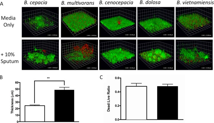
To confirm our findings under conditions more reflective of the CF lung, we repeated our experiments using a maximum of 1,000 μg/ml of tobramycin (mean peak sputum tobramycin concentrations) (13) in the presence of pooled 10% sputum supernatant. When the biofilm growth of all B. cepacia complex strains in the absence of antibiotics was examined in the presence of sputum supernatant, the addition of sputum to medium was found to significantly increase the thickness of the biofilm but did not influence the ratio of dead to live cells within the biofilm (Fig. 3).
FIG 3.
Effects of sputum on biofilm formation after 48 h of growth. (A) Representative images of Burkholderia cepacia, Burkholderia multivorans, Burkholderia cenocepacia, Burkholderia dolosa, and Burkholderia vietnamiensis biofilms grown in LB medium alone (top panel) or 10% (vol/vol) pooled sputum supernatants (bottom panel) for 48 h prior to staining with the Filmtracer biofilm viability kit and confocal imaging. (B) Average thickness of all B. cepacia complex strains grown in the presence (white bars) of absence (black bars) of 10% sputum supernatants for 48 h. (C) Dead-to-live ratio of B. cepacia complex biofilms grown in the presence (white bars) or absence (black bars) of 10% sputum for 48 h.
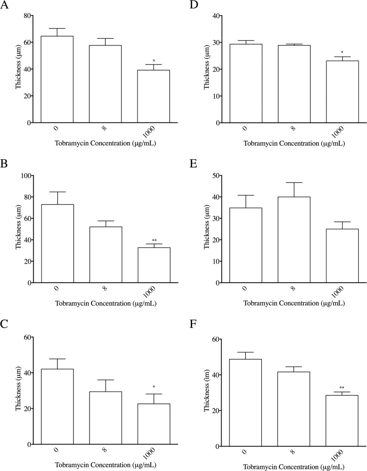
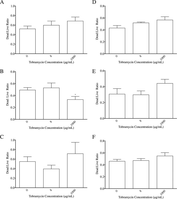
Under these conditions, biofilm thickness was significantly reduced with 1,000 μg/ml of tobramycin for B. cepacia complex species with the exception of B. vietnamiensis (Fig. 4). With respect to killing ability in the presence of sputum, significant changes were seen only at 1,000 μg/ml of tobramycin for B. multivorans, with a significant decrease in the dead-to-live ratio compared to control (Fig. 5).
FIG 4.
Average thickness (in micrometers) of biofilms in the presence of 10% sputum supernatant with 0, 8, and 1,000 μg/ml of tobramycin after 24 h of growth of isolates of Burkholderia cepacia (A), Burkholderia multivorans (B), Burkholderia cenocepacia (C), Burkholderia dolosa (D), and Burkholderia vietnamiensis (E) and all strains combined (F). *, P < 0.05; **, P < 0.001 compared to control (0 μg/ml) using the Kruskal-Wallis test with Dunn's multiple comparison posttest.
FIG 5.
Dead-to-live ratio of biofilms in the presence of 10% sputum supernatant with 0, 8, and 1,000 μg/ml of tobramycin after 24 h of growth of isolates of Burkholderia cepacia (A), Burkholderia multivorans (B), Burkholderia cenocepacia (C), Burkholderia dolosa (D), and Burkholderia vietnamiensis (E) and all strains combined (F). *, P < 0.05; **, P < 0.001 compared to control (0 μg/ml) using the Kruskal-Wallis test with Dunn's multiple comparison posttest.
DISCUSSION
This study demonstrated that high concentrations of tobramycin, such as those delivered via aerosolization, can decrease the thickness of biofilms as well as kill bacterial cells within a biofilm, even in the presence of sputum, across a range of species within the B. cepacia complex, representative of species typically isolated from CF patients.
B. cepacia complex bacteria are traditionally considered to be intrinsically resistant to aminoglycosides through a number of different resistance mechanisms, including the resistance-nodulation-division (RND) efflux systems (14). Confirming this fact, in our series of experiments, we found a near-complete absence of effect of tobramycin at a concentration of 8 μg/ml (representing systemic drug concentrations) on biofilm thickness and viability. In fact, lower tobramycin concentrations were found to increase the ratio of live to dead cells with a mature B. cepacia biofilm; previous investigations have demonstrated that subinhibitory concentrations of antimicrobials can induce biofilm formation (15). These findings suggest that intravenous administration of aminoglycosides for the treatment of B. cepacia complex pulmonary infections is not likely to be effective therapy.
Aerosolization of antibiotics, however, can achieve significantly higher intrapulmonary concentrations of drug, up to 2,000 μg/ml in the case of tobramycin, with the use of newer inhalation devices such as the podhaler (7). Our earlier investigations showed that tobramycin concentrations of 100 μg/ml were sufficient to inhibit both planktonic and biofilm growth of the majority of B. cepacia complex species isolated from CF patients (8). Previous studies using multiple combination bactericidal antibiotic testing also showed that high-dose tobramycin was the most effective at killing B. cepacia complex species in vitro (16). Our current study further demonstrates that high tobramycin concentrations, beyond 100 μg/ml, can also significantly reduce the thickness of mature, established biofilms and even kill cells within the biofilm, for certain B. cepacia complex species. Although there are genetic determinants of biofilm antibiotic susceptibility (17), such that even thin biofilms can be tolerant of antibiotics (18), it is reasonable to suppose that higher drug concentration leads to improved penetration within the biofilm (10), increased killing of bacterial cells, and subsequent decreased thickness of the biomass. Indeed, overall in our model, we observed that when all species were combined, as drug concentration increased, biofilm thickness decreased and the proportion of dead to live cells increased in a dose-dependent manner. However, at the level of an individual species, decreased thickness did not necessarily directly correlate with increased killing in a given experiment, as thickness was frequently decreased to the point where there were too few bacterial cells to kill, especially at the higher antibiotic concentrations. This model, however, allowed us to study both effects of tobramycin concurrently. In addition, this current biofilm model provided additional information that did not necessarily correlate with the simple biofilm inhibitory concentrations (BICs) measured using the Calgary Biofilm device, which are typically lower than the concentrations required to kill mature biofilms (19), such as those in this study. These experimental outcomes therefore represent complementary methods of studying antimicrobial effects against bacterial biofilms.
Lastly, the effects of high-dose tobramycin on B. cepacia complex biofilms were confirmed under conditions relevant to the CF lung environment. Although administration of tobramycin inhalation powder can achieve a maximum sputum concentration of 2,000 μg/ml, it is more realistic to expect sputum concentrations in the range of 1,000 μg/ml, given the wide standard deviations associated with this measure (13, 20). In addition, bacteria grown in the presence of sputum have been shown to display a distinctive morphology, forming aggregates with increased antimicrobial resistance, which are characteristic of bacterial biofilms within the CF lung (21). Similarly, the addition of 10% sputum supernatant also increased bacterial biofilm thickness in our experiments. Even with these additional barriers, however, high-dose tobramycin (1,000 μg/ml) was still able to significantly reduce biofilm thickness for the majority of the species within B. cepacia complex, although increased killing was observed only for B. multivorans. Eradication of mature B. cepacia complex pulmonary infections may thus be difficult to achieve in CF; in vitro studies have demonstrated that persister cells can often survive even after high-dose tobramycin treatment of B. cenocepacia biofilms, giving rise to new infections (22). However, chronic suppression of B. cepacia complex biofilm growth may be possible and may result in clinical benefits to CF patients, as has been shown with chronic suppressive treatment of Pseudomonas aeruginosa pulmonary infections in CF (23). Alteration of the tobramycin formulation, either through high-efficiency encapsulation into neutrally charged liposomes (24) or through combination with a matrix-degrading compound (25), may improve its antimicrobial activity against B. cepacia complex biofilms. This current model system represents an innovative way of visualizing the antimicrobial activity of novel therapeutics against bacterial biofilms.
This study had several limitations. Only one isolate from the selected species within the B. cepacia complex was tested. As there are likely variations between strains, this significantly limits any interspecies comparisons that can be made. However, we selected the most commonly isolated species from CF patients within the complex, with representative tobramycin MICs and BICs. In addition, biofilm cultures were grown for a maximum of 48 h before exposure to antibiotics, limiting the development of biofilm maturity. Biofilm thickness, however, was within the range of previously reported bacterial biofilm thickness in CF lung infections (26). Finally, this experimental biofilm model did not fully replicate the anoxic, polymicrobial, immune cell-filled environment of the CF lung. Host factors, such as neutrophils, have been shown to increase antimicrobial resistance of P. aeruginosa aggregates (27, 28). Although we did expose the bacterial biofilms to sputum from actual CF patients, these supernatants would have included only secreted products and not cellular materials.
In conclusion, tobramycin at concentrations greater than 100 μg/ml, as achievable through aerosolization, can significantly decrease the thickness of, and in certain instances kill bacterial cells within, B. cepacia complex biofilms. Although CF sputum supernatant significantly increases the thickness of these biofilms, high-dose tobramycin is still effective in decreasing its biomass but is limited in terms of its cidal activity, presenting a barrier to eradication. Clinical trials of tobramycin inhalation powder in CF patients with chronic B. cepacia complex infection are under way to determine whether high-dose tobramycin is effective in decreasing sputum bacterial density, thereby offering potential suppressive treatment for this patient population.
Funding Statement
This work was supported by funds from the Research Institute at The Hospital for Sick Children and by a grant from the Canadian Institutes of Health Research (CIHR MT13337) to P. Lynne Howell (Senior Scientist, Program in Molecular Structure and Function, The Hospital for Sick Children).
REFERENCES
- 1.Cystic Fibrosis Foundation. 2011. Patient data registry report. CF Foundation, Bethesda, MD. [Google Scholar]
- 2.Cystic Fibrosis Canada. 2013. Canadian patient data registry report. CF Canada, Toronto, Canada. [Google Scholar]
- 3.Corey M, Farewell V. 1996. Determinants of mortality from cystic fibrosis in Canada, 1970-1989. Am J Epidemiol 143:1007–1017. doi: 10.1093/oxfordjournals.aje.a008664. [DOI] [PubMed] [Google Scholar]
- 4.Tablan OC, Chorba TL, Schidlow DV, White JW, Hardy KA, Gilligan PH, Morgan WM, Carson LA, Martone WJ, Jason JM. 1985. Pseudomonas cepacia colonization in patients with cystic fibrosis: risk factors and clinical outcome. J Pediatr 107:382–387. doi: 10.1016/S0022-3476(85)80511-4. [DOI] [PubMed] [Google Scholar]
- 5.Waters V. 2012. New treatments for emerging cystic fibrosis pathogens other than Pseudomonas. Curr Pharm Des 18:696–725. doi: 10.2174/138161212799315939. [DOI] [PubMed] [Google Scholar]
- 6.Schwab U, Abdullah LH, Perlmutt OS, Albert D, Davis CW, Arnold RR, Yankaskas JR, Gilligan P, Neubauer H, Randell SH, Boucher RC. 2014. Localization of Burkholderia cepacia complex bacteria in cystic fibrosis lungs and interactions with Pseudomonas aeruginosa in hypoxic mucus. Infect Immun 82:4729–4745. doi: 10.1128/IAI.01876-14. [DOI] [PMC free article] [PubMed] [Google Scholar]
- 7.Konstan MW, Flume PA, Kappler M, Chiron R, Higgins M, Brockhaus F, Zhang J, Angyalosi G, He E, Geller DE. 2011. Safety, efficacy and convenience of tobramycin inhalation powder in cystic fibrosis patients: the EAGER trial. J Cyst Fibros 10:54–61. doi: 10.1016/j.jcf.2010.10.003. [DOI] [PMC free article] [PubMed] [Google Scholar]
- 8.Ratjen A, Yau Y, Wettlaufer J, Matukas L, Zlosnik JE, Speert DP, LiPuma JJ, Tullis E, Waters V. 2015. In vitro efficacy of high-dose tobramycin against Burkholderia cepacia complex and Stenotrophomonas maltophilia isolates from cystic fibrosis patients. Antimicrob Agents Chemother 59:711–713. doi: 10.1128/AAC.04123-14. [DOI] [PMC free article] [PubMed] [Google Scholar]
- 9.Ceri H, Olson ME, Stremick C, Read RR, Morck D, Buret A. 1999. The Calgary Biofilm Device: new technology for rapid determination of antibiotic susceptibilities of bacterial biofilms. J Clin Microbiol 37:1771–1776. [DOI] [PMC free article] [PubMed] [Google Scholar]
- 10.Tseng BS, Zhang W, Harrison JJ, Quach TP, Song JL, Penterman J, Singh PK, Chopp DL, Packman AI, Parsek MR. 2013. The extracellular matrix protects Pseudomonas aeruginosa biofilms by limiting the penetration of tobramycin. Environ Microbiol 15:2865–2878. doi: 10.1111/1462-2920.12155. [DOI] [PMC free article] [PubMed] [Google Scholar]
- 11.Clinical and Laboratories Standards Institute. 2012. Performance standards for antimicrobial susceptibility testing: 22nd informational supplement M100-S22. CLSI, Wayne, PA. [Google Scholar]
- 12.Wu K, Yau YC, Matukas L, Waters V. 2013. Biofilm compared to conventional antimicrobial susceptibility of Stenotrophomonas maltophilia isolates from cystic fibrosis patients. Antimicrob Agents Chemother 57:1546–1548. doi: 10.1128/AAC.02215-12. [DOI] [PMC free article] [PubMed] [Google Scholar]
- 13.Geller DE, Konstan MW, Smith J, Noonberg SB, Conrad C. 2007. Novel tobramycin inhalation powder in cystic fibrosis subjects: pharmacokinetics and safety. Pediatr Pulmonol 42:307–313. doi: 10.1002/ppul.20594. [DOI] [PubMed] [Google Scholar]
- 14.Bazzini S, Udine C, Sass A, Pasca MR, Longo F, Emiliani G, Fondi M, Perrin E, Decorosi F, Viti C, Giovannetti L, Leoni L, Fani R, Riccardi G, Mahenthiralingam E, Buroni S. 2011. Deciphering the role of RND efflux transporters in Burkholderia cenocepacia. PLoS One 6(4):e18902. doi: 10.1371/journal.pone.0018902. [DOI] [PMC free article] [PubMed] [Google Scholar]
- 15.Hoffman LR, D'Argenio DA, MacCoss MJ, Zhang Z, Jones RA, Miller SI. 2005. Aminoglycoside antibiotics induce bacterial biofilm formation. Nature 436:1171–1175. doi: 10.1038/nature03912. [DOI] [PubMed] [Google Scholar]
- 16.Aaron SD, Ferris W, Henry DA, Speert DP, Macdonald NE. 2000. Multiple combination bactericidal antibiotic testing for patients with cystic fibrosis infected with Burkholderia cepacia. Am J Respir Crit Care Med 161(4 Part 1):1206–1212. [DOI] [PubMed] [Google Scholar]
- 17.Mah TF, Pitts B, Pellock B, Walker GC, Stewart PS, O'Toole GA. 2003. A genetic basis for Pseudomonas aeruginosa biofilm antibiotic resistance. Nature 426:306–310. doi: 10.1038/nature02122. [DOI] [PubMed] [Google Scholar]
- 18.Gupta K, Marques CN, Petrova OE, Sauer K. 2013. Antimicrobial tolerance of Pseudomonas aeruginosa biofilms is activated during an early developmental stage and requires the two-component hybrid SagS. J Bacteriol 195:4975–4987. doi: 10.1128/JB.00732-13. [DOI] [PMC free article] [PubMed] [Google Scholar]
- 19.Bjarnsholt T, Ciofu O, Molin S, Givskov M, Hoiby N. 2013. Applying insights from biofilm biology to drug development—can a new approach be developed? Nat Rev Drug Discov 12:791–808. doi: 10.1038/nrd4000. [DOI] [PubMed] [Google Scholar]
- 20.Konstan MW, Geller DE, Minic P, Brockhaus F, Zhang J, Angyalosi G. 2011. Tobramycin inhalation powder for P. aeruginosa infection in cystic fibrosis: the EVOLVE trial. Pediatr Pulmonol 46:230–238. doi: 10.1002/ppul.21356. [DOI] [PMC free article] [PubMed] [Google Scholar]
- 21.Staudinger BJ, Muller JF, Halldorsson S, Boles B, Angermeyer A, Nguyen D, Rosen H, Baldursson O, Gottfreethsson M, Guethmundsson GH, Singh PK. 2014. Conditions associated with the cystic fibrosis defect promote chronic Pseudomonas aeruginosa infection. Am J Respir Crit Care Med 189:812–824. doi: 10.1164/rccm.201312-2142OC. [DOI] [PMC free article] [PubMed] [Google Scholar]
- 22.Van Acker H, Sass A, Bazzini S, De Roy K, Udine C, Messiaen T, Riccardi G, Boon N, Nelis HJ, Mahenthiralingam E, Coenye T. 2013. Biofilm-grown Burkholderia cepacia complex cells survive antibiotic treatment by avoiding production of reactive oxygen species. PLoS One 8(3):e58943. doi: 10.1371/journal.pone.0058943. [DOI] [PMC free article] [PubMed] [Google Scholar]
- 23.Ramsey BW, Pepe MS, Quan JM, Otto KL, Montgomery AB, Williams-Warren J, Vasiljev KM, Borowitz D, Bowman CM, Marshall BC, Marshall S, Smith AL. 1999. Intermittent administration of inhaled tobramycin in patients with cystic fibrosis. Cystic Fibrosis Inhaled Tobramycin Study Group. N Engl J Med 340:23–30. [DOI] [PubMed] [Google Scholar]
- 24.Messiaen AS, Forier K, Nelis H, Braeckmans K, Coenye T. 2013. Transport of nanoparticles and tobramycin-loaded liposomes in Burkholderia cepacia complex biofilms. PLoS One 8(11):e79220. doi: 10.1371/journal.pone.0079220. [DOI] [PMC free article] [PubMed] [Google Scholar]
- 25.Messiaen AS, Nelis H, Coenye T. 2014. Investigating the role of matrix components in protection of Burkholderia cepacia complex biofilms against tobramycin. J Cyst Fibros 13:56–62. doi: 10.1016/j.jcf.2013.07.004. [DOI] [PubMed] [Google Scholar]
- 26.Bjarnsholt T, Alhede M, Eickhardt-Sorensen SR, Moser C, Kuhl M, Jensen PO, Hoiby N. 2013. The in vivo biofilm. Trends Microbiol 21:466–474. doi: 10.1016/j.tim.2013.06.002. [DOI] [PubMed] [Google Scholar]
- 27.Caceres SM, Malcolm KC, Taylor-Cousar JL, Nichols DP, Saavedra MT, Bratton DL, Moskowitz SM, Burns JL, Nick JA. 2014. Enhanced in vitro formation and antibiotic resistance of nonattached Pseudomonas aeruginosa aggregates through incorporation of neutrophil products. Antimicrob Agents Chemother 58:6851–6860. doi: 10.1128/AAC.03514-14. [DOI] [PMC free article] [PubMed] [Google Scholar]
- 28.Chatzimoschou A, Simitsopoulou M, Antachopoulos C, Walsh TJ, Roilides E. 2015. Antipseudomonal agents exhibit differential pharmacodynamic interactions with human polymorphonuclear leukocytes against established biofilms of Pseudomonas aeruginosa. Antimicrob Agents Chemother 59:2198–2205. doi: 10.1128/AAC.04934-14. [DOI] [PMC free article] [PubMed] [Google Scholar]


