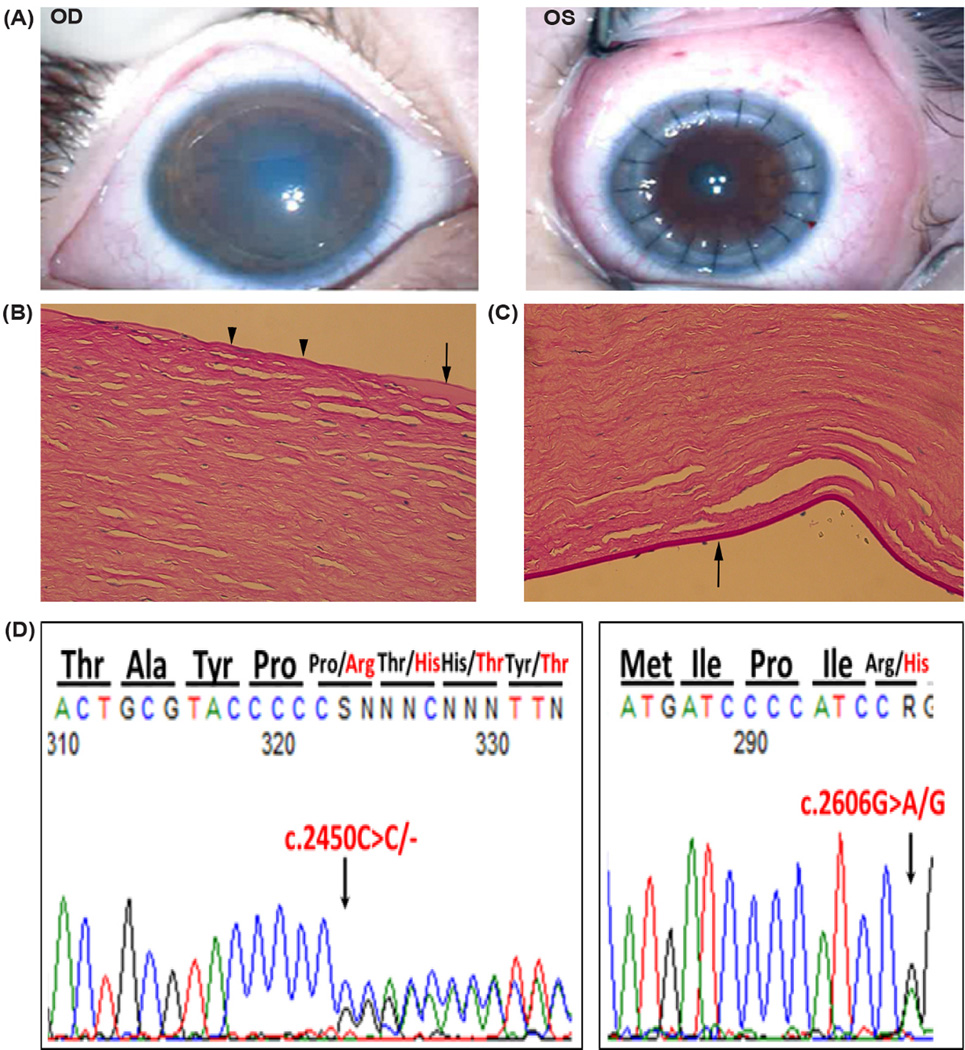
Figure 1.
Case A Patient. A. Photographs obtained with operating microscope 2 months status post penetrating keratoplasty to the left eye shows a cloudy edematous cornea in the right eye; and a clear and compact PKP graft in the left eye. Histopathological findings of keratoplasty button from Case A. Light microscopy showed features of CHED included B) focal loss in Bowman’s layer centrally (arrowheads), areas of thickened Bowman’s layer (arrow); and C) thin Descemet’s membrane with only a few remaining endothelial cells (arrow). D) DNA sequence chromatogram of proband revealing a novel de novo mutation (c.2450delC) in the paternal allele resulting in a frameshift mutation in exon 18 of the SLC4A11 gene. Co-inheritance of a maternal missense mutation (c.2606G>A) in exon 18 leads to the generation of the compound heterozygous mutation.

