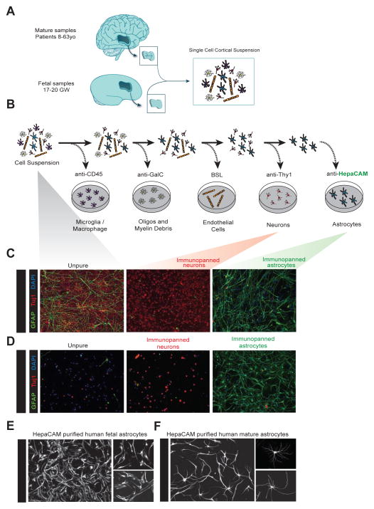
Figure 1.
Acute purification of fetal and mature human astrocytes. A. Juvenile and adult (8 to 63 years old) patient temporal lobe cortex tissue and fetal (17–20 gestational week) brain tissue is first dissociated into single cell suspensions. B. Schematics of immunopanning purification of cell types from human brain samples. C and D. Unpurified brain cells (left), Thy1-purified neurons (middle), and HepaCAM-purified astrocytes (right) from fetal (C) and mature (D) brains stained at 7div for neurons (TuJ1, red), astrocytes (GFAP, green), and nuclei of all cells (DAPI, blue). Scale bars: 100μm. E and F. Cultured human fetal (E) and mature (F) astrocytes grown in culture for 7 days and stained with GFAP. Scale bars: 100μm, insets 50 μm.

