Abstract
Background
Chronic aortic regurgitation (AR) patients demonstrate left ventricular (LV) remodeling with increased LV mass and volume but may have a preserved LV ejection fraction (EF). We hypothesize that in chronic AR, global longitudinal systolic and diastolic function will be reduced despite a preserved LV EF.
Methods
We studied with Doppler echocardiography 27 normal subjects, 87 patients with chronic AR with a LV EF > 50% (AR + PEF), 66 patients with an EF < 50% [AR + reduced LV ejection fraction (REF)] and 82 patients with hypertensive heart disease. LV volume, transmitral spectral and tissue Doppler were obtained. Myocardial velocities and their timing and longitudinal strain of the proximal and mid wall of each of the 3 apical views were obtained.
Results
As compared to normals, global longitudinal strain was reduced in AR + PEF (13.8 ± 4.0%) and AR + REF (11.4 ± 4.7%) vs. normals (18.4 ± 3.6%, both p < 0.001). As an additional comparison group for AR + PEF, global longitudinal strain was reduced as compared to patients with hypertensive heart disease (p = 0.032). The average peak diastolic annular velocity (e') was decreased in AR + PEF (6.9 ± 3.3 cm/s vs. 13.4 ± 2.6 cm/s, p < 0.001) and AR + REF (4.8 ± 2.1 cm/s, p < 0.001). Peak rapid filling velocity/e' (E/e') was increased in both AR + PEF (14.4 ± 6.2 vs. 6.2 ± 1.3, p < 0.001) and AR + REF (18.8 ± 6.4, p < 0.001 vs. normals). Independent correlates of global longitudinal strain (r = 0.6416, p < 0.001) included EF (p < 0.0001), E/e' (p < 0.0001), and tricuspid regurgitation velocity (p = 0.0176).
Conclusion
With chronic AR, there is impaired longitudinal function despite preserved EF. Moreover, global longitudinal strain was well correlated with noninvasive estimated LV filling pressures and pulmonary systolic arterial pressures.
Keywords: Aortic valve insufficiency, Left ventricular function, Left ventricular remodeling
Introduction
Chronic aortic regurgitation (AR) is a left ventricular (LV) volume overload lesion with a long latency period prior to symptom development. Prior to symptoms, patients may exhibit normal exercise tolerance associated with LV remodeling and a preserved ejection fraction (EF).1),2) The development of symptoms heralds a progressive downhill course marked by angina, heart failure, arrhythmias, syncope, and reduced survival. LV systolic performance involves not only contraction across its minor axis but both longitudinal and circumferential shortening.3),4) In patients with hypertensive heart disease, heart failure with preserved EF, and aortic stenosis, reduced longitudinal and mid-wall circumferential shortening occur despite normal radial endocardial fiber shortening.5),6),7),8)
We hypothesized that in chronic AR, there will be a reduction in longitudinal shortening despite a preserved EF due to normal endocardial minor axis shortening. The increase in LV mass with eccentric remodeling results in reduced LV relaxation and elevated LV filling pressures. Reduced EF with chronic AR would likely further exacerbate the above abnormalities.
The purpose of this study is to assess longitudinal systolic and diastolic function in patients with chronic AR with both preserved and reduced LV systolic function and assess the relationship to alterations in cardiac structure.
Methods
We conducted a single center retrospective review of all patients receiving an echocardiogram and found to have chronic moderate or greater AR as assessed by the American Society of Echocardiography criteria9) between 2004-2007. Patients deemed to have acute AR, coronary artery disease based on electrocardiogram evidence of a previous myocardial infarction or akinesis of 2 wall segments on echocardiography, or moderate or greater valvular disease were excluded. The study was approved (expedited review) by the institutional review board of the University of Florida College of Medicine-Jacksonville.
Patients
There were 182 patients with moderate or greater AR from 2004-2007. We were able to identify 153 patients (100 moderate and 53 severe chronic AR) with an adequate echocardiogram allowing for the calculation of AR severity, the determination of LV size, thickness, and function, left atrial volume, assessment of diastolic function with transmitral Doppler and tissue Doppler, and tissue Doppler indices of dyssynchrony and strain. Patients were divided into AR groups with preserved LV EF (AR + PEF; 87 patients with EF ≥ 50%) and AR with reduced LV EF (AR + REF; 66 patients with EF < 50%). A group of 27 subjects with no evidence of cardiac disease on the basis of history, physical examination, and echocardiogram were included as a comparison normal group. Hypertensive patients well controlled without LV hypertrophy (echocardiogram and electrocardiogram) were included in this group. The 27 patients were selected from a larger group of normal subjects and were age and sexed matched to the chronic AR groups. An additional comparison group consisting of 82 patients age and sexed matched with evidence of hypertensive heart disease with normal LV EF (> 50%), LV hypertrophy by echocardiography (LV mass index > 115 g/m2 in males and > 95 g/m2 in females), and no evidence of coronary or other valvular heart disease (echocardiography) was selected during the same time period for comparison with the AR + PEF group.
For each patient selected for inclusion, the medical records were examined for the patient's age, sex, laboratory results and medications at the time that the echocardiogram was performed. Patients were deemed to have hypertension if their blood pressure exceeded 140/90 or were taking antihypertensive medications. Patients were deemed to have diabetes mellitus if their fasting blood glucose was > 126 mg/dL, post-prandial glucose was > 200 mg/dL, or were taking anti-diabetic medications. Patients were deemed to have hyperlipidemia if their total cholesterol or fasting triglycerides were elevated or were taking medications to reduce cholesterol or triglycerides. Review of the medical records at the time of the echocardiogram was performed to determine the New York Heart Association functional class.
Echocardiography
Two dimensional Doppler echocardiography including M-mode, spectral and color flow Doppler, and tissue Doppler were obtained in all patients using a Vivid 7 echocardiograph (GE-Medical, Milwaukee, WI, USA). Only those studies in which all parameters described below could be measured were utilized (153 of 181). The systolic blood pressure, diastolic blood pressure, and pulse pressure were also recorded at the time of echocardiography.
From the M-mode tracings of the left atrium, we measured the left atrial dimension in mid systole using leading to leading edge technique. Using 2 dimensional echocardiography, the aortic root and LV outflow tract (LVOT) was measured from the parasternal long axis view at end diastole or during peak systole (LVOT). End diastolic dimension, end systolic dimension, septal and inferolateral wall thickness at end diastole and end systole and LV mass (indexed to body surface area) were measured using the American Society of Echocardiography standards.10) Relative wall thickness was calculated as twice the posterior wall thickness in end diastole divided by the LV end diastolic dimension. LV end diastolic (at the R wave) and end systolic volumes (smallest visual LV volume near the T wave) were measured utilizing the apical biplane Simpson's rule and indexed to body surface area. Biplane left atrial volumes were measured from the apical 4 and 2 chamber views using the area-length formula and indexed to body surface area.10)
From the transmitral spectral Doppler, we obtained the E, A, and deceleration time. The diastolic filling period was calculated from the onset to the end of transmitral spectral Doppler wave form. LV ejection time was measured from the onset of aortic flow to the end of aortic flow. The isovolumic relaxation time was measured by determining the time from the Q wave to the onset of mitral flow minus the Q wave to the end of aortic flow. Isovolumic contraction time was calculated as the time interval from the end of mitral flow to the onset of aortic flow.
Spectral tissue Doppler tracings of the mitral annulus in the apical 4 chamber view were measured using a 5 × 5 mm sample volume. The average of the peak systolic velocity (s') and early diastolic velocity (e') from the septal and lateral annulus were obtained. Peak rapid filling velocity/e' (E/e') was calculated as a measure of LV filling pressures.11) From color flow Doppler, tricuspid regurgitation (TR) jets were visualized in multiple views with the peak velocity being obtained. Color flow Doppler of the AR jet was visualized in the parasternal long axis view. The height of the jet in the outflow tract was indexed to the LVOT during the maximal jet. A ratio was > 0.25 was termed moderate and > 0.65 was termed severe. Regurgitant volume was determined using the pulsed Doppler recordings of the velocity time interval across the aortic valve and pulmonic valve. A regurgitant volume > 30 cc was termed moderate and > 60 cc was termed severe. Moderate AR was noted in 100 patients and severe AR (both of the above indices) was noted in 53 patients.9)
Color tissue Doppler was obtained in the apical 2, 3, and 4 chamber views at a frame rate 60-90 frames per second. A sample volume of 10 × 5 mm was placed in the proximal and mid-portion of each of the 6 walls in the 3 apical views. From the derived tracings of velocity, the time from the Q wave to the s' and e' myocardial velocities were obtained for each of the 6 sample volumes. The standard deviation of the Q wave to peak systolic and peak early diastolic wall velocities for all 12 segments was obtained as a measure of systolic and diastolic dyssynchrony.12) From the same 12 sample volumes, we obtained peak systolic strain in all 12 segments and averaged them as a measure of global longitudinal strain.
Inter-observer and intra-observer variability
Ten patient with chronic AR and 10 patients with normal LV function were randomly chosen and were reanalyzed 1 month following the initial analysis with regard to jet width/LVOT width, systolic and diastolic dyssynchrony, and global longitudinal strain by 2 observers. The mean difference between observers for jet width/LVOT width was 0.08 ± 0.02; for systolic dyssynchrony (6 ± 3 msec), for diastolic dyssyncrony (4 ± 2 msec), and for global longitudinal strain (1.4 ± 0.7%). The intra-observer variability for jet width/LVOT width was 0.05 ± 0.02; for systolic dyssynchrony (5 ± 2 msec), for diastolic dyssyncrony (4 ± 2 msec); and for global longitudinal strain (1.1 ± 0.5%).
Statistics
Data were expressed as mean ± standard deviation for continuous data that was normally distributed. For data that was not normally distributed, the median and inter-quartile ranges were computed. Differences among the 3 groups were determined by 1 way analysis of variance or 1 way analysis of variance using ranks. If the F value was < 0.05, the differences between individual groups was determined by multicomparison t tests (Tukey's). Categorical data was expressed as a percentage of the group having that attribute. Differences in percentages among groups was determined using chi-square. If the p < 0.05, then a multi-comparison technique was utilized to determine where the significant differences existed (COMPROP-SAS, Cary, NC, USA). Linear regression was performed to determine the relationship between global longitudinal strain and other variables. Forward stepwise regression was utilized to determine the independent predictors of global longitudinal strain. All variables with a p value of < 0.10 were entered in the forward stepwise regression. Statistics were performed using Sigma Stat (Sigma Plot 12, San Jose, CA, USA) and SAS (Cary, NC, USA).
Results
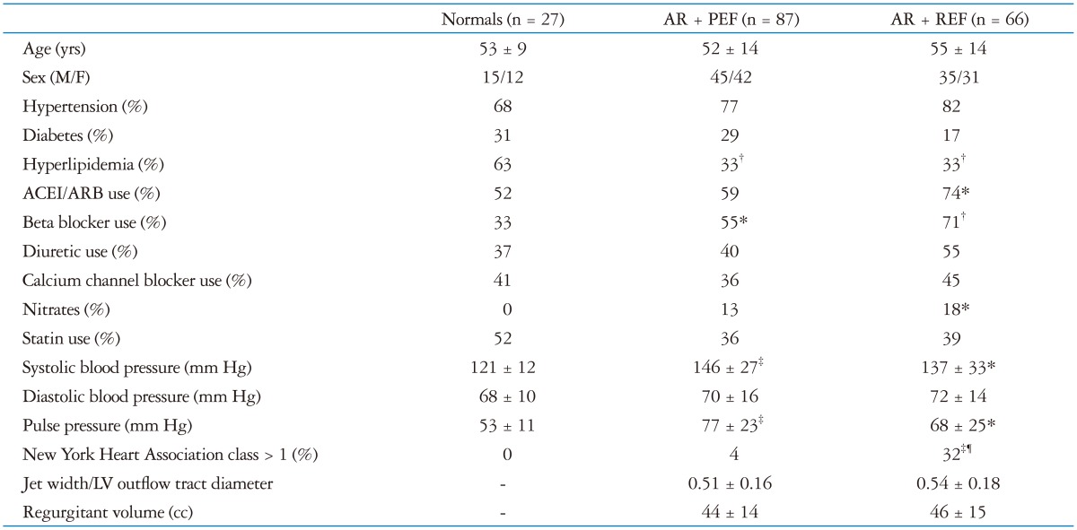
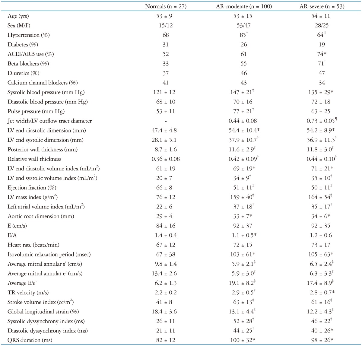
Table 1 summarizes patient characteristics in normals who are age and sex matched, AR + PEF patients, and AR + REF patients. The incidence of hyperlipidemia was lower in both AR groups. Angiotensin converting enzyme inhibitors/angiotensin receptor blockers use was more frequent in the AR + REF group as compared to normals and AR + PEF. Beta blockers were also more frequently used in both AR groups than in normals. Both systolic blood pressure and pulse pressure were greater in both AR groups. New York Heart Association functional class > 1 was more frequent in the AR + REF group than in normal subjects and patients with AR + PEF. The degree of AR was similar in both AR groups.
Table 1. Patient characteristics.
*p < 0.05, †p < 0.01, ‡p < 0.001 vs. normals; §p < 0.05, ∥p < 0.01, ¶p < 0.0001; AR + PEF vs. AR + REF. AR + PEF: chronic aortic regurgitation and preserved LV ejection fraction, AR + REF: chronic aortic regurgitation and reduced LV ejection fraction, ACEI/ARB: use of angiotensin converting enzyme inhibitor or angiotensin receptor blockers, LV: left ventricular
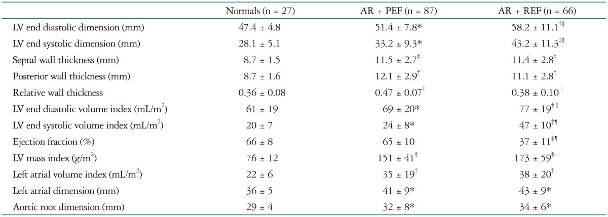
Table 2 summarizes the results for left atrial and LV size and function. Both AR groups demonstrated greater LV dimensions, wall thicknesses, LV volumes, and LV mass index. The AR + REF demonstrated greater dimensions and volume indexes than the PEF group. Relative wall thickness was greater in the AR + PEF group (concentric hypertrophy) than in both normal subjects and AR + REF (eccentric hypertrophy) groups. Left atrial volume index and aortic root size were increased in both AR groups.
Table 2. LV and left atrial diameters and volumes.
*p < 0.05, †p < 0.01, ‡p < 0.001 vs. normals; §p < 0.05, ∥p < 0.01, ¶p < 0.0001; AR + PEF vs. AR + REF. AR + PEF: chronic aortic regurgitation and preserved LV ejection fraction, AR + REF: chronic aortic regurgitation and reduced LV ejection fraction, LV: left ventricular
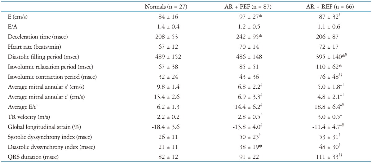
Table 3 summarizes Doppler indices of transmitral flow, transaortic flow, and mitral annular velocities. Patients with AR + PEF demonstrated higher E velocities than normal subjects and AR + REF. Diastolic filling period was shorter in AR + REF while the isovolumic contraction and relaxation periods were prolonged as compared to normal subjects. Mitral annular e' and s' velocities were reduced in AR + PEF and further reduced in AR + REF. Consequently the E/e' ratio was increased in AR + PEF and further increased in AR + REF as compared to normal subjects. TR velocities were increased in both AR groups.
Table 3. Transmitral spectral Doppler and tissue annular Doppler parameters.
*p < 0.05, †p < 0.01, ‡p < 0.001 vs. normals; §p < 0.05, ∥p < 0.01, ¶p < 0.0001; AR + PEF vs. AR + REF. AR + PEF: chronic aortic regurgitation and preserved left ventricular ejection fraction, AR + REF: chronic aortic regurgitation and reduced left ventricular ejection fraction, A: peak atrial filling velocity, E: peak mitral raid filling velocity, s': peak systolic mitral tissue Doppler velocity, e': peak rapid filling mitral annular velocity, TR: tricuspid regurgitation
Table 3 also summarizes the results of tissue Doppler parameters of longitudinal strain and dyssynchromy. Global longitudinal strain was significantly lower (less negative) in the AR + PEF group (despite normal EF) with further reductions (less negative) noted in the AR + REF group as compared to normal subjects which was consistent with New York Heart Association class symptoms > 1. Fig. 1 depicts the distribution of global longitudinal strain in normal subjects and both AR groups. There is a clear separation between normal subjects and AR + PEF despite similar LV EF with further reductions (less negative) noted in the AR + REF. Increased systolic and diastolic dyssynchrony were noted in both AR + PEF and AR + REF groups. Fig. 2 demonstrates systolic dyssynchrony in a patient with AR + PEF. QRS duration was prolonged in the AR + REF group as compared to AR + PEF and normal subjects.
Fig. 1. Individual patient values for global longitudinal strain are plotted for patients with normal function, patients with chronic AR and preserved LV ejection fraction (AR + PEF), and patients with chronic AR with reduced LV ejection fraction (AR + REF). There is a clear difference in the individual patient values for normals vs. both groups of AR patients. AR + PEF: chronic aortic regurgitation and preserved LV ejection fraction, AR + REF: chronic aortic regurgitation and reduced LV ejection fraction, LV: left ventricular.
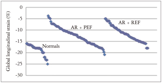
Fig. 2. Derived tissue Doppler recordings from the proximal septum and lateral wall are shown for a patient with moderate to severe chronic AR + PEF. The time to reach peak velocity was 110 msec later for the proximal lateral wall demonstrating systolic dyssynchrony. AR + PEF: chronic aortic regurgitation and preserved left ventricular ejection fraction.
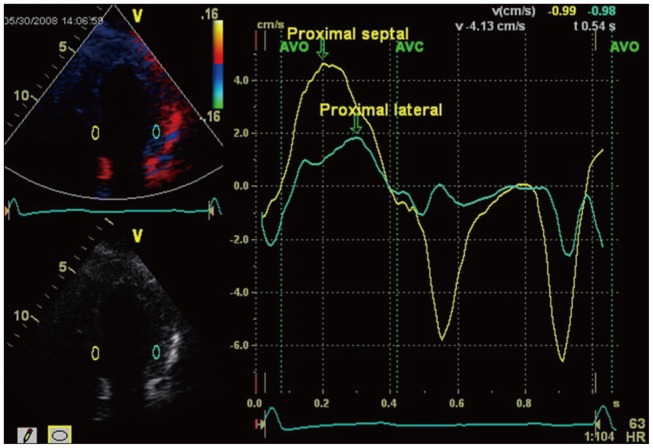
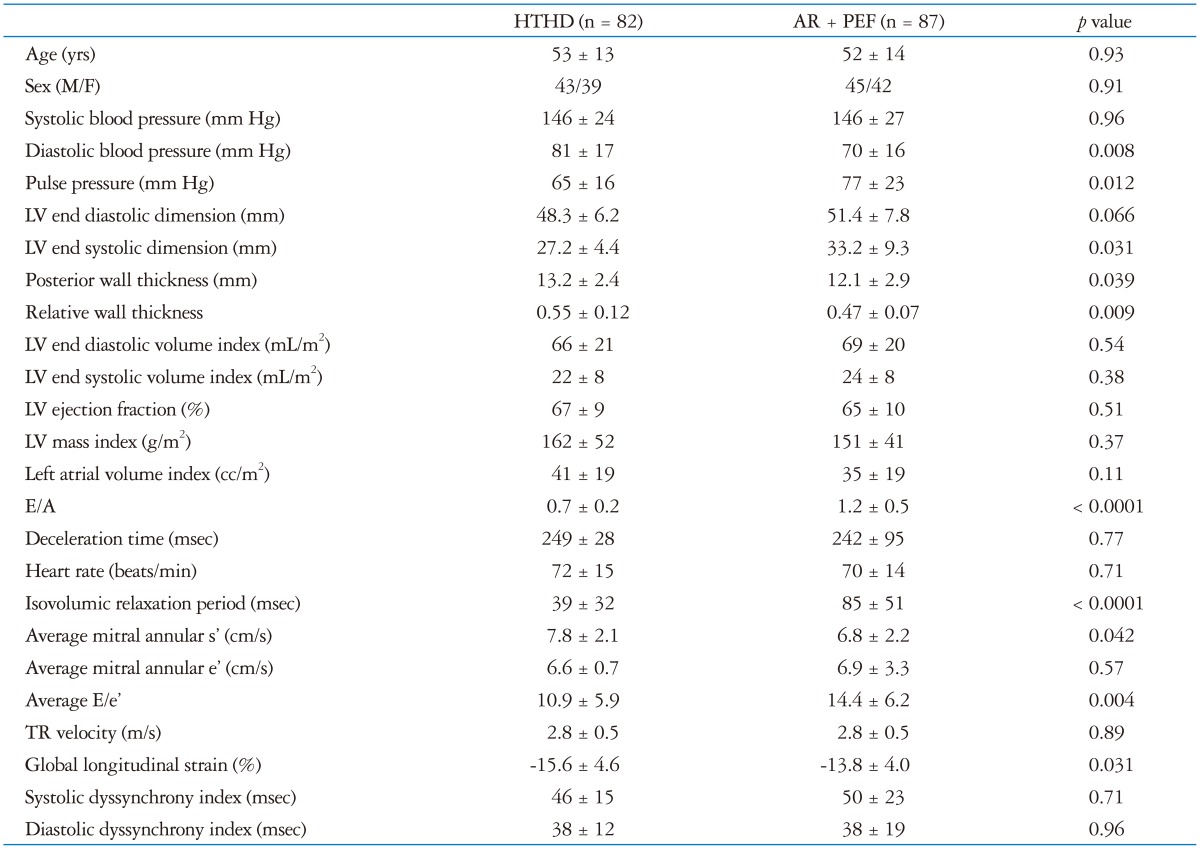
Table 4 compares patients with hypertensive heart disease with patients with AR + PEF. This comparison represents an attempt to determine whether an additional reduction in global longitudinal strain is associated with AR in addition to LV hypertrophy. Medication use and incidence of diabetes and hyperlipidemia were similar between the 2 groups (data not shown). Pulse pressure was greater in AR + PEF due to a lower diastolic blood pressure. Relative wall thickness was lower in AR + PEF but both groups still had concentric LV hypertrophy. This was due a nonsignificantly larger LV end diastolic dimension and a less thick posterior wall despite similar LV mass index in the AR + PEF group. Diastolic function indicated a higher E/peak atrial filling velocity in AR + PEF with a greater E/e' despite a longer isovolumic relaxation time and similar TR velocities. Mitral annular peak systolic velocity was lower, and global longitudinal strain was less negative (reduced) in AR + PEF. LV dyssynchrony was similar.
Table 4. Hypertensive heart disease vs. chronic AR with preserved ejection fraction.
HTHD: hypertensive heart disease, AR + PEF: chronic aortic regurgitation and preserved LV ejection fraction, LV: left ventricular A: peak atrial filling velocity, E: peak mitral raid filling velocity, s': peak systolic mitral tissue Doppler velocity, e': peak rapid filling mitral annular velocity, TR: tricuspid regurgitation
Table 5 summarizes the results for normal subjects, moderate AR, and severe AR. Both AR groups demonstrate differences from normal subjects with regard to systolic blood pressure, LV and left atrial volumes, diastolic function indices, global longitudinal strain, and dyssynchrony indexes. The only difference between the moderate and severe AR group resided in a lower incidence of hypertension in the severe group that was similar to normal subjects and a higher pulse pressure in the moderate AR group.
Table 5. Moderate vs. severe chronic aortic regurgitation.
*p < 0.05, †p < 0.01, ‡p < 0.001 vs. normals; §p < 0.05, ∥p < 0.01, ¶p < 0.0001; AR + PEF vs. AR + REF. AR + PEF: chronic aortic regurgitation and preserved LV ejection fraction, AR + REF: chronic aortic regurgitation and reduced LV ejection fraction, ACEI/ARB: use of angiotensin converting enzyme inhibitor or angiotensin receptor blockers, LV: left ventricular, A: peak atrial filling velocity, E: peak mitral raid filling velocity, s': peak systolic mitral tissue Doppler velocity, e': peak rapid filling mitral annular velocity, TR: tricuspid regurgitation
Forward stepwise regression indicated that global longitudinal strain was best correlated with the LV EF (p < 0.0001), E/e' (p < 0.0001), and the TR velocity (p = 0.0061). The overall correlation (r = 0.6416) was only moderate in strength.
Discussion
In this study, we demonstrated that in patients with AR + PEF there was significant longitudinal systolic dysfunction as characterized by reduced global longitudinal strain (less negative), reduced peak systolic mitral annular velocities, and systolic dyssynchrony. Diastolic function was abnormal with reduced peak early diastolic annular velocities, increased isovolumic relaxation times, increased E/e' ratio, increased TR velocities, elevated left atrial volume index, and diastolic dyssynchrony. Chronic AR + PEF patients surprisingly demonstrated significant concentric LV hypertrophy associated with larger LV volume index. Independent correlates of global longitudinal strain included LV EF, E/e', and TR velocity. Using a hypertensive cardiovascular group as a comparator, global longitudinal strain was still reduced. There were only 16 patients in the AR + PEF group without a history of hypertension and 12 without LV hypertrophy. The numbers were too few to perform meaningful statistical assessment as to whether AR + PEF without hypertension or LV hypertrophy resulted in reduced in global longitudinal strain. In AR + REF patients, there was eccentric hypertrophy with a further increase in LV volume indexes, and a similar increase in left atrial volume index. Greater abnormalities in longitudinal systolic function were noted in AR + REF likely related to a lower EF. Increased abnormalities in diastolic function as demonstrated by even greater reductions in the peak early diastolic mitral annular velocities and a greater increase in the E/e' and isovolumic relaxation time. There appear to be little difference between patient with moderate vs. severe AR with the exception of a lower incidence of hypertension in AR + REF and higher pulse pressure in AR + PEF.
Previous literature
Reduced longitudinal function in patients with aortic stenosis,5) heart failure with preserved EF,6) and in hypertensive heart disease7) has been previously described using mitral annular tissue velocities,13) tissue Doppler strain and strain rate,14),15) and speckle tracking.16) Similarly, in chronic AR patients, reduced longitudinal function has been noted using tissue Doppler17) and velocity vector imaging18) in patients with moderate or greater AR. Of note, in additional to global longitudinal (or regional) strain, abnormalities have been noted in global circumferential strain19) and radial strain20) in patients with severe AR. Using three-dimensional strain assessment, global longitudinal, global circumferential, and global area strain was noted to be reduced in patients with AR + PEF.21) Increased arterial pressure22) has been noted to be inversely related to global longitudinal strain. Finally, Park et al.23) has demonstrated that global longitudinal strain was a predictor of mortality in patient with chronic aortic regurgitation.
Our retrospective analysis indicates similar findings of reduced global longitudinal strain and reduced longitudinal systolic velocities (mitral annular) in a cohort of patients with preserved and reduced LV EF with moderate or severe AR. There were no differences in longitudinal function with moderate vs. severe AR. Furthermore, when AR + PEF was compared to a patient group with concentric hypertrophy (hypertensive cardiovascular disease), global longitudinal strain was reduced. Our study was larger and specifically segmented patients by EF which differs from previous studies. We did not find that arterial pressure was a predictor of global longitudinal strain. The inclusion of E/e' and TR velocity as independent predictors of global longitudinal strain are interesting findings. Both these indices might suggest that LV filling pressures are elevated11) but the use of E/e' as an estimate of LV filling pressure in AR has not been previously validated. We suspect that LV filling pressures might be elevated due to TR velocities averaging 2.8-3 m/s, increased left atrial volume indexes, and impaired relaxation. As few of these patients underwent left heart catheterization at the time of their echocardiogram, we are unable to provide additional insight.
An additional finding not previously noted was an increase in measures of systolic and diastolic dyssynchrony in both AR groups. Increases in these indices likely reflect abnormal isovolumic indices and was also seen in patient with hypertensive cardiovascular disease and likely related also to increased LV mass index.
Limitations
As this is a retrospective study, not all patents receiving an echocardiogram during the defined time period were selected for inclusion. Selection criteria resulted in 23 patients being excluded. It would be speculative to determine the effect of their inclusion on the data. Also, as patients were referred for echocardiography, there is a referral bias.
Unlike speckle tracking derived strain, tissue Doppler requires that the apical views be as parallel as possible to the imaging beam. The appropriate size of the sample volume used has never been established. We chose a 5 mm wide and 10 mm long sample volume to provide a long enough sample where the value would reflect the average of the area which would be similar to what is obtained with speckle tracking. Our intra-observer and inter-observer variability for tissue Doppler strain was both sufficiently small enough to allow the observed differences between groups to be meaningful but were larger than published values for speckle tracking.4) The rigid evaluation approach may have contributed to smaller values for intra-observer and inter-observer variability. The decision to divide AR patients based on EF was arbitrary. EF was chosen since valvular heart disease guidelines9) use severe AR and EF < 50% as an indication for aortic valve replacement. Separating groups on the basis of the degree of AR has limitations in the precision of the series of measurement to determine the extent of AR.9) Finally, there are always unknown and unmeasured differences not accounted for between groups.
Clinical implications
Reduced longitudinal systolic function as characterized by reduced global longitudinal strain and peak mitral annular systolic velocities in the setting of preserved LV EF indicates that EF may be a misleading indicator of LV systolic function in moderate or greater chronic AR. Heart failure in the setting of chronic AR is likely to portend a worse prognosis and yet the LV EF may be near normal9) in some patients. LV remodeling with increased LV volumes and mass result in a ventricular shape that is not conducive to longitudinal shortening and lengthening. The result may be a spherical left ventricle that may eject > 50% but does so with increased LV filling pressures and with impaired relaxation.
Conclusion
In this single center retrospective study, patient with AR + PEF demonstrated reduced global longitudinal strain associated with abnormal diastolic function with impaired relaxation and possibly elevated LV filling pressures in a remodeled LV with increased volume and mass. These findings are more accentuated than in patients with hypertensive cardiovascular disease who also demonstrate reduced longitudinal dysfunction with concentric hypertrophy. Patient with AR + REF demonstrated either similar findings or further significant reduction in the above indices.
References
- 1.Ishii K, Hirota Y, Suwa M, Kita Y, Onaka H, Kawamura K. Natural history and left ventricular response in chronic aortic regurgitation. Am J Cardiol. 1996;78:357–361. doi: 10.1016/s0002-9149(96)00295-0. [DOI] [PubMed] [Google Scholar]
- 2.Bonow RO, Lakatos E, Maron BJ, Epstein SE. Serial long-term assessment of the natural history of asymptomatic patients with chronic aortic regurgitation and normal left ventricular systolic function. Circulation. 1991;84:1625–1635. doi: 10.1161/01.cir.84.4.1625. [DOI] [PubMed] [Google Scholar]
- 3.Geyer H, Caracciolo G, Abe H, Wilansky S, Carerj S, Gentile F, Nesser HJ, Khandheria B, Narula J, Sengupta PP. Assessment of myocardial mechanics using speckle tracking echocardiography: fundamentals and clinical applications. J Am Soc Echocardiogr. 2010;23:351–369. quiz 453-5. doi: 10.1016/j.echo.2010.02.015. [DOI] [PubMed] [Google Scholar]
- 4.Hoit BD. Strain and strain rate echocardiography and coronary artery disease. Circ Cardiovasc Imaging. 2011;4:179–190. doi: 10.1161/CIRCIMAGING.110.959817. [DOI] [PubMed] [Google Scholar]
- 5.Sengupta SP, Caracciolo G, Thompson C, Abe H, Sengupta PP. Early impairment of left ventricular function in patients with systemic hypertension: new insights with 2-dimensional speckle tracking echocardiography. Indian Heart J. 2013;65:48–52. doi: 10.1016/j.ihj.2012.12.009. [DOI] [PMC free article] [PubMed] [Google Scholar]
- 6.Kraigher-Krainer E, Shah AM, Gupta DK, Santos A, Claggett B, Pieske B, Zile MR, Voors AA, Lefkowitz MP, Packer M, McMurray JJ, Solomon SD PARAMOUNT Investigators. Impaired systolic function by strain imaging in heart failure with preserved ejection fraction. J Am Coll Cardiol. 2014;63:447–456. doi: 10.1016/j.jacc.2013.09.052. [DOI] [PMC free article] [PubMed] [Google Scholar]
- 7.Kearney LG, Lu K, Ord M, Patel SK, Profitis K, Matalanis G, Burrell LM, Srivastava PM. Global longitudinal strain is a strong independent predictor of all-cause mortality in patients with aortic stenosis. Eur Heart J Cardiovasc Imaging. 2012;13:827–833. doi: 10.1093/ehjci/jes115. [DOI] [PubMed] [Google Scholar]
- 8.Cuspidi C, Negri F, Giudici V, Sala C, Mancia G. Impaired midwall mechanics and biventricular hypertrophy in essential hypertension. Blood Press. 2010;19:234–239. doi: 10.3109/08037051003606413. [DOI] [PubMed] [Google Scholar]
- 9.Nishimura RA, Otto CM, Bonow RO, Carabello BA, Erwin JP, 3rd, Guyton RA, O'Gara PT, Ruiz CE, Skubas NJ, Sorajja P, Sundt TM, 3rd, Thomas JD American College of Cardiology/American Heart Association Task Force on Practice Guidelines. 2014 AHA/ACC guideline for the management of patients with valvular heart disease: executive summary: a report of the American College of Cardiology/American Heart Association Task Force on Practice Guidelines. J Am Coll Cardiol. 2014;63:2438–2488. doi: 10.1016/j.jacc.2014.02.537. [DOI] [PubMed] [Google Scholar]
- 10.Lang RM, Badano LP, Mor-Avi V, Afilalo J, Armstrong A, Ernande L, Flachskampf FA, Foster E, Goldstein SA, Kuznetsova T, Lancellotti P, Muraru D, Picard MH, Rietzschel ER, Rudski L, Spencer KT, Tsang W, Voigt JU. Recommendations for cardiac chamber quantification by echocardiography in adults: an update from the American Society of Echocardiography and the European Association of Cardiovascular Imaging. J Am Soc Echocardiogr. 2015;28:1–39.e14. doi: 10.1016/j.echo.2014.10.003. [DOI] [PubMed] [Google Scholar]
- 11.Nagueh SF, Appleton CP, Gillebert TC, Marino PN, Oh JK, Smiseth OA, Waggoner AD, Flachskampf FA, Pellikka PA, Evangelista A. Recommendations for the evaluation of left ventricular diastolic function by echocardiography. J Am Soc Echocardiogr. 2009;22:107–133. doi: 10.1016/j.echo.2008.11.023. [DOI] [PubMed] [Google Scholar]
- 12.Yu CM, Sanderson JE, Marwick TH, Oh JK. Tissue Doppler imaging a new prognosticator for cardiovascular diseases. J Am Coll Cardiol. 2007;49:1903–1914. doi: 10.1016/j.jacc.2007.01.078. [DOI] [PubMed] [Google Scholar]
- 13.Seo JS, Kim DH, Kim WJ, Song JM, Kang DH, Song JK. Peak systolic velocity of mitral annular longitudinal movement measured by pulsed tissue Doppler imaging as an index of global left ventricular contractility. Am J Physiol Heart Circ Physiol. 2010;298:H1608–H1615. doi: 10.1152/ajpheart.01231.2009. [DOI] [PubMed] [Google Scholar]
- 14.Mele D, Censi S, La Corte R, Merli E, Lo Monaco A, Locaputo A, Ceconi C, Trotta F, Ferrari R. Abnormalities of left ventricular function in asymptomatic patients with systemic sclerosis using Doppler measures of myocardial strain. J Am Soc Echocardiogr. 2008;21:1257–1264. doi: 10.1016/j.echo.2008.08.004. [DOI] [PubMed] [Google Scholar]
- 15.Kuznetsova T, Herbots L, Richart T, D'hooge J, Thijs L, Fagard RH, Herregods MC, Staessen JA. Left ventricular strain and strain rate in a general population. Eur Heart J. 2008;29:2014–2023. doi: 10.1093/eurheartj/ehn280. [DOI] [PubMed] [Google Scholar]
- 16.Cho GY, Marwick TH, Kim HS, Kim MK, Hong KS, Oh DJ. Global 2-dimensional strain as a new prognosticator in patients with heart failure. J Am Coll Cardiol. 2009;54:618–624. doi: 10.1016/j.jacc.2009.04.061. [DOI] [PubMed] [Google Scholar]
- 17.Marciniak A, Sutherland GR, Marciniak M, Claus P, Bijnens B, Jahangiri M. Myocardial deformation abnormalities in patients with aortic regurgitation: a strain rate imaging study. Eur J Echocardiogr. 2009;10:112–119. doi: 10.1093/ejechocard/jen185. [DOI] [PubMed] [Google Scholar]
- 18.Tayyareci Y, Yildirimturk O, Aytekin V, Demiroglu IC, Aytekin S. Subclinical left ventricular dysfunction in asymptomatic severe aortic regurgitation patients with normal ejection fraction: a combined tissue Doppler and velocity vector imaging study. Echocardiography. 2010;27:260–268. doi: 10.1111/j.1540-8175.2009.01053.x. [DOI] [PubMed] [Google Scholar]
- 19.Mizarienė V, Bučytė S, Zaliaduonytė-Pekšienė D, Jonkaitienė R, Janėnaitė J, Vaškelytė J, Jurkevičius R. Components of left ventricular ejection and filling in patients with aortic regurgitation assessed by speckle-tracking echocardiography. Medicina (Kaunas) 2012;48:31–38. [PubMed] [Google Scholar]
- 20.Knutsen AK, Ma N, Taggar AK, Brady BD, Cupps BP, Pasque MK. Heterogeneous distribution of left ventricular contractile injury in chronic aortic insufficiency. Ann Thorac Surg. 2012;93:1121–1127. doi: 10.1016/j.athoracsur.2011.12.067. [DOI] [PMC free article] [PubMed] [Google Scholar]
- 21.Li CM, Li C, Bai WJ, Zhang XL, Tang H, Qing Z, Li R. Value of three-dimensional speckle-tracking in detecting left ventricular dysfunction in patients with aortic valvular diseases. J Am Soc Echocardiogr. 2013;26:1245–1252. doi: 10.1016/j.echo.2013.07.018. [DOI] [PubMed] [Google Scholar]
- 22.Mizariene V, Grybauskiene R, Vaskelyte J, Jonkaitiene R, Pavilioniene J, Jurkevicius R. Strain value in the assessment of left ventricular function and prediction of heart failure markers in aortic regurgitation. Echocardiography. 2011;28:983–992. doi: 10.1111/j.1540-8175.2011.01483.x. [DOI] [PubMed] [Google Scholar]
- 23.Park SH, Yang YA, Kim KY, Park SM, Kim HN, Kim JH, Jang SY, Bae MH, Lee JH, Yang DH. Left ventricular strain as predictor of chronic aortic regurgitation. J Cardiovasc Ultrasound. 2015;23:78–85. doi: 10.4250/jcu.2015.23.2.78. [DOI] [PMC free article] [PubMed] [Google Scholar]


