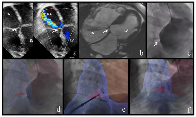
A 16 year old with double outlet right ventricle, D-malposed great vessels and a subpulmonary ventricular septal defect (VSD) status-post surgical VSD patch closure and arterial switch procedure at two months of age, reported progressive exercise intolerance. He was found to have moderate right atrial enlargement, mild dilation of right and left ventricles and a persistent residual left ventricle to right atrium (LV-RA) intracardiac shunt on echocardiographic assessment, Figure 1a and Movie 1, (similar physiology to a Gerbode type defect). Cardiac magnetic resonance imaging (MRI) delineated the LV-RA shunt, Figure 1b and Movie 1, (steady-state free precession cine) with estimated Qp:Qs of 1.4:1 (velocity-encoded MRI). Cardiac MRI derived left ventricular end diastolic volume was 132 ml/m2 (z-score = +3.6) with normal ejection fraction (EF) of 55% and right ventricular end diastolic volume was 136 ml/m2 (z-score = +2.2) with EF of 45%.1 Given his symptoms and progressive right heart dilation he was referred for percutaneous device closure.
Figure 1.
Image Fusion Guided Device Closure of Left Ventricle to Right Atrium Shunt. A. Pre-intervention trans-thoracic echocardiogram, 4-chamber view in color compare mode showing left ventricle to right atrium (LV-RA) shunt (white arrow). B. Pre-intervention steady-state free precession cardiac MRI (Siemens 1.5 Tesla) with noncontrast angiographic sequences, assisted by respiratory compensation and EKG-based cardiac gating. The magnetic resonance image shows right heart enlargement and LV-RA shunt (white arrow). The defect diameter is 3 mm and velocity-encoded MRI Qp:Qs is 1.4:1. C. The baseline conventional left ventriculogram shows the LV-RA shunt (white arrow). D. XFM [x-ray fused with MRI] overlay baseline left ventriculogram (blue = right ventricle, light pink = left ventricle, dark pink = LV-RA shunt) highlights the intracardiac shunt. E. Following XFM guided wire crossing of the LV-RA defect, this panel shows XFM guided defect closure with a muscular VSD occluder. The device is deployed in the defect and still attached to the delivery cable. F. The post-intervention left ventriculogram with XFM overlay shows successful device closure of the LV-RA defect.
Invasive hemodynamic assessment yielded Qp:Qs of 1.5:1 (Fick) and normal pulmonary vascular resistance. A three-dimensional map of the cardiac chambers and LV-RA shunt was manually extracted from the cardiac MRI using previously described techniques.2 Live X-ray, Figure 1c and Movie 1, was fused with this three-dimensional MRI map (XFM) (MR fusion and overlay, Siemens, Forchheim, Germany) with automatic correction for gantry and table position to guide closure of the LV-RA shunt with an Amplatzer Congenital Muscular VSD Occluder (St. Jude Medical, Minneapolis, MN, USA). XFM roadmaps optimized gantry angle and simplified wire crossing of the defect without requiring additional iodinated contrast, Figure 1d–f and Movie 1. One month afterwards, the patient was asymptomatic with greatly improved exercise ability. A transthoracic echo was performed one year post-procedure with the following results: no residual shunt, normal right atrial size (2D dimension = 3.9 cm versus 5.3 cm pre-procedure), mildly dilated right and left ventricles (M-MODE RVDD 2.1 cm, LVIDd 5.4 cm, LVIDs 3.6 cm versus pre-procedure M-MODE RVDD 2.3 cm, LVIDd 5.0 cm, LVIDs 3.1 cm) with normal biventricular systolic function.
XFM simplifies complex interventions by providing three-dimensional procedural guidance in the familiar X-ray working environment.2,3 It may also reduce radiation and contrast exposure.4
Supplementary Material
Acknowledgments
Funding Sources: This work is funded by the National Heart Lung and Blood Institute (HHSN268201500001C and Z01-HL006039).
Footnotes
Disclosures: Children’s National Medical Center and Siemens Medical have a master research agreement.
References
- 1.Buechel EV, Kaiser T, Jackson C, Schmitz A, Kellenberger CJ. Normal right and left ventricular volumes and myocardial mass in children measured by steady state free precession cardiovascular magnetic resonance. J Cardiovasc Magn Reson. 2009;11:19. doi: 10.1186/1532-429X-11-19. [DOI] [PMC free article] [PubMed] [Google Scholar]
- 2.Ratnayaka K, Raman VK, Faranesh AZ, Sonmez M, Kim JH, Gutierrez LF, Ozturk C, McVeigh ER, Slack MC, Lederman RJ. Antegrade percutaneous closure of membranous ventricular septal defect using X-ray fused with magnetic resonance imaging. JACC Cardiovasc Interv. 2009;2:224–230. doi: 10.1016/j.jcin.2008.09.014. [DOI] [PMC free article] [PubMed] [Google Scholar]
- 3.Glöckler M, Halbfaβ J, Koch A, Achenbach S, Dittrich S. Multimodality 3D-roadmap for cardiovascular interventions in congenital heart disease--a single-center, retrospective analysis of 78 cases. Catheter Cardiovasc Interv. 2013;82:436–42. doi: 10.1002/ccd.24646. [DOI] [PubMed] [Google Scholar]
- 4.Abu-Hazeem AA, Dori Y, Whitehead KK, Harris MA, Fogel MA, Gillespie MJ, Rome JJ, Glatz AC. X-ray magnetic resonance fusion modality may reduce radiation exposure and contrast dose in diagnostic cardiac catheterization of congenital heart disease. Catheter Cardiovasc Interv. 2014;84:795–800. doi: 10.1002/ccd.25473. [DOI] [PubMed] [Google Scholar]
Associated Data
This section collects any data citations, data availability statements, or supplementary materials included in this article.


