Abstract
Background
Angiomyolipoma is a benign mesenchymal tumor composed of variable amounts of smooth muscle, adipose tissue and thick-walled blood vessels, and usually named PEComas (perivascular epithelioid cell tumors). PEComas share overlapping histopathological features with epithelioid cells along a perivascular distribution and characteristic immunohistochemistry with coexpression of myoid and melanocytic markers (HMB-45 /or Melan-A). We report the first case of primary orbital angiomyolipoma with negative melanocytic marker.
Case presentation
An 80-year-old Asian woman had a 2-year history of progressive swelling in the left upper eyelid. External examination revealed 3 cm of relative proptosis of the left eye and a palpable mass in the left superonasal orbit. Computed tomographic scan demonstrated a circumscribed, heterogeneous orbital mass. Excision biopsy was done and the histological finding demonstrated the orbital mass was composed of mature adipocytes, intermingled with spindle or oval-shaped cells, and accompanied by thick-walled blood vessels. Immunohistochemically, tumor cells were positive for CD34 and HHF-35, but negative for cytokeratin, HMB-45 and Melan-A. The diagnosis of angiomyolipoma was made. No recurrence was noted at 2-year follow-up.
Conclusion
In our case, the HMB-45 negativity may be explained by the rarity of the epithelioid cells, and the HMB-45 positivity is often weaker or absent in spindle cells. Angiomyolipoma, although rare, should be added to the differential diagnosis of space-occupying orbital lesion.
Keywords: Orbit, Angiomyolipoma, Perivascular epithelioid cell tumors, HMB-45
Background
Angiomyolipoma, originally thought to be a hamartoma, is a benign mesenchymal tumor composed of variable amounts of smooth muscle, adipose tissue and thick-walled blood vessels, and usually named PEComas (perivascular epithelioid cell tumors). It occurs most commonly in the kidney as a sporadic case or as part of the tuberous sclerosis complex [1]. We presented the first case of primary orbital angiomyolipoma with negative melanocytic markers.
Case presentation
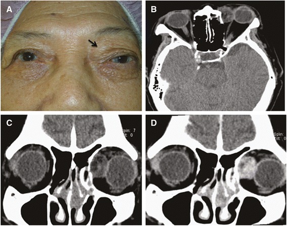
An 80-year-old woman had a 2-year history of progressive fullness in the left upper eyelid. External examination revealed 3 mm of relative proptosis of the left eye and a nontender palpable firm mass in the left superonasal orbit (Fig. 1a). The remainder of the ocular examination was within normal limit. Past medical history was otherwise unremarkable. Computed tomographic scan demonstrated a circumscribed, heterogeneous orbital mass displacing the left globe laterally (Fig. 1b, c and d). Surgical removal of the tumor was performed through anterior orbitotomy in an en bloc fashion. At the time of surgery, the 1.8 × 1.8 × 1.3 cm yellowish mass was encapsulated and solid (Fig. 2a). Histologically, the orbital mass was composed of mature adipocytes, intermingled with spindle or oval-shaped cells with eosinophilic cytoplasm, accompanied by thick-walled blood vessels (Fig. 2b). Immunohistochemically, tumor cells were positive for CD34 and HHF-35 (Fig. 2c and d), but negative for cytokeratin, HMB-45 and Melan-A. These findings confirmed the diagnosis of angiomyolipoma. Systemic check-up was unremarkable. No recurrence was noted at 2-year follow-up.
Fig. 1.

Preoperative photograph shows a palpable mass on left superonasal orbit (a, arrow). Axial (b) and coronal (c) planes of computed tomographic scan show a circumscribed soft tissue mass on the left superonasal orbit with heterogenic density containing areas of fat attenuation. The mass demonstrates heterogenous enhancement (d)
Fig. 2.

Grossly, the tumor is a 1.8 × 1.8 × 1.3 cm yellowish, encapsulated, solid mass (a). Microscopic features of the tumor shows it is comprised of spindle to ovoid-shaped muscle cells interspersed with irregular vascular channels and mature adipocytes (b, hematoxylin and eosin stain, original magnification, ×100). Immunohistochemical staining reveals that the proliferating vessels are positive for CD34 (c) and smooth muscle cells are positive for HHF-35 (d) (original magnification, ×100)
Primary orbital angiomyolipoma is a rare entity of orbital tumor. Until now, only 4 cases of ocular perivascular epithelioid cell tumor (PEComa) have been reported, and all had positive melanocytic markers [2–4]. All reported 4 cases of ocular PEComas were female and their tumor location was eyelid (2 cases), ciliary body (1 case), and orbit (1 case) respectively. PEComas often share overlapping histopathological features with epithelioid cells along a perivascular distribution and characteristic immunohistochemistry with coexpression of myoid and melanocytic markers (HMB-45 /or Melan-A) [5]. Current case is unique in that the tumor lacked reactivity for melanin-associated antigens HMB-45 and Melan-A, which is similar to some angiomyolipomas from skin, head and neck [6–9]. The HMB-45 negativity may be explained by the rarity of the epithelioid cells in these cases, and the HMB-45 positivity is often weaker or absent in spindle cells [9]. In addition, these angiomyolipomas are usually relative small, contrary to what happens to kidney and liver tumors, which are often large. However, because of the small number of reported cases, whether these HMB-negative angiomyolipoma is a new variant of PEComas require further investigation. Differential diagnosis should include giant cell angiofibroma which is a highly vascular tumor comprising a spindle-cell proliferation with numerous multinucleated giant cells and pseudovascular spaces, and immunohistochemically positive for CD34, CD99, and vimentin [10].
Approximately one third of renal angiomyolipomas occur in patients with tuberous sclerosis. However, this association has been rarely reported in extrarenal angiomyolipoma, including of ocular angiomyolipoma. Because most angiomyolipomas contain varied amounts of adipose tissue, image features of fat attenuation at unenhanced CT may help in diagnosis. Although most angiomyolipomas show a benign course, some reports have suggested that histologically atypical angiomyolipomas are potentially malignancy. Therefore, wide excision and regular follow-up are warranted.
Conclusion
In summary, we report a case of primary orbital angiomyolipoma, which showed different immunohistochemical features from prior reported ocular PEComa. Although rare, angiomyolipoma should be added to the differential diagnosis of space-occupying orbital lesion.
Consent
Written informed consent was obtained from the patient for publication of this case report and any accompanying images. A copy of the written consent is available for review by the editor of this journal.
Acknowledgement
This study was partially supported by a grant (V104-C-092) from Taipei Veterans General Hospital, Taipei, Taiwan, and a grant (104-2314-B-075 -056 -MY2) from Ministry of Science and Technology, Taiwan.
Abbreviations
- PEComas
Perivascular epithelioid cell tumors
- HHF35
Muscle actin antibody
- HMB-45
Melanosome specific antigen
- Melan-A
Melanoma antigen
- CT
Computed tomography
Footnotes
Competing interests
The authors declare that they have no competing interests.
Authors’ contributions
CYL and HCK drafted this manuscript, collected the data, and reviewed the literature. WKY and SCK reviewed the literature. CCT interpreted the data, and critically reviewed the manuscript. CJLL critically reviewed the manuscript finally. All authors read and approved the final manuscript.
Contributor Information
Che-Yu Lin, Email: alex740328@gmail.com.
Chieh-Chih Tsai, Phone: 886-2-28757325, Email: cctsai1234@yahoo.com.
Hui-Chuan Kau, Email: hckau1234@yahoo.com.
Wei-Kuang Yu, Email: wkyu3@vghtpe.gov.tw.
Shu-Ching Kao, Email: sckao@vghtpe.gov.tw.
Catherine Jui-Ling Liu, Email: jlliu@vghtpe.gov.tw.
References
- 1.Neumann HP, Schwarzkopf G, Henske EP. Renal angiomyolipomas, cyst, and cancer in tuberous sclerosis complex. Semin Pediatr Neurol. 1998;5:269–75. doi: 10.1016/S1071-9091(98)80005-3. [DOI] [PubMed] [Google Scholar]
- 2.Iyengar P, Deangelis DD, Greenberg M, Taylor G. Perivascular epithelioid cell tumor of the orbit: a case report and review of the literature. Pediatr Dev Pathol. 2005;8:98–104. doi: 10.1007/s10024-004-5055-0. [DOI] [PubMed] [Google Scholar]
- 3.Guthoff R, Guthoff T. Perivascular epithelioid cell tumor of the orbit. Arch Ophthalmol. 2008;126:1009–11. doi: 10.1001/archopht.126.7.1009. [DOI] [PubMed] [Google Scholar]
- 4.Furusato E, Cameron JD, Newsom RW, Fujishiro T, Kojima T, Specht CS, et al. Ocular perivascular epithelioid cell tumor: report of 2 cases with distinct clinical presentations. Hum Pathol. 2010;41:768–72. doi: 10.1016/j.humpath.2009.12.006. [DOI] [PubMed] [Google Scholar]
- 5.Schoolmeester JK, Howitt BE, Hirsch MS, Dal Cin P, Quade BJ, Nucci MR. Perivascular epithelioid cell neoplasm (PEComa) of the gynecologic tract: clinicopathologic and immunohistochemical characterization of 16 cases. Am J Surg Pathol. 2014;38:176–88. doi: 10.1097/PAS.0000000000000133. [DOI] [PubMed] [Google Scholar]
- 6.Alvarez Alvarez C, Fernández Sanromán J, Fernández Castilla M, Badiola A. Sporadic oral angiomyolipoma. Case report. Med Oral Patol Oral Cir Bucal. 2007;12:E391–3. [PubMed] [Google Scholar]
- 7.Watanabe K, Suzuki T. Mucocutaneous angiomyolipoma. A report of 2 cases arising in the nasal cavity. Arch Pathol Lab Med. 1999;123:789–92. doi: 10.5858/1999-123-0789-MA. [DOI] [PubMed] [Google Scholar]
- 8.da Silva AA, Carlos R, Contreras E, de Almeida OP, Lopes MA, Vargas PA. Angiomyolipoma of the upper lip: case report and review of the literature. Med Oral Patol Oral Cir Bucal. 2007;12:E101–4. [PubMed] [Google Scholar]
- 9.Foschini MP, Corti B, DaCol M, Cenzi M, Zanella MD, Barbazza R. Angiomyolipoma of the parotid gland: a case report. Oral Surg Oral Med Oral Pathol Oral Radiol Endod. 1999;87:738–41. doi: 10.1016/S1079-2104(99)70172-1. [DOI] [PubMed] [Google Scholar]
- 10.Zoumalan CI, Egbert PR, Warwar RE, McCulley TJ. Orbital giant cell angiofibroma recurring as a solitary fibrous tumor. Ophthal Plast Reconstr Surg. 2008;24:325–7. doi: 10.1097/IOP.0b013e31817dd6f5. [DOI] [PubMed] [Google Scholar]


