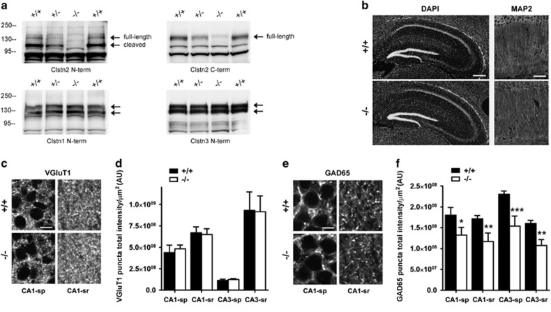
Figure 1.
Clstn2 −/− mice show reductions of an inhibitory but not excitatory synaptic marker. (a) Western blot analysis of post-nuclear brain samples reveals loss of calsyntenin-2 in Clstn2−/− mice but no change in the levels of other calsyntenins. The signal remaining with the C-terminal antibody (arrow) likely represents other calsyntenins (Supplementary Figure S1). Note the gene dosage effect on calsyntenin-2 expression in the heterozygous brain. (b) Clstn2−/− mice showed no gross anatomical differences from WT, as shown here in hippocampal regions with DAPI nuclear stain, and in CA1 regions with immunofluorescence staining for the dendritic marker microtubule associated protein 2 (MAP2). Scale bars, 200 μm (left) and 50 μm (right). (c, d) VGluT1 puncta immunofluorescence was unaltered in hippocampal regions of Clstn2−/− mice. Two-way repeated measures (RM) ANOVA, no genotype effect, F(1,16)=0.0041, P=0.95. Scale bar, 10 μm. (e, f) GAD65 puncta immunofluorescence was reduced in all regions analyzed in Clstn2−/−mice. Two-way RM ANOVA, genotype effect F(1,8)=92.0, P<0.0001 and *P<0.05, **P<0.01 and ***P<0.001 by Bonferroni post hoc test comparing with WT for the same region. Scale bar, 10 μm. sp, stratum pyramidale; sr, stratum radiatum; AU, arbitrary units.

