

Abstract
Patient: Male, 36
Final Diagnosis: Multiple concomitant right upper limb fractures
Symptoms: —
Medication: —
Clinical Procedure: Open reduction and internal fixation of right upper limb fractures
Specialty: Orthopedics and Traumatology
Objective:
Rare co-existance of disease or pathology
Background:
This report is about unusual multiple upper extremity concomitant injuries in an adult after a fall from a height. To the best of our knowledge this is the first reported case of concomitant ipsilateral occurrence of multiple common injuries, uncommonly occurring together in a single traumatic episode.
Case Report:
A 36-year-old right-handed man fell through a skylight to the floor about 4 meters below. He presented with multiple concomitant injuries in his right upper extremity: elbow dislocation with radial head fracture associated with distal radius, ulnar styloid, and scaphoid fractures.
Conclusions:
The probable mechanism of injury along with the surgical treatment of these previously undescribed injuries is discussed to emphasize the need to clinically examine the whole upper extremity in severe injuries. The awareness of such an association for early recognition is paramount for excellent clinical results.
MeSH Keywords: Dislocations; Elbow Joint; Fractures, Bone; Radius Fractures; Scaphoid Bone
Background
The elbow joint is the second most commonly dislocated joint in adults after the shoulder joint [1]. Usually the mechanism of injury is a fall on an outstretched hand, which leads to disruption of the ligaments and soft tissues around the elbow, which result in loss of the static stabilizers of the ulnohumeral joint [1,2]. Posterior elbow dislocation is associated with other injuries in around 10–15% of cases, radial head fracture being one of them [1].
Distal radius fracture is the most common fracture treated by orthopedic surgeons [3]. The most common carpal bone fracture is scaphoid fracture and other upper limb fractures are associated with it in 5–10% of the cases [4]. Like distal radius fracture, scaphoid fracture usually results from a fall on the extended wrist or a forced dorsiflexion injury of the wrist [5]. Various combinations of upper extremity injuries have been reported in the literature [4,6–8]; however, to the best of our knowledge the combination presented in this paper has never been reported. This case is reported to stress the need to search for possible missed injuries in a severely injured patient.
Case Report
A 36-year-old right-handed man fell about four meters through a skylight while trying to repair his own satellite.
On arrival to the emergency room (ER), his right elbow was in an awkward position supported by his left wrist. He could not recall how he impacted the ground. He denied any history of loss of consciousness. There was no history of previous trauma to his right upper limb or use of long – term medication. He gave history of smoking 60 cigarettes per day.
His right elbow was markedly deformed with gross swelling. There were mild superficial abrasions over the dorsal side of his right mid forearm and the ulnar side of his right wrist. Elbow and wrist were tender to touch with inability to move the right elbow. There was mild crepitation noted over his right wrist. His neurovascular examination was normal.
Radiographic examination of the right elbow showed posterolateral elbow dislocation with radial head fracture (Figure 1A, 1B). Wrist radiographs showed a dorsally displaced metaphyseal distal radius fracture with mild metaphyseal comminution and ulnar styloid fracture (Figure 2A, 2B).
Figure 1.

Initial X-rays in Emergency Department, anteroposterior (A) and lateral (B) views of right elbow showing posterolateral elbow dislocation with radial head fracture.
Figure 2.

Initial x-rays in Emergency Department, anteroposterior (A) and lateral (B) views of right wrist showing comminuted fracture distal radius.
In the ER a trial of elbow closed reduction under conscious sedation failed. His elbow and wrist were immobilized in an above-elbow posterior slab. Informed consent was taken and the patient was prepared for an urgent operative intervention.
Under general anesthesia with the patient in supine position, the elbow was reduced under image intensifier (I I) control. Examining the elbow range of motion under I I revealed widening of the ulnohumeral joint and a mildly displaced radial head fracture. The presence of joint widening and a displaced radial head fracture were the indication for an open ligament repair with stabilization or excision of the radial head fracture [9]. Examination of the wrist under I I revealed a middle third scaphoid fracture (Figure 3A, 3B), which was missed in the initial assessment in ER, in addition to the distal radius fracture with the tip of ulnar styloid fracture.
Figure 3.

Intraoperative anteroposterior (A) and lateral (B) wrist views under image intensifier showing fracture distal radius, middle third scaphoid, and ulnar styloid tip.
The elbow was approached through a posterior approach just lateral to the tip of the olecranon with elevation of a lateral-based full fasciocutaneous flap. The fractured radial head-piece was excised (Figure 4), as it was a thin osteocartilaginous piece that was difficult to stabilize.
Figure 4.

Intraoperative anteroposterior elbow view under image intensifier showing radial head fracture.
A complete avulsion of the lateral collateral ligament (LCL) from the lateral humeral condyle was noticed along with partial avulsion of the common extensor origin. The LCL and the extensor origin were repaired using suture anchors (GII Quick Anchor plus, DePuy Synthes, Mitek Sport Medicine) (Figure 5A, 5B).
Figure 5.

Anteroposterior (A) and lateral (B) elbow views showing congruent elbow joint reduction, post repair of the lateral collateral ligament, extensor origin, and excision of the radial head fragment.
The distal radius fracture was stabilized through a volar approach using a volar distal radius plate and screws (Volar 2.4 mm LCP Distal Radius System, DePuy Synthes). The scaphoid was stabilized with a mini screw (2.4 mm Cortex Screw, DePuy Synthes) (Figure 6A, 6B); the distal radioulnar joint was found stable so it was decided not to stabilize the ulnar styloid fracture. The elbow was found to be stable with concentric reduction under I I after the repair, so a long arm cast including the thumb with the forearm in pronation was applied.
Figure 6.
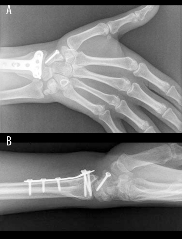
Anteroposterior (A) and lateral (B) wrist views showing healed distal radius and scaphoid fractures. Note: the ulnar styloid was not stabilized.
The cast was kept on for three weeks and then a custom-made hinged elbow brace extending to the wrist and thumb was applied where he was allowed to flex and extend his elbow while keeping the forearm in pronation for another six weeks. Patient started to perform elbow and forearm range of motion exercises, including forearm rotation, after brace removal.
Scaphoid cast was kept on for a total of 12 weeks to protect the scaphoid and distal radius (Figure 6A, 6B).
He was closely followed up by a physiotherapist to recover his wrist and elbow range of motion. An occupational therapist was involved to help the patient regain his hand function and grip power. The patient was followed up for about 18 months, during which he regained almost near normal range of motion compared to the normal side (Figure 7A–7F].
Figure 7.

Clinical appearance of right upper limb range of motion 16 months postoperatively showing excellent range of motion; elbow extension (A) and flexion (B), wrist dorsiflexion (C) and palmar flexion (D), forearm supination (E) and pronation (F).
Discussion
One can only speculate on the exact mechanism of injury in this case since the injury was not witnessed and the patient could not recall it. Posterolateral elbow dislocation may occur secondary to an elbow joint valgus injury combined with axial load and supination [10,11]. Widening of the ulnohumeral joint following closed reduction of a dislocated elbow may be a result of an entrapped osteochondral fragment or soft tissue interposition in which surgical intervention is indicated [9,12]. Joint widening may also be a sign of persistent subluxation, which will eventually lead to recurrent instability and arthritis if not treated [12]. Keeping the forearm in pronation after elbow reduction adds to the stability of the repair when the LCL complex is disrupted, like in the current case [13]. Early reduction of elbow dislocation followed by rigid stabilization of the associated fractures can result in early return to work and better outcome.
Concomitant fractures of distal radius and scaphoid are due to high-energy trauma; the combination of both fractures is uncommon and the combination of both with elbow dislocation and fractured head of the radius is even less common. If one or both fractures are displaced, then internal stabilization of both fractures followed by early rehabilitation program will give the most favorable outcome [4,8,14,15]. The important message here, especially for the junior staff dealing with trauma cases, is that although we are taught during training that to reduce the incidence of missed associated injuries when we find one injury we need to look for associated injuries, and usually there are known associations of injuries that occur together, in severe high-energy trauma cases one should look for associated injuries and should not stop after finding one concomitant injury, but should complete a thorough examination of the patient and obtain the appropriate x-rays to rule out the occurrence of more injuries in the same limb or other body parts. The reported association in this report is one more association to look for in similar cases.
Conclusions
It could be the nature of this high- energy fall onto an outstretched upper extremity that contributed to this patient’s rare combination of injuries. To the best of our knowledge, this is the first case report with these concomitant injuries in one upper extremity. Knowledge of this unusual presentation with a high index of suspicion is important in recognizing such a combination.
Footnotes
Competing interests:
The authors declare that they have no competing interests.
References:
- 1.Linscheid RL, Wheeler DK. Elbow dislocations. JAMA. 1965;194(11):1171–76. [PubMed] [Google Scholar]
- 2.Haddad ES, Manktelow AR, Sarkar JS. The posterior Monteggia: a pathological lesion? Injury. 1996;27(27):101–2. doi: 10.1016/0020-1383(95)00187-5. [DOI] [PubMed] [Google Scholar]
- 3.Court-Brown CM, Caesar B. Epidemiology of adult fractures: A review. Injury. 2006;37(8):691–97. doi: 10.1016/j.injury.2006.04.130. [DOI] [PubMed] [Google Scholar]
- 4.Bjornsen LP. Bilateral combined fractures of the scaphoid and the distal radius in a 13 year-old male. Acta Orthopaedica Belgica. 2009;74(6):856–59. [PubMed] [Google Scholar]
- 5.Herbert TJ, Fisher WE. Management of the fractured scaphoid using a new bone screw. J Bone Joint Surg Br. 1984;66(1):114–23. doi: 10.1302/0301-620X.66B1.6693468. [DOI] [PubMed] [Google Scholar]
- 6.Najeb Y, Essadki B, Latifi M, Fikry T. Bipolar dislocation of the forearm. Chir Main. 2007;26(1):62–64. doi: 10.1016/j.main.2006.09.003. [DOI] [PubMed] [Google Scholar]
- 7.El Andaloussi Y, Bendriss A, Bennouna D, Ouarab M. Concomitant fractures of the radius and scaphoid with an elbow dislocation. Chir Main. 2008;27(4):180–82. doi: 10.1016/j.main.2008.07.006. [DOI] [PubMed] [Google Scholar]
- 8.Najura I, Fujioka H, Nabeshima Y. Simultaneous fractures of the scaphoid, proximal and distal end of the radius: a case report. Hand Surgery. 2010;15(2):123–25. doi: 10.1142/S0218810410004643. [DOI] [PubMed] [Google Scholar]
- 9.Armstrong AD. Acute, recurrent and chronic elbow instability. In: Galatz Leesa M., editor. Orthopedic Knowledge Update, Shoulder and Elbow3. 3rd. American Academy of Orthopedic Surgeons; 2008. pp. 461–67. [Google Scholar]
- 10.O’Driscoll SW, Morrey BF, Korinek S, An KN. Elbow subluxation and dislocation. A spectrum of instability. Clin Orthop Relat Res. 1992;280:186–97. [PubMed] [Google Scholar]
- 11.Sojbjerg JO, Helmig P, Kjaersgaard-Andersen P. Dislocation of the elbow: an experimental study of the ligamentus injuries. Orthopedics. 1989;12(3):461–63. doi: 10.3928/0147-7447-19890301-18. [DOI] [PubMed] [Google Scholar]
- 12.Coonrad RW, Roush TF, Major NM, Basamania CJ. The drop sign, a radiographic warning sign of elbow instability. J Shoulder Elbow Surg. 2005;14:312–17. doi: 10.1016/j.jse.2004.09.002. [DOI] [PubMed] [Google Scholar]
- 13.Dunning CE, Zarzour ZD, Patterson SD, et al. Muscle forces and pronation stabilize the lateral ligament deficient elbow. Clin Orthop Relat Res. 2001;388:118–24. doi: 10.1097/00003086-200107000-00018. [DOI] [PubMed] [Google Scholar]
- 14.Rutgers M, Mudgal CS, Shin R. Combined fractures of the distal radius and scaphoid. J Hand Surg Eur Vol. 2008;33(4):478–83. doi: 10.1177/1753193408090099. [DOI] [PubMed] [Google Scholar]
- 15.Slade JF, III, Taksali S, Safanda J. Combined fractures of the scaphoid and distal radius: a revised treatment rationale using percutaneous and arthroscopic techniques. Hand Clin. 2005;21(3):427–41. doi: 10.1016/j.hcl.2005.03.004. [DOI] [PubMed] [Google Scholar]