Summary
So-called atypical fractures have been related to prolonged treatment with bisphosphonates. Although there remain unanswered questions with respect to their etiology and physiopathology, it does appear to be a causal relationship. There are many references in the literature about this problem in patients in whom these drugs have been used to treat osteoporosis, but few reports in patients who have received this therapy for the management of osteogenesis imperfecta. The Authors describe a case of a young male patient with osteogenesis imperfecta with a number of historical fractures, and who received treatment with these drugs, initially parenterally and subsequently orally, presenting as a complication of the treatment, an atypical diaphyseal femoral fracture. The characteristics of the fracture are consistent with the updated diagnostic criteria of the American Society for Bone and Mineral Research. The clinical case, its treatment, both surgically and metabolically with teriparatide, and its development over a year, are analysed. The case is notable for, on the one hand, the significance of the presence of this type of fracture in a young patient with this disease, and on the other, because of the administration of teriparatide outside its established clinical indications, with twin objectives: to improve the bone structure of the patient’s underlying disease, and to counteract the harmful effects which bisphosphonates may have on this bone.
Keywords: atypical femoral fracture, bisphosphonates, osteogenesis imperfecta, teriparatide
Introduction
The bisphosphonates are considered to be the standard treatment for osteoporosis, with alendronate and risedronate being the drugs of first choice. Their recognised efficacy in the reduction of both vertebral and hip fractures, their position as preferred drugs in osteoporosis in men and osteoporosis induced by glucocorticoids, combined with their presence in the market for a number of years, means that they are used widely and extensively in the treatment of this disease (1). In recent years, a series of secondary effects related with their prolonged use have been reported, among which is the possible development of so-called atypical fractures, whose location and radiological appearance differ from classical fractures with an osteoporotic profile (2). Although a number of questions remains, the essential underlying physiopathology appears to be the marked and sustained suppression of bone remodelling which this group of drugs may generate, with alterations in mineralisation and the accumulation of micro-fractures which may trigger a reduction in bone strength (3). In addition, bisphosphonates inhibit vascular proliferation and also induce apoptosis in endothelial cells, which may contribute to a reduction of bone regeneration capacity (4).
On the other hand, the bisphosphonates have changed the clinical course of other metabolic diseases, as is the case with osteogenesis imperfecta. These drugs increase bone mineral density, reduce the incidence of fractures and therefore their secondary bone deformities, also improving functional parameters such as mobility and autonomy. They also reduce bone pain and ultimately improve the overall quality of life of patients (5). However, a recent meta-analysis shows limited evidence of the effect of these drugs in the prevention of fractures in this disease, in spite of their demonstrable effect of increasing bone mineral density (6). As far as fracture reduction is concerned, it seems that, in effect, fracture prevention rate is not as important when treating children with this illness, compared to rates from treatment carried out in adults (7). The fact that many of these patients have received therapy with bisphosphonates, some of them over several years of treatment, means that they are at risk of suffering this type of fracture. However, there are few references in the literature to the presence of an atypical fracture in the context of treatment for this disease (8–11).
In this paper, the clinical case is presented of a young male patient diagnosed with osteogenesis imperfecta and who received treatment with bisphosphonates for several years, developing over time an atypical diaphyseal femoral fracture which was treated surgically by means of an intramedullary nailing and with later pharmacological treatment with teriparatide, with a satisfactory outcome at one year follow-up.
Case report
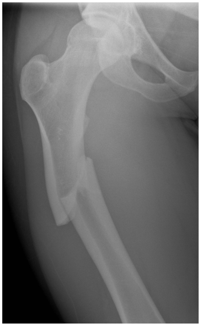
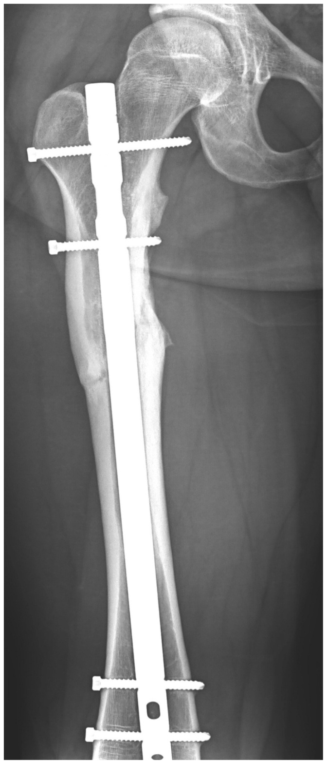
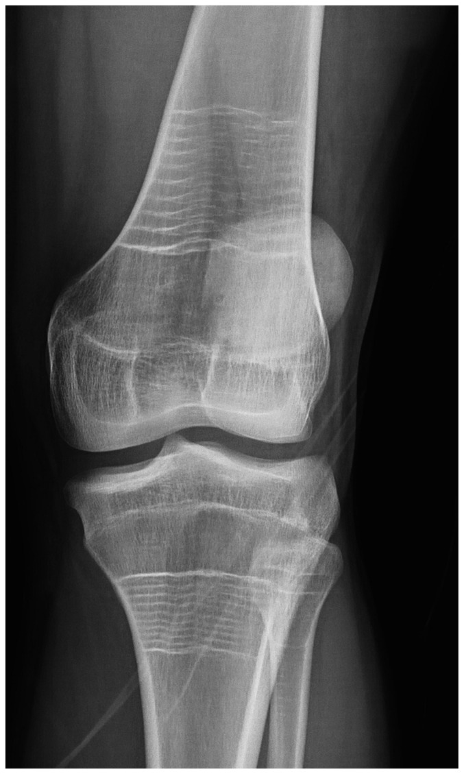
21-year-old male with a diagnosis of osteogenesis imperfecta type IV-B. History of a tibial fracture and a number of femoral fractures between the ages of 6 months and 3 years (2 in the left femur and 2 in the right) treated conservatively. He received from the age of 10 to 13 years four-monthly infusions over three consecutive days of intravenous pamidronate (disodium pamidronate at a dose of 1mg/kg/dose). From the age of 13, treatment was initiated with alendronate at a weekly dose of 40 mg, up to the age of 16, at which point the dose was reduced to 20 mg weekly and maintained until the age of 18. A series of analytical and densitometric checks were carried out. The last measurement of bone mineral density was made three years ago with values within the normal range. For the last three years the patient has not received any specific treatment. He attended the casualty department after suffering a fall from standing height, presenting as a consequence a diaphyseal fracture of his right femur, with atypical fracture data consistent with the updated criteria of the American Society for Bone and Mineral Research (12). He met all the main criteria and in terms of minor criteria he had generalised diaphyseal cortical thickening (Figure 1). He presented no prodromal symptoms, without showing bilateralism. Osteosynthesis of the fracture was carried out and treatment was initiated with teriparatide, with a good development and consolidation of the fracture over time. An X-ray taken a year after the intervention showed the correct consolidation of the fracture (Figure 2). Distally, the so-called zebra lines can be seen (Figure 3), the radiological results of the effect of the bisphosphonates on the immature skeleton (13). A study was carried out of bone markers P1NP and NTx with an initial peak in both in their progressive decrease over one year of development. The patient is asymptomatic, without after-effects and with good tolerance and adherence to the treatment.
Figure 1.
Initial fracture.
Figure 2.
Status of the fracture after 1 year.
Figure 3.
Current appearance of the zebra lines.
Discussion
Osteogenesis imperfecta (OI) is a heterogeneous group of diseases which have in common a change in the genes which code for collagen type I. Seven forms of OI have been described, five of which are of known genetic origin and two with the OI phenotype without the characteristic mutations in the genes involved in the disease (14), being in most cases autosomal dominant or the novo heterocygous mutations in one of the two genes that codify procollagen I, COL1A1 and COL1A2 peptide chains. Currently have been described mutations in different genes, such us in interferon-induced transmembrane protein 5 (IFITM5), although this is unusual (15). The IV-B type has an autosomal dominant pattern of inheritance, with frequent fractures, especially in childhood, slight to moderate limb deformities, initially blue sclera which normalise with growth, short stature, ligamentous laxity and alterations of dentinogenesis. It is considered to be a moderate form of OI.
When analysing the original causes of the fracture it should certainly be borne in mind that the underlying disease may itself be a clear triggering factor, with stress fractures in patients with osteogenesis imperfecta reported in the literature (16). However, the patient had been treated for 8 years with bisphosphonates, first intravenously and later orally, its effects being seen in the bone with the development of the aforementioned zebra lines. Although the last measurement of bone mineral density was found to be within normal limits and there have been no fractures during this period, which invites us to think that the drugs have been effective and have exerted their preventative role in the occurrence of fractures, we believe that these drugs may have had an influence. Furthermore, there have been eight years of therapy, with which the probability of suffering an atypical fracture is increased (2). In addition, the radiological characteristics of the fracture are the classic ones described in the literature as secondary to the use of these drugs, and meet the criteria for its diagnosis established by the American Society for Bone and Mineral Research (12).
The use of teriparatide for the treatment of osteogenesis imperfecta, a drug which is an analogue of the parathyroid hormone with anabolic actions on the bone indicated for the treatment of osteoporosis, is not reflected in its data sheet. There are studies in which an increase in bone mineral density and in markers for bone formation and resorption are seen after treatment over 18 months with this drug in adult patients with OI type I (17). Recently, in a double blind randomised placebo controlled study in adult patients with OI, teriparatide showed an improvement in bone mineral density both in the lumbar spine and in the hip, as well as in volumetric bone mineral density in the vertebrae and in vertebral strength estimated after 18 months of treatment (18). Teriparatide has also been reported as a drug adjuvant to bone consolidation, although most of the studies in the literature which reflect its beneficial effects in the development of fracture calluses are experimental or series of cases (19), it not being possible to make a recommendation of its use in this respect, nor to reflect this use in its data sheet. Furthermore, the drug appears to reduce the negative effects the bisphosphonates have on the bone (20), for which reason it has also been recommended in some works as the drug of choice for the treatment of atypical fractures. At present, and according to the last recommendations of the American Society for Bone metabolism (12), there is not sufficient evidence to establish a systematic recommendation for its use, this being reserved for those cases in which bone consolidation is not achieved with conservative treatment, after the cessation of bisphosphonates and the maintenance of an appropriate balance of calcium and vitamin D.
In the management of our patient we assumed that the fracture occurred due to the effects of the bisphosphonates in the bone, although there is a possibility that it was a secondary effect of his underlying disease. Some Authors describe this type of fractures as a pediatric variant of atypical femoral fracture seen in adults with prolonged bisphosphonates use (21). It seems to be an influence of bisphosphonates effect on that type of bone. Actually however, there are only a few cases described and most of them in older patients. We believe, like other Authors (10), that the use of teriparatide following surgery has helped to correct the microstructural damage potentially caused by the drug, has also helped the completion of the process of consolidation successfully and on time, and, on the other hand, has improved the morphometric and structural parameters of a young patient with osteogenesis imperfecta as an underlying disease which sustains a greater probability of new fractures in the future. Further studies are needed to confirm the benefits of this treatment for this type of bone.
Footnotes
Conflict of interest for
- Iñigo Etxebarria-Foronda: none.
- Pedro Carpintero: none.
References
- 1.Etxebarria-Foronda I, Caeiro-Rey JR, Larrainzar-Garijo R, Vaquero-Cervino E, Roca-Ruiz L, Mesa-Ramos M, Merino Pérez J, Carpintero-Benitez P, Fernández Cebrián A, Gil-Garay E. SECOT-GEIO guidelines in osteoporosis and fragility fracture. An update. Rev Esp Cir Ortop Traumatol. 2015;59(6):373–393. doi: 10.1016/j.recot.2015.05.007. [DOI] [PubMed] [Google Scholar]
- 2.Caeiro-Rey JR, Etxebarria-Foronda I, Mesa-Ramos M. Atypical fractures associated with the long term use of bisphosphonates. The current situation. Rev Esp Cir Ortop Traumatol. 2011;55:392–404. [Google Scholar]
- 3.Compston J. Pathophysiology of atypical femoral fractures and osteonecrosis of the jaw. Osteoporos Int. 2011;22:2951–61. doi: 10.1007/s00198-011-1804-x. [DOI] [PubMed] [Google Scholar]
- 4.Carulli C, Innocenti M, Brandi ML. Bone vascularization in normal and disease. Front Endocrinol. 2013;4:106. doi: 10.3389/fendo.2013.00106. [DOI] [PMC free article] [PubMed] [Google Scholar]
- 5.Glorieux FH. Experience with bisphosphonates in osteogenesis imperfecta. Pediatrics. 2007;119:S163–S165. doi: 10.1542/peds.2006-2023I. [DOI] [PubMed] [Google Scholar]
- 6.Hald JD, Evangelou E, Langdahl BL, Ralston SH. Bisphosphonates for the prevention of fractures in osteogenesis imperfecta: meta-analysis of placebo-controlled trials. J Bone Miner Res. 2015;30:929–33. doi: 10.1002/jbmr.2410. [DOI] [PubMed] [Google Scholar]
- 7.Shi CG, Zhang Y, Yuan W. Efficacy of Bisphosphonates on Bone Mineral Density and Fracture Rate in Patients With Osteogenesis Imperfecta: A Systematic Review and Meta-analysis. Am J Ther. 2015 doi: 10.1097/MJT.0000000000000236. [Epub ahead of print] [DOI] [PubMed] [Google Scholar]
- 8.Meier RP, Ing Lorenzini K, Uebelhart B, Stern R, Peter RE, Rizzoli R. Atypical femoral fracture following bisphosphonate treatment in a woman with osteogenesis imperfecta. A case report. Acta Orthop. 2012;83:548–50. doi: 10.3109/17453674.2012.729183. [DOI] [PMC free article] [PubMed] [Google Scholar]
- 9.Manolopoulos KN, West A, Gittoes N. The paradox of prevention. Bilateral atypical subtrochanteric fractures due to bisphosphonates in osteogenesis imperfecta. J Clin Endocrinol Metab. 2013;98:871–2. doi: 10.1210/jc.2012-4195. [DOI] [PubMed] [Google Scholar]
- 10.Holm J, Eiken P, Hyldstrup L, Jensen JE. Atypical femoral fracture in an osteogenesis imperfecta patient succesfully treated with teriparatide. Endocr Pract. 2014;20:e187–90. doi: 10.4158/EP14141.CR. [DOI] [PubMed] [Google Scholar]
- 11.Carpintero P, Del Fresno J, Ruiz Sanz J, Jaenal P. Atypical fracture in a child with osteogenesis imperfecta. Joint Bone Spine. 2015;82:287–8. doi: 10.1016/j.jbspin.2014.10.014. [DOI] [PubMed] [Google Scholar]
- 12.Shane E, Burr D, Abrahamsen B, Adler RA, Brown TD, Cheung AM, Cosman F, Curtis JR, Dell R, Dempster DW, Ebeling PR, Einhorn TA, Genant HK, Geusens P, Klaushofer K, Lane JM, McKiernan F, McKinney R, Ng A, Nieves J, O’Keefe R, Papapoulos S, Howe TS, van der Meulen MC, Weinstein RS, Whyte MP. Atypical subtrochanteric and diaphyseal femoral fractures: second report of a task forcé of the American Society for Bone and Mineral Research. J Bone Miner Res. 2014;29:1–23. doi: 10.1002/jbmr.1998. [DOI] [PubMed] [Google Scholar]
- 13.Etxebarria-Foronda I, Gorostiola-Vidaurrazaga L. Zebra lines: Radiological repercussions of the action of bisphosphonates on the immature skeleton. Rev Osteoporos Metab Miner. 2013;1:39–41. [Google Scholar]
- 14.Escribano-Rey RJ, Duart-Clemente J, Martínez de la Llana O, Beguiristain-Gúrpide JL. Rev Esp Cir Ortop Traumatol. 2014;58:114–9. doi: 10.1016/j.recot.2013.11.007. [DOI] [PubMed] [Google Scholar]
- 15.Hanagata N. IFITM5 mutations and osteogenesis imperfect. J Bone Miner Metab. 2015 doi: 10.1007/s00774-015-0667-1. [DOI] [PubMed] [Google Scholar]
- 16.Shabat S. Osteogenesis imperfecta-induced migratory stress fractures in a military recruit. Clin Exp Rheumatol. 2000;18:654. [PubMed] [Google Scholar]
- 17.Gatti D, Rossini M, Viapiana O, Povino MR, Liuzza S, Fracassi E, Idol-azzi L, Adami S. Teriparatide treatment in adult patients with osteogenesis imperfecta type I. Calcif Tissue Int. 2013;93:448–52. doi: 10.1007/s00223-013-9770-2. [DOI] [PubMed] [Google Scholar]
- 18.Orwoll ES, Shapiro J, Veith S, Wang Y, Lapidus J, Vanek C, Reeder JL, Keaveny TM, Lee dC, Mullins MA, Nagamani SC, Lee B. Evaluation of teriparatide treatment in adults with osteogenesis imperfecta. J Clin Invest. 2014;124:491–8. doi: 10.1172/JCI71101. [DOI] [PMC free article] [PubMed] [Google Scholar]
- 19.Zhang D, Potty A, Vyas P, Lane J. The role of recombinant PTH in human fracture healing: a systematic review. J Orthop Trauma. 2014;28:57–62. doi: 10.1097/BOT.0b013e31828e13fe. [DOI] [PubMed] [Google Scholar]
- 20.Dobnig H, Stepan JJ, Burr DB, Li J, Michalská D, Sipos A, Petto H, Fahrleitner-Pammer A, Pavo I. Teriparatide reduces bone microdamage accumulation in postmenopausal women previously treated with alendronate. J Bone Miner Res. 2009;24:1998–2006. doi: 10.1359/jbmr.090527. [DOI] [PubMed] [Google Scholar]
- 21.Hegazy A, Kenawey M, Sochett E, Tile L, Cheung AM, Howard AW. Unusual Femur Stress Fractures in Children With Osteogenesis Imperfecta and Intramedullary Rods on Long-term Intravenous Pamidronate Therapy. J Pediatr Orthop. 2015 doi: 10.1097/BPO.0000000000000552. Epub ahead of print. [DOI] [PubMed] [Google Scholar]


