Summary
We report a case of a patient who presented with inability to walk for 2 days which was acute in onset, with no history or preceeding trauma. On examination it was found to have stable vitals. Both lower limbs were in the flexed, abducted & externally rotated attitude, with no sensory motor deficit. Radiological investigations revealing osteoporosis and bilateral fracture neck of femur and blood investigations indicated severe vitamin D deficiency.
Keywords: spontaneous bilateral fracture neck of femur, vitamin D
Introduction
Vitamin D deficiency is now recognized as a pandemic. The major cause of vitamin D deficiency is the lack of appreciation that sun exposure in moderation is the major source of vitamin D for most humans. Very few foods naturally contain vitamin D, and foods that are fortified with vitamin D are often inadequate to satisfy either a child’s or an adult’s vitamin D requirement. Vitamin D deficiency causes rickets in children and will precipitate and exacerbate osteopenia, osteoporosis, and fractures in adults (1). Bilateral fracture neck femur is rare and only few cases are reported. The possible reasons of fracture that have been identified are trauma, osteoporosis and seizure (2).
Case report
A 78-year-old ex-military officer presented to us at the out-patient department with a history of acute onset inability to walk since 2 days. On examination was found to have a high blood pressure of 160/90mmHg. There was no history of trauma, seizures, kochs contact, long term drug intake, chronic smoking, or of noticing any swellings in the neck. Patient was detected to be hypertensive and diabetic on admission.
Both lower limbs on examination were in the flexed, abducted, and externally rotated attitude, no neurological or vascular deficit was detected.
Routine blood investigations done were: Hb 9.7g%; total count 7800cells/cmm; serum bilirubin- 1.5mg%, Alkaline Phosphatase- 74 U/L; serum Prostate specific antigen- 0.92 ng/ml (N- <4.00).
Serum vitamin D levels were found to be <16ng/dl (normal being >25ng/dl). Serum calcium 8.9meq/L, and serum levels of parathormone being 47.40pg/ml (15.00–68.00pg/ml). Arterial blood gas report was found to be normal.
Bone Mineral Density (BMD) calculated via DXA scan was found to be −3.6. The WHO classification was used to define osteopenia (T score between −1 and −2.5) and osteoporosis (T score < −2.5).
Bone scan using Tc99 MDP was done and only mild diffuse uptake was found in D-L spine and Pelvis suggestive of only degenerative disease and no metastatic involvement.
Radiological investigations done were as follows:
X-ray skull (lateral view), spine and base of skull revealed osteoporosis of bones under review with anterior wedging/compression of upper lumbar vertebrae with the vault of the skull being normal.
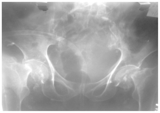
Pre op X-ray pelvis with both hips revealed bilateral fracture neck of femur
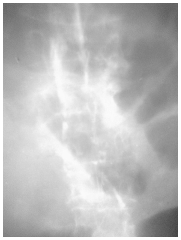
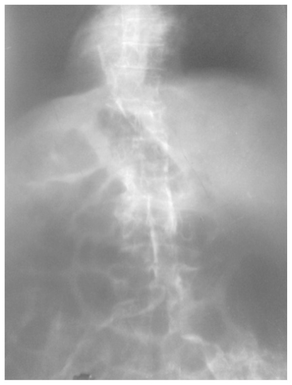
X-ray dorsolumbar spine lateral view was suggestive of severe multiple osteoporotic wedge compression fractures.
A CT thorax revealed bilateral pleural effusion with features suggestive of aspiration pneumonia and multiple compression fractures of the dorsolumbar vertebrae with retropulsion.
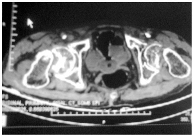
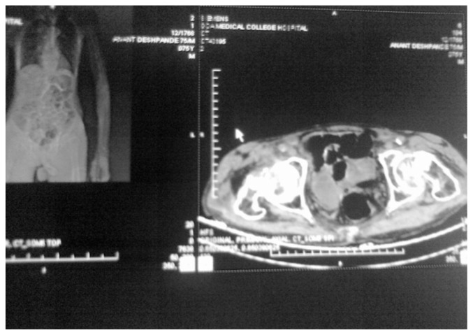
CT pelvis revealed osteoporosis and bilateral fracture neck of femur.
Patient was diagnosed as a bilateral fracture neck of femur secondary to osteoporosis and decision to operate was taken.
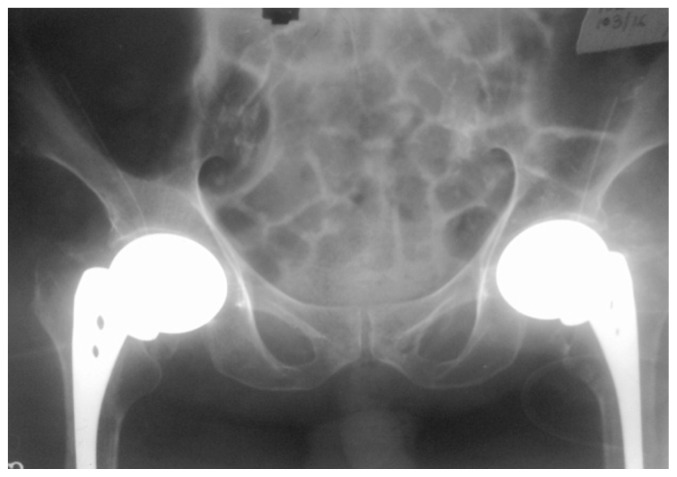
Patient was operated under regional epidural anaesthesia. Hemi Arthroplasty with Bilateral Austin-Moore prosthesis was performed in a lateral position with one hip at a time. Patient also received injectable vitamin D supplementation postoperatively.
Postoperatively patient was kept for two weeks in hospital. Immediate bed side mobilisation was started the next day of surgery. Patient was also given subcutaneous enoxaparin for Deep vein thrombosis (DVT) prophylaxis. Prior to discharge at the end of two weeks stitch removal was done after confirming healthy wound status.
At next OPD visit, two weeks later, patient was started on full weight bearing using a walker for support. Four weeks later patient was able to walk without support and had no other complaints or complications post op. For the next 6 months patient followed up at our OPD and showed considerable progress after which patient was lost to follow up.
Discussion
Isolated cases of bilateral fracture neck of femur have been described in literature as well as a case series of 33 cases was studied and published in the Journal of Bone and Joint surgery. Such injuries usually occur secondary trauma, osteoporosis and seizures (2).
Before the 1960s, such injuries were seen in association with powerful muscular contractions induced by electroconvulsive therapies. Since then, this pattern of injury has been reported to be the result of high-energy trauma, high- voltage electrical injury, abnormal anatomy, chronic renal failure, and associated with primary and secondary bone disease such as osteomalacia. In the elderly population, simple trip and fall can lead to bilateral hip injury (3).
Other causes of this condition include insufficiency fractures and fatigue fractures. Insufficiency fractures occur in patients with intrinsic or iatrogenic osteopenia under normal loading conditions. Carter et al. reported a case of an insufficiency fracture in a 67-year-old female which was secondary to vitamin D deficiency and concurrent steroid use (4).
These fractures have been described in patients on long term corticosteroid treatment, those who have renal osteodystrophy, patients who have received fluoride treatment, amenorrheic athletes and those who have undergone pelvic irradiation. Fatigue fractures on the other hand occur as a result of excessive and usually repetitive strain on normal bones. These fractures are common in endurance athletes, military personnel and people with epilepsy. Fatigue fractures can also result from normal stress forces through abnormal anatomy. Femoral neck fractures have been described post bilateral total knee replacement due to the resultant alteration in normal leg axis, and also due to bilateral protrusio acetabuli in a patient with Marfan’s syndrome (5).
Case report published in the Journal of Bone and Joint Surgery implicated irradiation as another cause of spontaneous bilateral femur neck fractures. It has been suggested that an obliterative endarteritis caused by irradiation was a predisposing factor to non-union, but inaccurate reduction and incomplete fixation may have been the determining causes (6).
Atraumatic bilateral fracture femur has been reported with long term bisphosphonate use (7, 8).
With the earliest cases being reported in the 1960s, Electroconvulsive therapy has been one of the commonest clinical setting which leads to simultaneous bilateral fracture femur. As many as 27 cases were reported as early as the 1960s published in the Journal of Bone and Joint surgery along with few cases having bilateral hip dislocation as an associated component. Unilateral or bilateral fracture femur has also been seen in patients with seizure disorders (9).
The mainstay of treatment in this age group remains hemiarthroplasty or total hip replacement. In this case the patient was high risk for anaesthesia due to his other medical co-morbidities, decision of doing uncemented hemiarthroplasty was taken. The patient underwent hemiarthroplasty with insertion of Austin Moore prosthesis bilaterally.
Figure 1.
Pre op X-ray pelvis with both hips revealed bilateral fracture neck of femur.
Figure 2.
X-ray dorsolumbar spine (lateral view) showing multi level wedge compression fractures plus severe osteopenia.
Figure 3.
X-ray dorsolumbar spine (AP view) showing multi level wedge compression fractures plus severe osteopenia.
Figure 4.
CT image of pelvis revealing fracture neck of femur bilaterally and severe osteoporosis.
Figure 5.
CT image of pelvis revealing fracture neck of femur bilaterally and severe osteoporosis.
Figure 6.
Post bilateral hemiarthroplasty: X-ray pelvis with both hips.
References
- 1.Holick MF, Chen TC. Vitamin D deficiency: a worldwide problem with health consequences. Am J Clin Nutr. 2008;87(suppl):1080S–6S. doi: 10.1093/ajcn/87.4.1080S. [DOI] [PubMed] [Google Scholar]
- 2.Devkota P, Ahmad S. Bilateral impacted femoral neck fracture in a renal disease patient. Niger Med J. 2013 Sep-Oct;54(5):354–355. doi: 10.4103/0300-1652.122374. [DOI] [PMC free article] [PubMed] [Google Scholar]
- 3.McGoldrick NP, Dodds MK, Green C, Synnott K. Management of Simultaneous Bilateral Neck of Femur Fractures in an Elderly Patient. Geriatric Orthopaedic Surgery & Rehabilitation. 2013;4(3):71–73. doi: 10.1177/2151458513511626. [DOI] [PMC free article] [PubMed] [Google Scholar]
- 4.Carter T, Nutt J, Simons A. Bilateral femoral neck insufficiency fractures secondary to vitamin D deficiency and concurrent corticosteroid use - a case report. Arch Osteoporos. 2014;9(1):172. doi: 10.1007/s11657-014-0172-5. [DOI] [PubMed] [Google Scholar]
- 5.Gurdezi S, Trehan RK, Rickman M. Bilateral undisplaced insufficiency neck of femur fractures associated with short-term steroid use: a case report. Journal of Medical Case Reports. 2008;2:79. doi: 10.1186/1752-1947-2-79. [DOI] [PMC free article] [PubMed] [Google Scholar]
- 6.Clarence H, Heyman MD. Spontaneous bilateral fracture of the neck of the femur following irradiation. Journal of Bone and Joint Surgery. 1945 Oct;XXVII(4) [Google Scholar]
- 7.Goddard MS, Reid KR, Johnston JC, Khanuja HS. Atraumatic bilateral femur fracture in long-term bisphosphonate use. Orthopedics. 2009 Aug;32(8) doi: 10.3928/01477447-20090624-27. [DOI] [PubMed] [Google Scholar]
- 8.Patel VC, Lazzarini AM. Bilateral simultaneous femoral diaphyseal fractures in a patient with long-term ibandronate use. Orthopedics. 2010 Oct 11;33(10):775. doi: 10.3928/01477447-20100826-31. [DOI] [PubMed] [Google Scholar]
- 9.Powell HD. Simultaneous bilateral fracture neck of femur. Journal of Bone and Joint surgery. 1960 May;42 B(2) doi: 10.1302/0301-620X.42B2.236. [DOI] [PubMed] [Google Scholar]


