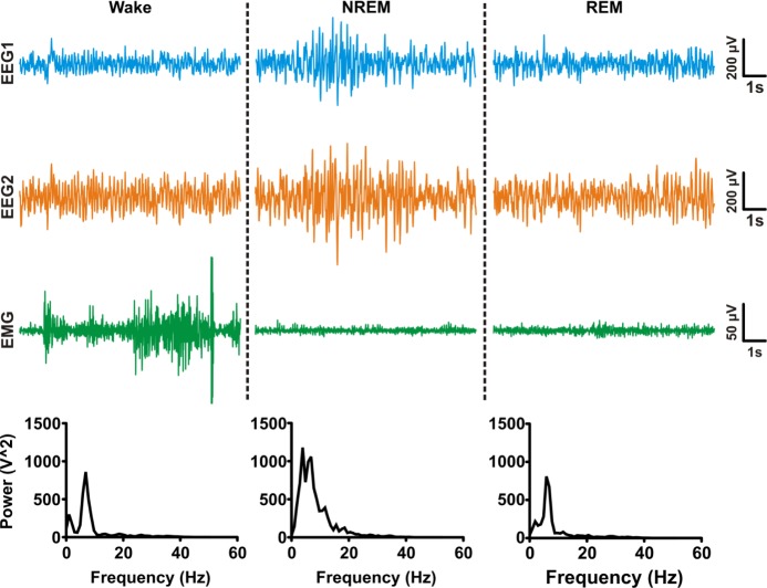
Figure 3.
Representative traces of behavioral states. Sleep architecture was scored using video-EEGEMG data in Sirenia Sleep Pro using a semiautomated method. Each automated EEG-EMG score was manually verified with time-synchronized video data. Traces from time-matched subdural EEG electrodes (blue and orange traces) and EMG electrode (green) show typical characteristics of wake (low amplitude EEG, high amplitude EMG), nonrapid eye movement (NREM) sleep (high amplitude EEG, low-medium amplitude EMG), and rapid eye movement (REM) sleep (low amplitude EEG and EMG). EEG rhythms for each example epoch are shown in corresponding spectrograms.

