

Abstract
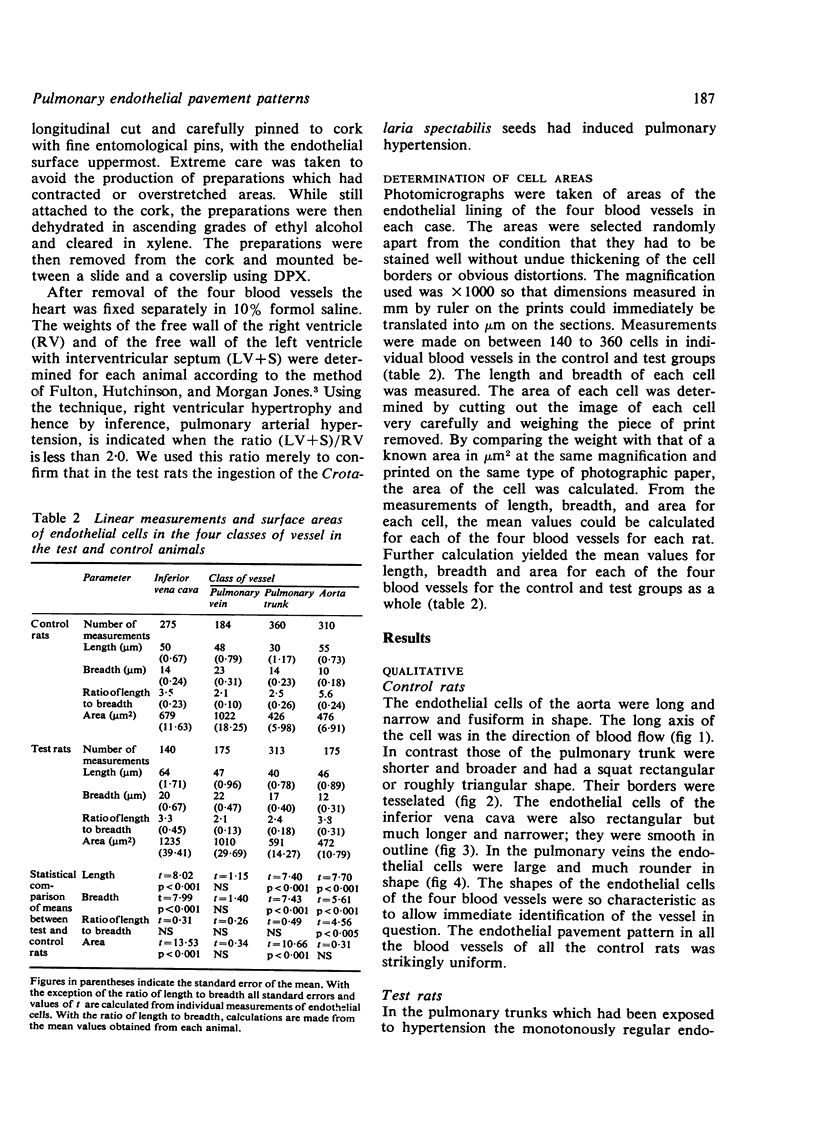
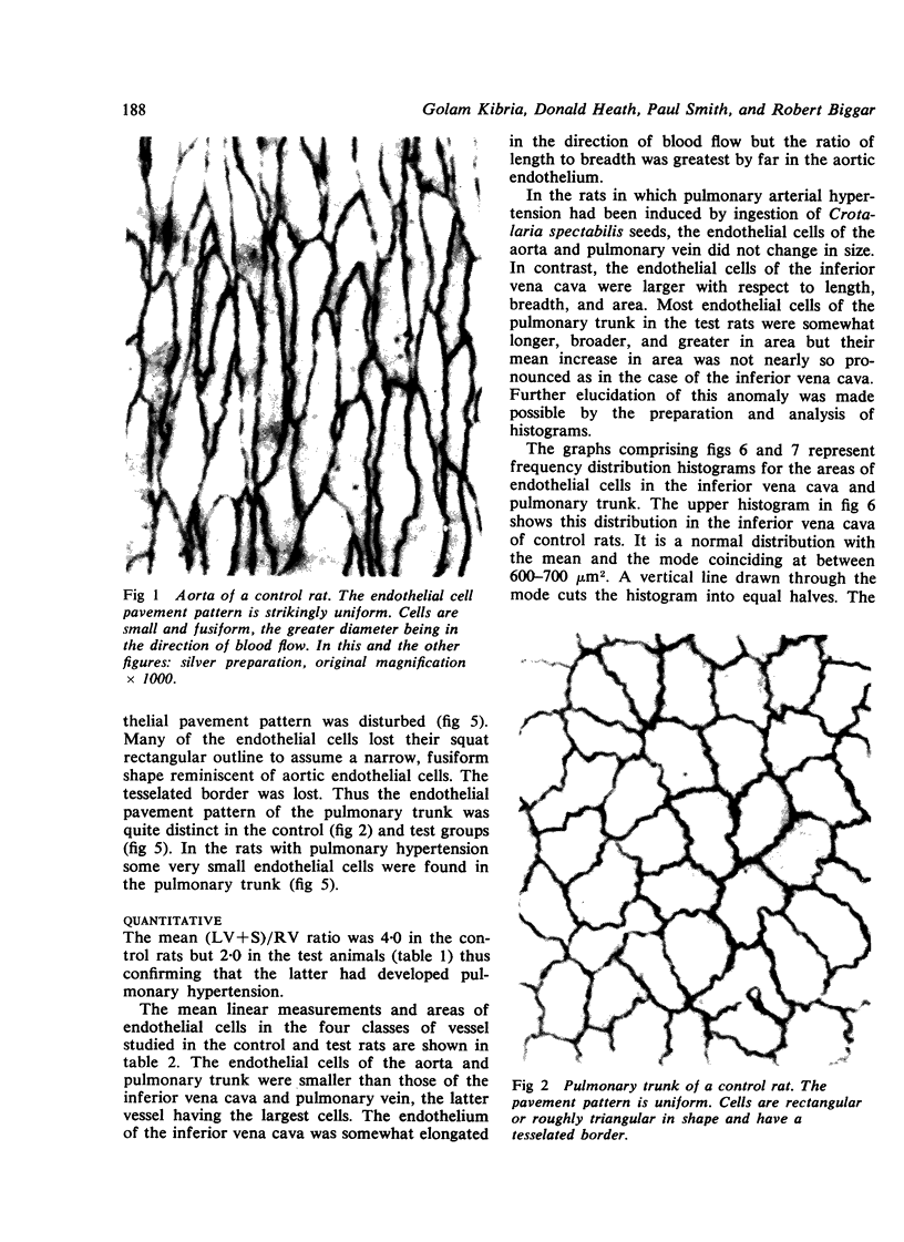
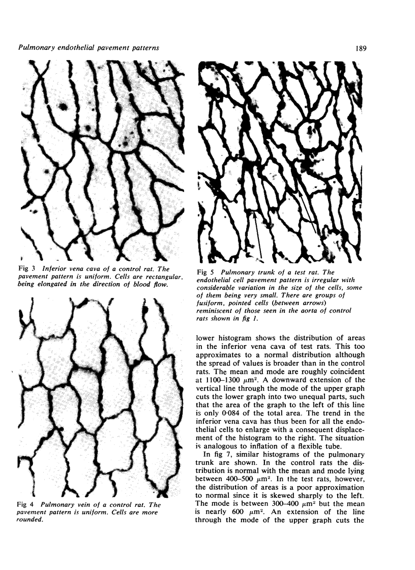
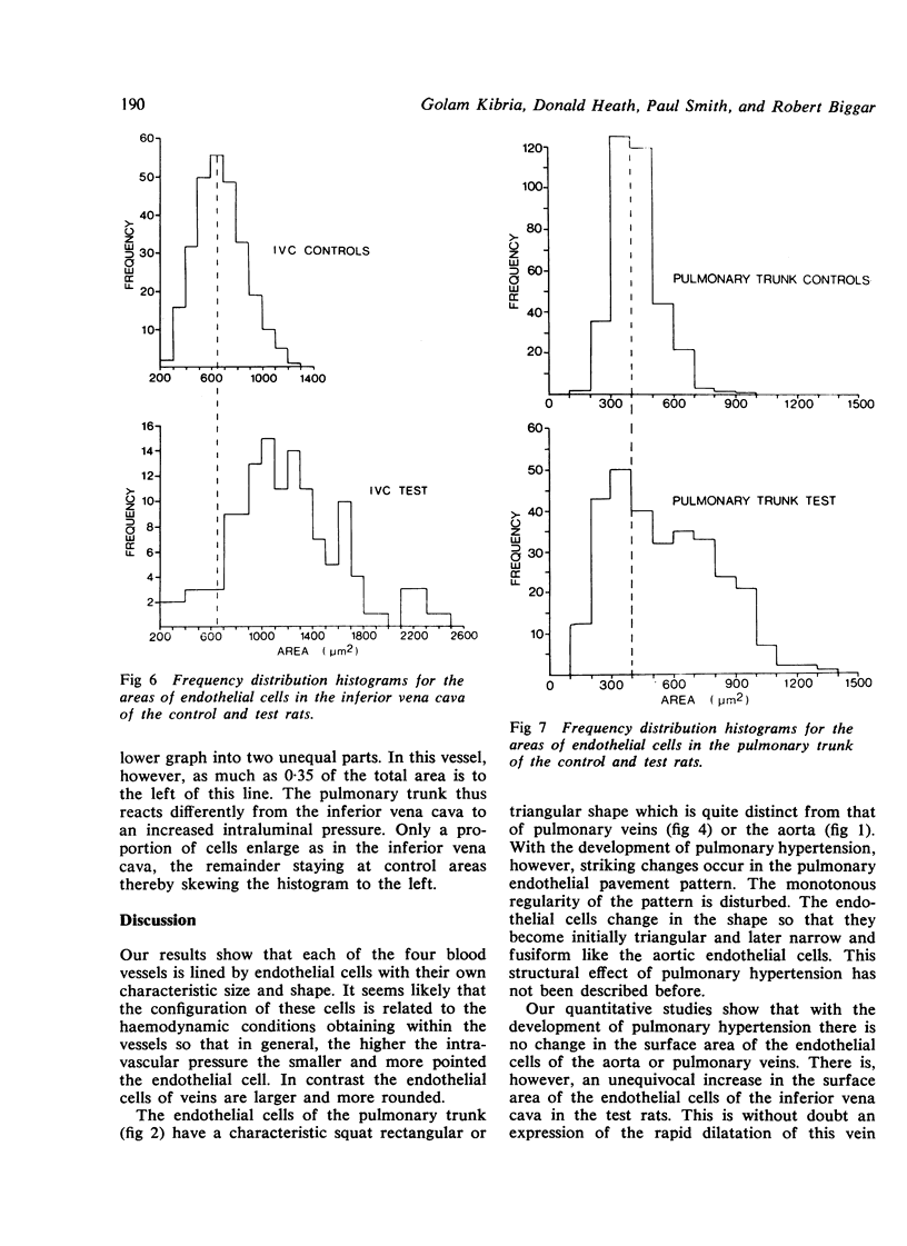
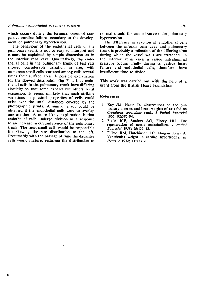
The appearance of the endothelial pavement pattern was studied in the pulmonary trunk, pulmonary veins, aorta, and inferior vena cava of the rat by means of silver staining of the cell borders. The endothelial cell in each of the four blood vessels was found to have its own distinctive shape, fusiform and pointed in the direction of blood flow in the case of the aorta and larger and more rectangular in the pulmonary trunk and pulmonary veins. Detailed quantitation of the dimensions and surface area of the endothelial cells in each blood vessel was carried out by a photographic technique. Pulmonary hypertension was induced in one group of rats by feeding them on Crotalaria spectabilis seeds. The endothelial pavement pattern in their pulmonary trunks became disrupted with many of the cells assuming a fusiform shape reminiscent of aortic endothelium. Many small, new endothelial cells formed in the pulmonary trunk suggesting division of cells to line the enlarging blood vessels. In contrast the endothelial cells of the inferior vena cava merely increased in size to cope with the dilatation of this vein.
Full text
PDF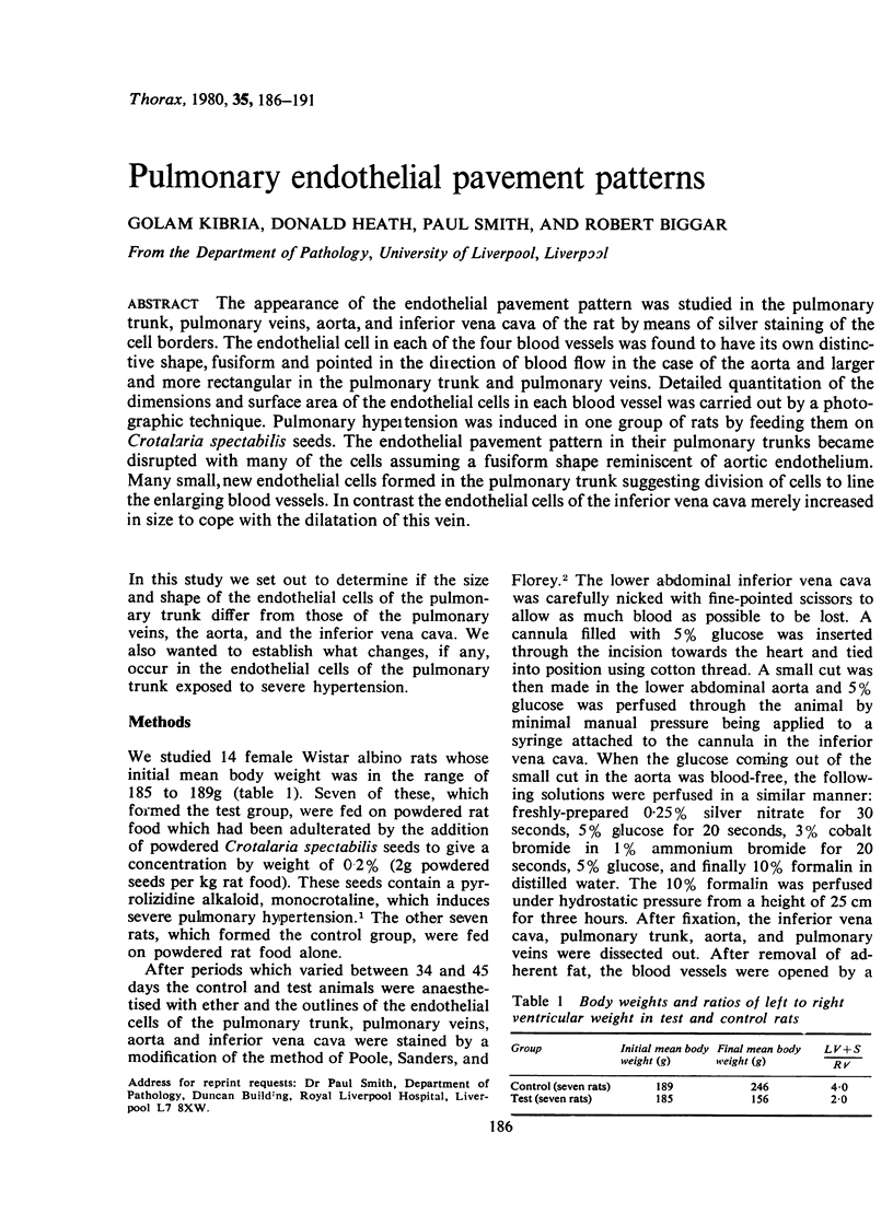
Images in this article
Selected References
These references are in PubMed. This may not be the complete list of references from this article.
- FULTON R. M., HUTCHINSON E. C., JONES A. M. Ventricular weight in cardiac hypertrophy. Br Heart J. 1952 Jul;14(3):413–420. doi: 10.1136/hrt.14.3.413. [DOI] [PMC free article] [PubMed] [Google Scholar]
- Kay J. M., Heath D. Observations on the pulmonary arteries and heart weight of rats fed on Crotalaria spectabilis seeds. J Pathol Bacteriol. 1966 Oct;92(2):385–394. doi: 10.1002/path.1700920216. [DOI] [PubMed] [Google Scholar]
- POOLE J. C., SANDERS A. G., FLOREY H. W. The regeneration of aortic endothelium. J Pathol Bacteriol. 1958 Jan;75(1):133–143. doi: 10.1002/path.1700750116. [DOI] [PubMed] [Google Scholar]