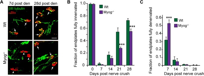
Fig. 5.
Myog stimulates endplate reinnervation. (A) Representative images and (B,C) quantification of innervated and denervated endplates in Wt and Myog−/− soleus muscle at different times post nerve crush. βIII tubulin+ regenerating motor nerve branches are green and αBtx+ endplates are red. Arrows point to partially or fully innervated endplates and arrowheads point to denervated endplates. Scale bar: 50 µm. Error bars are s.d.; n=3 for 7 days and n=4 for 14, 21 and 28 days post nerve crush. ***P<0.001 relative to Wt.

