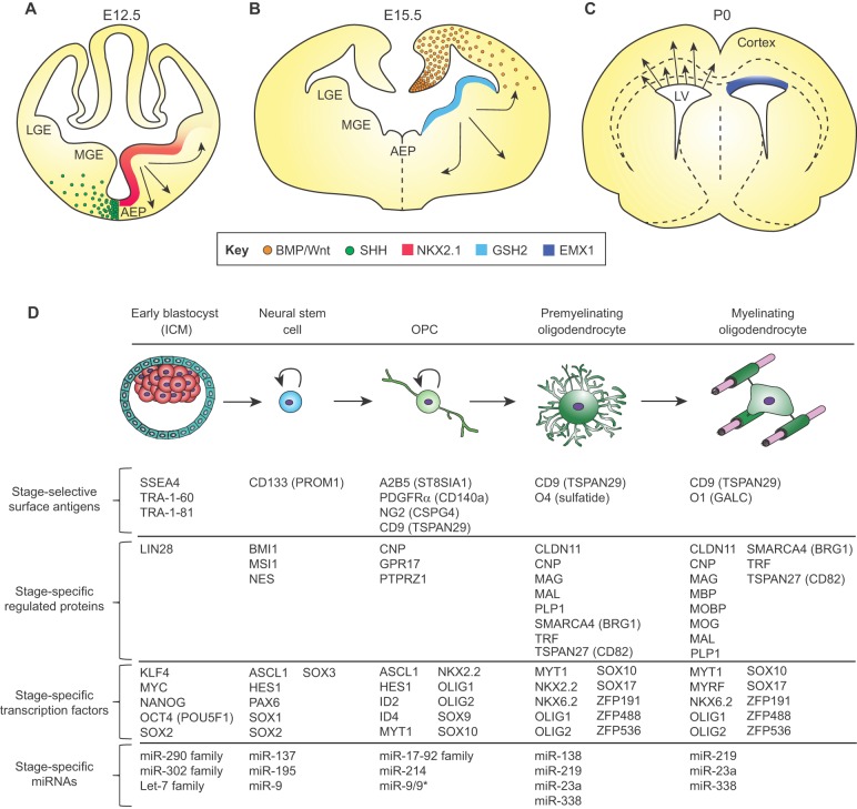
Fig. 2.
The transcription factor code and associated markers of oligodendroglial fate restriction. Oligodendrocytes arise from different regions of the developing forebrain (shown here) and spinal cord. (A) By E12.5 in the mouse, oligodendroglia are seen to arise ventrally from NKX2.1-expressing cells in the medial ganglionic eminence, which migrate to the dorsal forebrain. (B) By E15.5 in the mouse, cells generated under GSH2 control arise from the expanded ganglionic eminence and striatal anlagen to migrate broadly. (C) Around birth, the EMX1-expressing dorsal subcallosal ventricular wall shifts to OPC production. These distinct populations compete with one another as maturation proceeds, resulting in an admixture of OPCs and oligodendrocytes derived from different developmental germinal zones in the adult brain. See also Rowitch and Kriegstein (2010). (D) At the cellular level, either in vitro or in vivo, pluripotent stem cells progress through serial developmental stages: neuroepithelial stem cell, OPC, premyelinating and ultimately myelinating oligodendroglia. Such cardinal stages of oligodendroglial development are characterized by distinct signal effectors and transcription factors that regulate the expression of multiple stage-specific marker genes (Ahmed et al., 2009; Cahoy et al., 2008; Nishiyama et al., 2009; Sim et al., 2011; Young, 2011; Zhang, 2001; Zhang et al., 2014; Zuchero and Barres, 2013). At each stage, cells can be identified and isolated on the basis of temporally regulated surface antigens, which permit the selection of cells of defined mitotic and differentiation potential (Ishibashi et al., 2004; Nishiyama et al., 2009; Yuan et al., 2011; Zhang, 2001; Zhao et al., 2012). These discrete stages exhibit distinct miRNA profiles as well, that may serve to regulate translation, and thus sharpen phenotypic transitions during lineage progression (Barca-Mayo and Lu, 2012; Dugas et al., 2010; He et al., 2012; Lau et al., 2008; Mallanna and Rizzino, 2010; Shenoy and Blelloch, 2014; Shi et al., 2010; Zhao et al., 2010). AEP, anterior entopeduncular nucleus; ICM, inner cell mass; LGE, lateral ganglionic eminence; LV, lateral ventricle; MGE, medial ganglionic eminence.

