Abstract
Background and study aims: Colonic endoscopic submucosal dissection (ESD) is a challenging procedure because it is often difficult to maintain good visualization of the submucosal layer. To facilitate colonic ESD, we designed a novel traction method, namely traction-assisted colonic ESD using clip and line (TAC), and investigated its feasibility.
Patients and methods: We retrospectively analyzed 23 patients with large colonic superficial lesions who had undergone TAC. The main outcome was the procedural success rate of TAC, which we defined as successful, sustained application of clip and line to the lesion until the end of the procedure.
Results: The procedural success rate of TAC was 87 % (20/23). In all three unsuccessful cases, the lesions were in the proximal colon and the procedure times over 100 minutes. The overall mean procedure time was 61 min (95 % confidence interval, 18 – 172 min). We achieved en bloc resections of all lesions. There were no perforations or fatal adverse events.
Conclusions: TAC is feasible and safe for colonic ESD and may improve the ease of performing this procedure.
Introduction
Colorectal endoscopic submucosal dissection (ESD) is a promising, minimally invasive procedure that enables high en bloc resection rates regardless of lesion size 1. However, colorectal ESD, particularly colonic ESD, is a more challenging procedure than conventional endoscopic mucosal resection (EMR) because of its technical difficulties, longer procedure time, and higher risk of adverse events such as perforation 1 2. Therefore, colonic ESD is not a standard therapy, especially not in Western countries 3.
All endoscopic procedures should be performed under direct visualization but visualization of the operating site is recommended, in particular, when performing ESD to enable precision sufficient to avoid perforation and bleeding. However, a major issue often encountered with colorectal ESD is difficulty in maintaining an adequate view during submucosal dissection because the mucosa cannot be lifted as in open surgery. Recently, a traction method using an endoclip and line called “clip with line” 4 was developed for maintaining good visualization of the submucosal layer during esophageal and gastric ESD 4 5 6. This method made ESD easier and reduced the procedure time for submucosal dissection 6, but it is not applicable to colonic ESD because the colonoscope had to be withdrawn to mount the endoclip and line 4. To facilitate ESD and make it more widely available, particularly for colonic application, a simple and safe traction method is required.
We therefore designed a novel traction method – traction-assisted colonic ESD using clip and line (TAC) – 7 that does not require withdrawal and reinsertion of the colonoscope. We herein present a feasibility study of our method of colonic ESD.
Patients and methods
Patients
We retrospectively enrolled 23 patients with large colonic superficial lesions who had undergone ESD at the Osaka Medical Center for Cancer and Cardiovascular Diseases between October 2014 and March 2015. Colonic lesions larger than 20 mm that were difficult to remove en bloc with conventional EMR were included. Lesions predicted to be noninvasive tumors and cancers with minute (< 1000 μm) submucosal invasion that were thought to carry no risk of lymphovascular metastasis were removed by ESD using the TAC method. Lesions larger than 50 mm, showing a non-lifting sign or residual lesions after EMR were excluded. Lesions using variations of TAC in which different types of clip or line were used were also excluded ( Fig.1). Outcome measures were procedural success rate of TAC, en bloc resection rate, complete en bloc resection (R0 resection) rate, procedure time, and adverse events. Procedural success of TAC was defined as successful sustained application of clip and line to the lesion until the end of the procedure. R0 resection was defined as en bloc resection with no tumor identified at the lateral or vertical margins. Procedure time was measured from the start of the submucosal injection until removal of the lesion. The procedure speed and the area of submucosa dissected per unit time (mm2/min) were calculated by dividing the dissection time into the size of the resected specimen. Specimen sized was calculated using the formula π · the major axis · the minor axis/4. Histopathologic diagnoses were classified according to the Japanese classification8. This study was approved by the institutional review board and written informed consent for ESD was obtained from all patients.
Fig. 1.
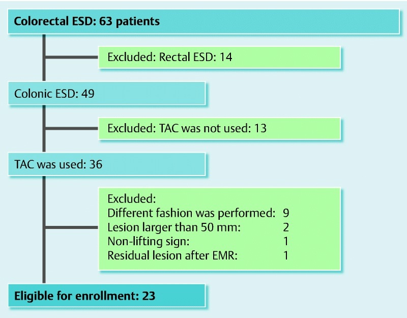
Flow diagram for enrollment in the study (procedures performed from October 2014 to March 2015).
ESD procedures using the novel “TAC” method
ESD was conducted using a colonoscope (PCF-Q260AZI or CF-Q260DI; Olympus, Tokyo, Japan) with a distal attachment cap (D-201-13404; Olympus). TAC was performed in a uniform, standardized fashion as previously described 7. Before the colonoscope was inserted, a polyester line measuring 0.2 mm in diameter and 3 m in length was inserted into its accessory channel by grasping the end of the line with hemostatic forceps (Coaglasper; FD-410LR, Olympus) and pulling it up through the working channel, and then tying the ends of the line together outside the colonoscope (Video 1). After the line had been set up, the colonoscope was inserted (Fig. 2, Fig. 3) and the actual procedure started. All ESD procedures were performed using a Flush knife (1.5-mm, DK2618JN15, Fujifilm Medical, Tokyo, Japan) and 0.9 % saline solution was used as water-jet fluid. An electrosurgical generator (VIO 300D; ERBE, Tübingen, Germany) was used for all ESD procedures.
Fig. 2.
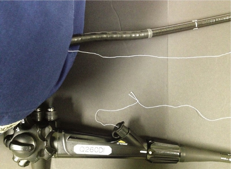
The silk line is tied outside the colonoscope and the colonoscope inserted.
Fig. 3.
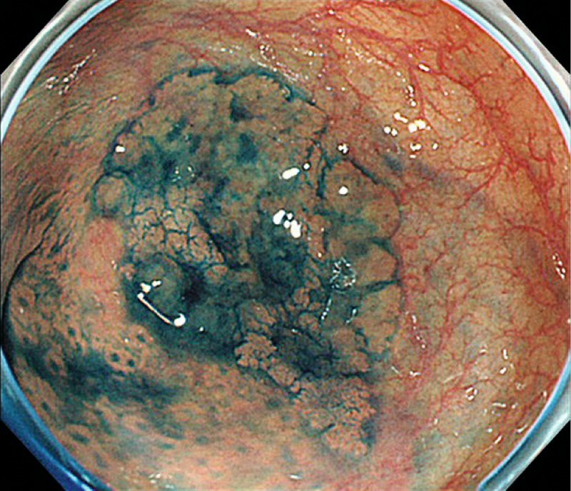
Colonoscopy image showing a slightly elevated 30-mm lesion in the cecum.
Video 1
Colonic ESD using the novel traction method TAC
First, 0.4 % hyaluronate sodium solution (MucoUp; Johnson & Johnson K. K., Tokyo, Japan) was injected into the submucosa, after which the mucosa was incised on the anal side of the lesion. When the line interfered with endoscopic view, we changed the position of a patient's body, resulting in good visualization of the cutting line of the mucosa. Next, the line was cut externally at the hand control end of the colonoscope and the accessory channel end of the line tied to the teeth of an endoclip (HX-610-090; Olympus) attached to an applicator (Fig. 4, Video 1). At this stage, it was important not to fully open the endoclip. The clip and line were then retracted into the applicator and the applicator inserted into the accessory channel. The endoclip was fully opened in the colon and used to grasp the anal side of the specimen (Fig. 5, Video 1), after which the line was pulled gently by hand (Fig. 6, Video 1) to provide good visibility of the submucosal layer.
Fig. 4.
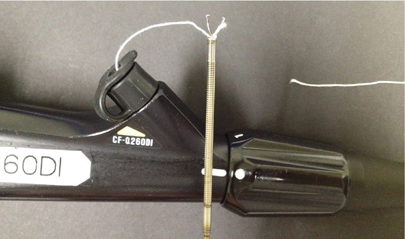
The accessory channel side of the line is tied to the teeth of an endoclip that has not been fully opened.
Fig. 5.
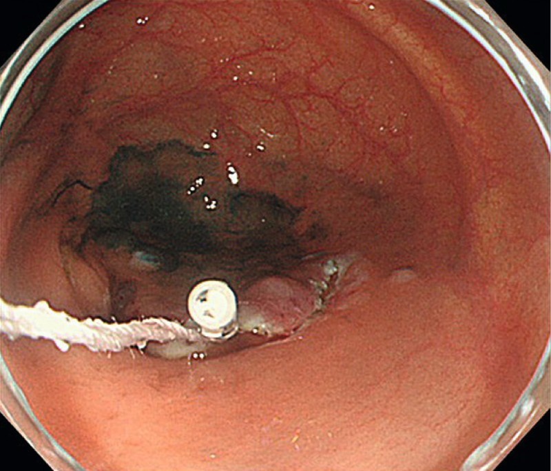
The anal side of a lesion is grasped with the endoclip and line.
Fig. 6.
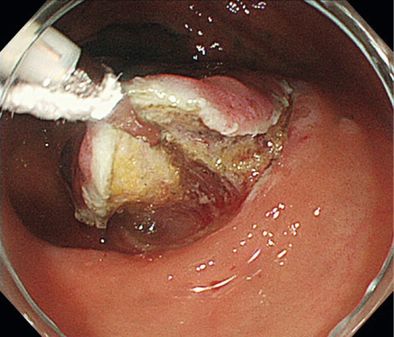
The submucosal layer is lifted, achieving good visibility and making it easy to dissect the submucosal layer by keeping the submucosal layer an appropriate distance from the colonoscope.
Then, a distal attachment cap placed at the tip of scope was used to add traction and create angulation, which resulted in good visibility of the submucosal layer of dissection. Once the submucosal layer of dissection was visualized, we released the hand from the line. After the circumferential mucosal incision was completed, the submucosal layer was dissected easily under direct visualization. If more traction was needed, we pulled the line by hand again.
Results
Patient and lesion characteristics are shown in Table 1. The study subjects were 12 men and 11 women with a median age of 72 years (range 49 – 88 years). Eighteen (78 %) lesions were located in the proximal colon, and 48 % of lesions were a granular type of laterally spreading tumor The median lesion size was 27 mm (range 20 – 44 mm).
Table 1. Patient characteristics.
| N = 23 | (%) | |
| Age, yr | 72 (49 – 88) | |
| Male/female, n | 12/11 | |
| Lesion location, n | ||
| Cecum | 7 | (30) |
| Ascending colon | 6 | (26) |
| Transverse colon | 5 | (22) |
| Descending colon | 1 | (4) |
| Sigmoid colon | 4 | (17) |
| Location group, n | ||
| Proximal colon (transverse colon or above) | 18 | (78) |
| Distal colon | 5 | (22) |
| Paris classification, n | ||
| 0-Is | 6 | (26) |
| 0-Is + IIa | 8 | (35) |
| 0-IIa | 6 | (26) |
| 0-IIc | 3 | (13) |
| Morphology, n | ||
| Granular | 11 | (48) |
| Non-granular | 7 | (30) |
| Unclassified | 5 | (22) |
| Lesion size, mm | 27 (20 – 44) |
NOTE: Values are expressed as median (range) unless otherwise noted.
Outcomes of ESD using the TAC method are shown in Table 2. A clip and line was attached to the lesion until the procedure ended, thus maintaining good visibility of the submucosal layer in 20/23 subjects and resulting in a procedural success rate for TAC of 87 %. In three cases, during submucosal dissection, the endoclip detached from lesions, all of which were in the proximal colon.The procedure times for these lesions were over 100 minutes whereas the overall mean procedure time was 61 min (95 % confidence interval [CI], 18 – 172 min.). The overall mean procedure speed was 16.7 mm2/min (95 % CI, 3.4 – 47.4 mm2/min.). En bloc resections of all lesions were achieved and R0 resections confirmed in 22/23 lesions (96 %). The lateral margin of the remaining lesion was unclear on histologic examination. Pathologic examination revealed deep submucosal invasion or lymphatic invasion in four lesions; additional surgical resection with lymph node dissection was recommended for these patients. Delayed bleeding occurred in one lesion and was treated successfully with endoscopic hemostasis. There were no perforations or fatal adverse events.
Table 2. Outcomes of treatment.
| N = 23 | (%) | |
| Successful rate of TAC | 20 | (87) |
| Resected specimen size, mm | 35 (23 – 60) | |
| Procedure time, min, mean (95 % confidence interval) | 61 (18 – 172) | |
| Procedure speed, mm2/min, mean (95 % confidence interval) |
16.7 (3.4 – 47.4) | |
| Histology, n | ||
| Tubular adenoma | 3 | |
| Tubulovillous adenoma | 4 | |
| Sessile serrated adenoma/polyp | 2 | |
| Intramucosal or minute submucosal invasive cancer | 11 | |
| Deeply submucosal invasive cancer | 3 | |
| En bloc resection, n | 23 | (100) |
| R0 resection, n | 22 | (96) |
| Adverse events, n | ||
| Delayed bleeding | 1 | (4) |
| Intraoperative perforation | 0 | (0) |
| Delayed perforation | 0 |
NOTE: Values are expressed as median (range) unless otherwise noted.
TAC, traction-assisted colonic ESD using clip and line
Minute submucosal invasive cancer: SM < 1000 μm
Deeply submucosal invasive cancer: SM ≥ 1000 μm
Discussion
The novel traction method TAC was successfully applied in most cases in our trial. This simple technique does not require withdrawal and reinsertion of the colonoscope and takes only a few minutes to attain good visualization of the submucosal layer during procedure. We found that this method is feasible for colonic ESD.
Several traction techniques, such as sinker-assisted ESD 9, S-O clip 10, cross-counter technique 11, and clip flap 12, have been developed for facilitating colorectal ESD. Although these techniques can provide useful traction during colorectal ESD, they are not used extensively because of their limitations. Sinker-assisted ESD, S-O clip, and cross-counter technique require special devices or equipment to obtain traction. Clip flap is simple and helpful; however, being an indirect traction method, it does not always provide sufficient traction or ability to lift the lesion. In addition, clip flap does not suppress movement of a lesion caused by respiration or arterial pulsation. To address these difficulties, Oyama et al developed the “clip with line” method for esophageal and gastric ESD and reported its safety and usefulness 4. This method has two advantages: first, adequate traction enables good visualization of the submucosal layer during submucosal dissection; and second, a clip and line fixes the specimens, thus preventing movement of the gastrointestinal wall caused by respiration or arterial pulsation. However, it is time consuming and painful because it withdrawal and reinsertion of the endoscope is required when this method is applied to colonic ESD. Therefore, we designed a novel “clip with line” fastening method for colonic ESD, which does not necessitate withdrawal and reinsertion of the colonoscope.
In a previous study, experienced endoscopists achieved a mean procedure time for colorectal ESD using Flush knife of 61 minutes (95 % CI 49 – 72) 13. Although in the current study half the colonic ESDs were performed by endoscopy fellows under direct supervision of experienced endoscopists, all lesions were resected en bloc and the mean procedure time was comparable with that in our previous study. The mean procedure time and procedure speed for the 20 cases in which TAC remained attached to the lesion until the end of the procedure was 53 min (95 % CI 18 – 72) and 17.9 mm2/min (95 % CI, 3.4 – 47.4 mm2/min.), respectively (data not shown). Of these 20 cases, only one took over 100 minutes. These results suggest that this novel traction method reduces the procedure time for colonic ESD. In this study, the endoclip detached from three lesions, all of which were in the proximal colon. Two of those procedures were performed by experienced endoscopists and one was performed by endoscopic fellow. The procedure times for these lesions were over 100 minutes. It seems that prolonged procedure times in the proximal colon can create so much friction on the clip and line that they detach from the specimen. With proximal colon lesions, we therefore recommend pulling gently on the line.
Even though we have shown that this novel traction method is feasible, this study does have several limitations. First, it was a small, retrospective, single-center study. Further prospective studies are warranted to assess the efficacy of this method for colorectal ESD. Second, the procedures were performed by four experienced endoscopists and four supervised endoscopy fellows, making it difficult to accurately assess what the en bloc resection rate would be in less experienced hands. Therefore, other outcome measures must be identified for checking the validity of our method.
In this feasibility study, we demonstrated that the TAC method is feasible and safe for colonic ESD. Further prospective randomized studies are needed to fully evaluate the usefulness of this method.
Footnotes
Competing interests: None
References
- 1.Takeuchi Y, Iishi H, Tanaka S. et al. Factors associated with technical difficulties and adverse events of colorectal endoscopic submucosal dissection: retrospective exploratory factor analysis of a multicenter prospective cohort. Int J Colorectal Dis. 2014;29:1275–1284. doi: 10.1007/s00384-014-1947-2. [DOI] [PubMed] [Google Scholar]
- 2.Lee E J, Lee J B, Lee S H. et al. Endoscopic submucosal dissection for colorectal tumors – 1000 colorectal ESD cases: one specialized institute’s experiences. Surg Endosc. 2013;27:31–39. doi: 10.1007/s00464-012-2403-4. [DOI] [PubMed] [Google Scholar]
- 3.Moss A, Williams S J, Hourigan L F. et al. Long-term adenoma recurrence following wide-field endoscopic mucosal resection (WF-EMR) for advanced colonic mucosal neoplasia is infrequent: results and risk factors in 1000 cases from the Australian Colonic EMR (ACE) study. Gut. 2015;64:57–65. doi: 10.1136/gutjnl-2013-305516. [DOI] [PubMed] [Google Scholar]
- 4.Oyama T. Counter traction makes endoscopic submucosal dissection easier. Clin Endosc. 2012;45:375–378. doi: 10.5946/ce.2012.45.4.375. [DOI] [PMC free article] [PubMed] [Google Scholar]
- 5.Ota M, Nakamura T, Hayashi K. et al. Usefulness of clip traction in the early phase of esophageal endoscopic submucosal dissection. Dig Endosc. 2012;24:315–318. doi: 10.1111/j.1443-1661.2012.01286.x. [DOI] [PubMed] [Google Scholar]
- 6.Koike Y, Hirasawa D, Fujita N. et al. Usefulness of the thread-traction method in esophageal endoscopic submucosal dissection: Randomized controlled trial. Dig Endosc. 2015;27:303–309. doi: 10.1111/den.12396. [DOI] [PubMed] [Google Scholar]
- 7.Yamasaki Y, Takeuchi Y, Hanaoka N. et al. A novel traction method using an endoclip attached to a nylon string during colonic endoscopic submucosal dissection. Endoscopy. 2015;47:E238–239. doi: 10.1055/s-0034-1391868. [DOI] [PubMed] [Google Scholar]
- 8.Japanese Society for Cancer of the Colon and Rectum editor. Japanese Classification of Colorectal Carcinoma 2nd ed Tokyo: Kanehara & Co., Ltd; 2009 [Google Scholar]
- 9.Saito Y, Emura F, Matsuda T. et al. A new sinker-assisted endoscopic submucosal dissection for colorectal tumors. Gastrointest Endosc. 2005;62:297–301. doi: 10.1016/s0016-5107(05)00546-8. [DOI] [PubMed] [Google Scholar]
- 10.Sakamoto N, Osada T, Shibuya T. et al. Endoscopic submucosal dissection of large colorectal tumors by using a novel spring-action S-O clip for traction (with video) Gastrointest Endosc. 2009;69:1370–1374. doi: 10.1016/j.gie.2008.12.245. [DOI] [PubMed] [Google Scholar]
- 11.Okamoto K, Muguruma N, Kitamura S. et al. Endoscopic submucosal dissection for large colorectal tumors using a cross-counter technique and a novel large-diameter balloon overtube. Dig Endosc. 2012;24:96–99. doi: 10.1111/j.1443-1661.2012.01264.x. [DOI] [PubMed] [Google Scholar]
- 12.Yamamoto K, Hayashi S, Saiki H. et al. Endoscopic submucosal dissection for large superficial colorectal tumors using the "clip-flap method". Endoscopy. 2015;47:262–265. doi: 10.1055/s-0034-1390739. [DOI] [PubMed] [Google Scholar]
- 13.Takeuchi Y, Uedo N, Ishihara R. et al. Efficacy of an endo-knife with a water-jet function (Flushknife) for endoscopic submucosal dissection of superficial colorectal neoplasms. Am J Gastroenterol. 2010;105:314–322. doi: 10.1038/ajg.2009.547. [DOI] [PubMed] [Google Scholar]


