



Abstract
Long non-coding RNAs (lncRNAs) CCAT1 and HOTAIR have been shown to play an important regulatory role in cancer biology, and CCAT1 and HOTAIR are upregulated in several cancers, however, its value in the diagnosis of colorectal cancer (CRC) is unclear. Therefore, the aim of this study is to evaluate the clinical significance of plasma CCAT1 and HOTAIR as a biomarker in the screening of CRC. In our study, we found that the levels of HOTAIR (P < 0.05) and CCAT1 (P < 0.05) were significantly higher in plasma of CRC patients than that of the healthy control. Moreover, the levels of lincRNA-p21 (P < 0.05) were obviously decreased in plasma of CRC patients as compared to those of healthy control. There was highly correlated for CCAT1 (R = 0.752, mean differences = -0.06 ± 1.20), HOTAIR (R = 0.739, mean differences = -0.26 ± 0.76) and lincRNA-p21 (R = 0.848, mean differences = -0.41 ± 0.89) in plasma and serum. By receiver operating characteristic curve (ROC) analysis, plasma CCAT1 provided the higher diagnostic performance for detection of CRC (the area under the ROC curve (AUC), 0.836; P < 0.001; sensitivity, 75.7%; specificity, 85.3%). Moreover, CCAT1 combining with HOTAIR could provide a more effective diagnosis performance (AUC, 0.954, P < 0.001, sensitivity, 84.3%; specificity, 80.2%). Most importantly, this combination was effective to detect CRC at an early stage (85%). In conclusion, our results demonstrated that increased plasma HOTAIR and CCAT1 could be used as a predictive biomarker for CRC screening, and that combination of HOTAIR and CCAT1 had a higher positive diagnostic rate of CRC than HOTAIR or CCAT1 alone.
Keywords: Colorectal carcinoma, long non-coding RNA, CCAT1, HOTAIR, tumor biomarker
Introduction
Colorectal cancer (CRC) is the third most common cancer and accounts for approximately 8%, more than 600,000 deaths of all cancer deaths, meanwhile around 1.2 million new cancer cases are diagnosed each year in the worldwide [1,2]. In China, the incidence and death rates of CRC have been rapidly increasing over the last few years, and the rates are much higher than the worldwide average [3]. In recent years, there are mounting progresses in clinical treatment for CRC, but the overall survival time of CRC patients have not improved dramatically. An important reason for that is the lack of molecular biomarkers. Recent biomarkers such as human cartilage glycoprotein 39 (YKL-40) [4], carbohydrate antigen 72-4 (CA72-4) [5] and carcino-embryonic antigen (CEA) [6] are the classic tumor markers commonly used in the management of patients with CRC. However, these tumor markers have limited utility in the early detection of CRC due to lack of sufficiently high diagnostic sensitivity and specificity. Therefore, the significance of exploration of new biomarkers with high sensitivity and specificity in early detection of CRC should be emphasized.
Eukaryotic genomes encode numerous long non-coding RNAs (also known as lncRNAs), which are defined as endogenous cellular RNAs with length longer than 200 nucleotides, but lack open reading frames of significant length [7]. Within 4 years, the number of identified lncRNA genes increase more than 8000, and estimates suggest the number of human lncRNAs ranging from 10,000 to 20,000 [8,9]. Although the function of most lncRNAs are still unknown, increasing numbers and accumulating evidences indicate that lncRNAs have been correlated to cancer development, invasion and metastasis in the malignant cell, including CRC [10]. Mounting evidences have showed that circulating lncRNA in plasma, serum or urine has been an emerging field for noninvasive diagnostic applications [7,11]. For example, plasma lncRNA-POU3F3 can serve as a potential biomarker for diagnosis of esophageal squamous cell carcinoma (ESCC), in particular for early tumor screening [7]. LncRNA-MALAT1 has been found to be significantly upregulated in plasma and urine of prostate cancer patients and can be used to discriminate cancer patients from healthy controls [11,12]. In gastric cancer patients, the levels of lncRNA-AA174084 in plasma are markedly lower on day 15 after surgery than preoperative level [13]. A similar study has found that plasma lncRNA-H19 levels are significantly higher in gastric cancer patients than in healthy controls and reduced in postoperative patients [14]. However, for all we know, no literature has been reported regarding the circulating lncRNAs for early screening of CRC patients.
LncRNA colon cancer-associated transcript 1 (CCAT1) is a recently discovered 2628 nucleotide lncRNA, which is located in the chromosome 8q24.21 and described as a “hot spot” with many genetic alternations in colon cancer [15,16]. Previous studies show that CCAT1 is significantly upregulated in tumor tissues of colon cancer patients compared with the healthy human tissues and promotes colon cancer cell proliferation and invasion [16,17]. In this study, by using lncRNA and disease database (http://210.73.221.6/lncrnadisease), other 12 lncRNAs, including H19, HOTAIR, MALAT1, MEG3, CRNDE, HULC, KCNQ1OT1, lincRNA-p21, NPTN-IT1, SNHG16, ATB and LSINCT5, were previously reported abnormal expression and selected as candidate diagnostic biomakers in colorectal cancer [18-30]. We were examined the expression levels of lncRNAs in plasma and serum, and their potential use as tumor biomarkers for CRC detection were evaluated. We hypothesized that these CRC-related lncRNAs might be released into the circulation during CRC initiation and could be utilized to detect and screen CRC.
Materials and methods
Patients and specimens
Thirty-two CRC tissues and matched adjacent non-tumor tissues were collected from the Second and Third Affiliated Hospital of Xinjiang Medical University between Jan 2012 and June 2014. All patients recruited in this study were not subjected to preoperative radiotherapy or chemotherapy and diagnosed with CRC based on histopathological evaluation. All collected tissue samples were immediately stored at liquid nitrogen until use. Human samples were obtained with written informed consent from all patients. The study was approved by the Ethics Committee of the Xinjiang Medical University, China.
Real-time PCR
Total RNA was extracted by Trizol reagent (Invitrogen, Carlsbad, CA, USA). Reaction mixture (20 μl) containing 2 μg of total RNA was reversely transcribed to cDNA by using PrimeScript RT-polymerase (Takara, Dalian, China). Quantitative PCR was performed on the cDNA using specific primers (Sangon, Shanghai, China). The first strand cDNAs served as the template for the regular polymerase chain reaction (PCR) performed using a DNA Engine (ABI 9700). Glyceraldehyde-phosphate dehydrogenase (GAPDH) as an internal control was used to normalize the data to determine the relative expression of the target genes. The reaction conditions were set according to the kit instructions. After completion of the reaction, the expression levels of lncRNAs were calculated using ΔCt method, where ΔCt = Cttarget-Ctreference, smaller ΔCt value indicates higher expression. Relative expression of lncRNAs was calculated using 2-ΔΔCt method normalized to endogenous control, with ΔCt = Cttarget-Ctreference, -ΔΔCt = -(sample ΔCt-control ΔCt). All the primers used in the present study were listed in Table 1.
Table 1.
Summary of CRC-related LncRNAs in human
| Symbol | Expression levels | Chromosome Location | Start | End | Species |
|---|---|---|---|---|---|
| H19 | undifferentiated | Chr 11 | 2016406 | 2019065 | Human |
| HOTAIR | upregulated | Chr 12 | 54356096 | 54362515 | Human |
| MALAT1 | undifferentiated | Chr 11 | 65265233 | 65273940 | Human |
| MEG3 | undifferentiated | Chr 14 | 101292445 | 101327363 | Human |
| CCAT1 | upregulated | Chr 8 | 128219629 | 128231724 | Human |
| CRNDE | upregulated | Chr 16 | 54952775 | 54963079 | Human |
| HULC | upregulated | Chr 6 | 8652442 | 8654079 | Human |
| KCNQ1OT1 | upregulated | Chr 11 | 2661768 | 2721228 | Human |
| lincRNA-p21 | downregulated | Chr 17 | 29057474 | 29078961 | Human |
| NPTN-IT1 | downregulated | Chr 15 | 73859365 | 73861635 | Human |
| SNHG16 | downregulated | Chr 17 | 76557764 | 76565348 | Human |
| ATB | upregulated | N/A | N/A | N/A | Human |
| LSINCT5 | upregulated | N/A | N/A | N/A | Human |
Statistical analysis
All statistical analyses were performed using SPSS version 18.0 software. Data were analyzed using independent two-tailed t test. Categorical data were analyzed using the two-side chi-square test. Overall survival was estimated by using Kaplan-Meier method, and univariate analysis was conducted by log-rank test. The Cox proportional hazards model was used in the multivariate analysis. Values of P < 0.05 were considered statistically significant.
Results
Identification of tumor tissues-enriched lncRNA implicated in CRC patients
Based on previous research, by using lncRNA and disease database, 13 lncRNAs, including CCAT1, H19, HOTAIR, MALAT1, MEG3, CRNDE, HULC, KCNQ1OT1, lincRNA-p21, NPTN-IT1, SNHG16, ATB and LSINCT5, were previously reported abnormal expression and selected as candidate diagnostic biomakers in CRC (Table 2). To further validated lncRNAs levels in CRC, the real-time PCR analysis was performed to determine the expression level of lncRNAs in 32 pairs of human CRC tissues and corresponding non-tumourous specimens. The results showed that 7 lncRNAs (HOTAIR, CCAT1, CRNDE, HULC, KCNQ1OT1, ATB and LSINCT5) were significantly higher in most of CRC tumor tissues than that observed in pair-matched adjacent non-tumourous tissues (Figure 1). Moreover, among them, lincRNA-p21, NPTN-IT1 and SNHG16 were obviously down-regulated in most of CRC tumor tissues as compared to those of non-tumourous group. However, the levels of H19, MALAT1 and MEG3 did not show any significantly different between CRC tumor tissues and corresponding non-tumourous specimens (Figure 1).
Table 2.
The PCR primers used in this study
| Symbol | Forward (5’-3’) | Reverse (5’-3’) |
|---|---|---|
| H19 | CTCCACGACTCTGTTTCC | CTCCACGACTCTGTTTCC |
| HOTAIR | AATAGACATAGGAGAACACTT | AATCTTAATAGCAGGAGGAA |
| MALAT1 | CCGCTGCTATTAGAATGC | CTTCAACAATCACTACTCCAA |
| MEG3 | TGGCATAGAGGAGGTGAT | AGACAAGTAAGACAAGCAAGA |
| CCAT1 | CATTGGGAAAGGTGCCGAGA | ACGCTTAGCCATACAGAGCC |
| CRNDE | TGGATGCTGTCAGCTAGTTCAC | TTCCAGTGGCATCCTCCTTATC |
| HULC | TCAGAGTTCCTGCATGGTCTGGTTC | GTTCCTGCATGGTCTGGTTCTCGTG |
| KCNQ1OT1 | GCATATCTGTCTTCCGTAT | CCTCTTCCTTCGTTCAAT |
| lincRNA-p21 | CCCGGGCTTGTCTTTTGTT | GAGTGGGTGGCTCACTCTTCTG |
| NPTN-IT1 | GACCGGTGCGAAGTGGCTTC | GTTCCCGACGGACCTTGCGAG |
| SNHG16 | GGCCGATGAGATGTGTAT | CCTGATGTGAGCGTAGCTGT |
| ATB | CCGATGGATGCGTGAGCTTT | GAGAGTGTAGATGCTGCAG |
| LSINCT5 | TAGACAACTTACTTAACCTCAT | TCCTTATCCACCTTATCCA |
| GAPDH | ATGGTGAAGGTCGGTGTGA | CCATGTAGTTGAGGTCAATGAG |
Figure 1.
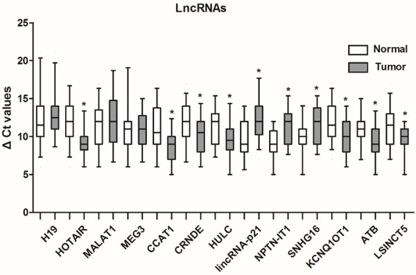
Identification of tumor tissues-enriched lncRNA implicated in CRC patients. 13 CRC-related lncRNAs are filtered from lncRNA and disease database. Real-time PCR analysis is performed to determine the expression levels of lncRNAs in 32 pairs of CRC tumor tissues and corresponding non-tumourous specimens. ΔCt method is used to calculate lncRNA expression, which is normalized to GAPDH, and smaller ΔCt value indicated higher expression. Values are expressed as mean ± SD, *P < 0.05 versus non-tumourous group.
HOTAIR, CCAT1 and lincRNA-p21 were detectable in plasma
To explore whether these CRC-related lncRNAs could reach the circulation at levels sufficient to be detectable, real-time PCR analysis was performed to determine the expression levels of HOTAIR, CCAT1 and lincRNA-p21 in 32 pairs of CRC tumor tissues and corresponding non-tumourous specimens. As shown in Figure 2A and 2B, the levels of HOTAIR (P < 0.05) and CCAT1 (P < 0.05) were significantly higher in plasma of CRC patients than that of the healthy control. Moreover, the levels of lincRNA-p21 (P < 0.05) were obviously decreased in plasma of CRC patients as compared to those of healthy control (Figure 2C). These results indicated that CRC-related lncRNAs could enter into the circulation, and their differentiated expression in plasma could be used as diagnostic biomarkers for CRC.
Figure 2.
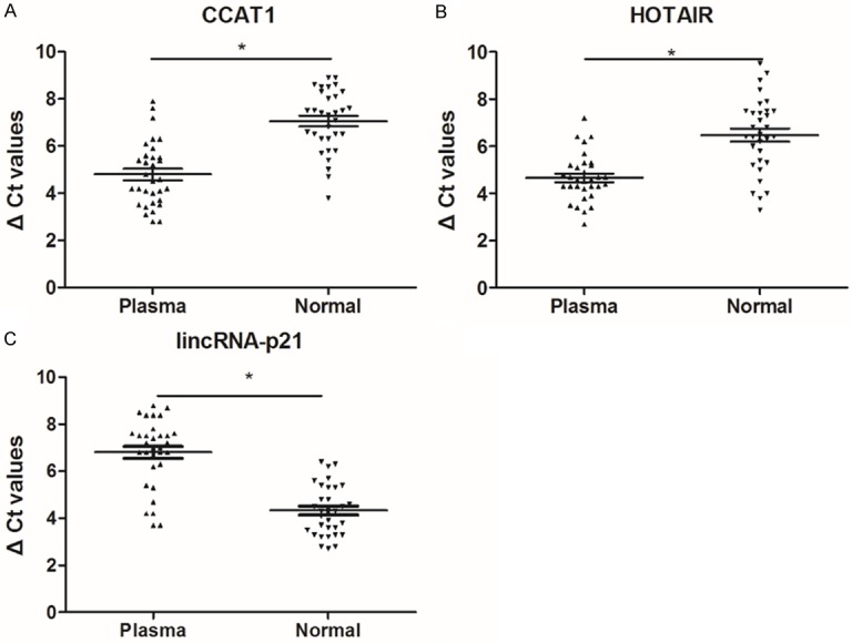
CCAT1, HOTAIR and lincRNA-p21 were detectable in plasma from CRC patients. CCAT1 (A), HOTAIR (B) and lincRNA-p21 (C) expression are examined by real-time PCR and normalized to GAPDH expression in plasma from CRC patients and healthy controls. Circulating lncRNAs expressions are calculated using ΔCt method. Values are expressed as mean ± SD, n = 32 in each group, *P < 0.05 versus healthy controls.
Correlation of lncRNAs levels between plasma and serum in CRC patients
To test whether there was a relationship of lncRNAs levels between plasma and serum in CRC patients, CCAT1, HOTAIR and lincRNA-p21 were measured in plasma and serum from the same individuals. As shown in Figure 3A-C, there was highly correlated for CCAT1 (R = 0.752, mean differences = -0.06 ± 1.20), HOTAIR (R = 0.739, mean differences = -0.26 ± 0.76) and lincRNA-p21 (R = 0.848, mean differences = -0.41 ± 0.89) in plasma and serum.
Figure 3.
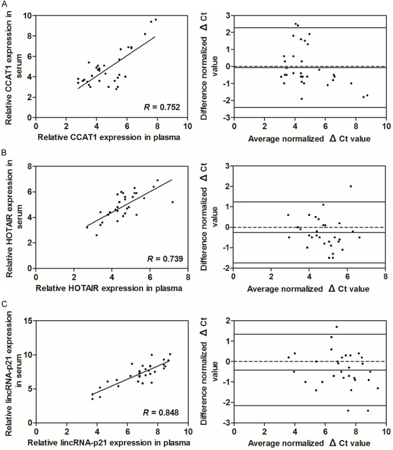
Correlation of lncRNAs levels between plasma and serum in CRC patients. Linear correlation plot of CCAT1 (A), HOTAIR (B) and lincRNA-p21 (C) in plasma and serum (left). There is a high correlation comparing the indicated lncRNAs levels between plasma and serum. Bland-Altman plot of the difference between plasma and serum CRC-related lncRNAs level versus their average. Horizontal solid lines in the middle represent the mean difference. Upper and lower solid lines represent the limits of agreement (95% confidence intervals) (right).
HOTAIR, CCAT1 and lincRNA-p21 levels in pre-operative and post-operative plasma samples
Since circulating lncRNAs were primarily released or leaked from tumor cells, they would revert to normal after the tumor has been resected. In the present study, the CCAT1, HOTAIR and lincRNA-p21 were carried out to investigate the differences in CRC-related lncRNAs in plasma pro-operative and 14 days post-operative. As expected, serum levels of CCAT1 and HOTAIR were significantly decreased after surgical treatment as compared to pre-operative (Figure 4A and 4B), and lincRNA-p21 was significantly increased after surgical treatment as compared to pre-operative (Figure 4C).
Figure 4.
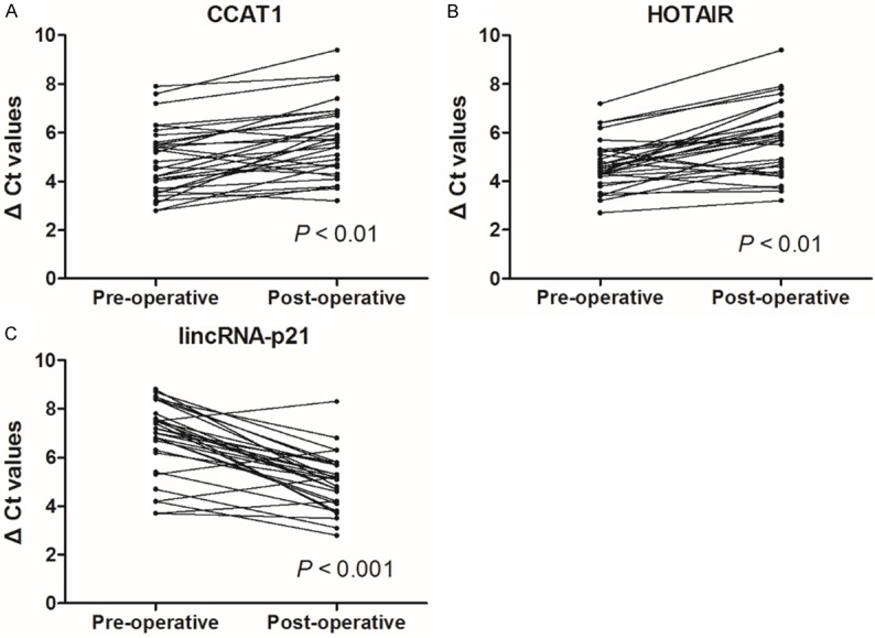
HOTAIR, CCAT1 and lincRNA-p21 levels in pre-operative and post-operative plasma samples. CRC-related lncRNAs CCAT1 (A), HOTAIR (B) and lincRNA-p21 (C) levels are examined by real-time PCR in post-operative samples as compared to pre-operative samples. Circulating lncRNAs expressions are calculated using ΔCt method. Values are expressed as mean ± SD, n = 32 in each group.
Evaluation of HOTAIR, CCAT1 or lincRNA-p21 in plasma as predictive CRC-related biomarkers
To investigate the characteristics of HOTAIR, CCAT1 or lincRNA-p21 as potential biomarkers for CRC, receiver operating characteristics (ROC) curves and the area under the ROC curves (AUC) were performed on data from all subjects, including 32 CRC patients and 32 healthy donors. The ROC curves illustrated strong separation between the CRC patients and control group, with an AUC of 0.836 (95% CI: 0.739-0.934; P < 0.001) for CCAT1, 0.777 (95% CI: 0.663-0.891; P < 0.001) for HOTAIR and 0.886 (95% CI: 0.802-0.970; P < 0.001) for lincRNA-p21 (Figure 5A-C). Intriguingly, there is increasing evidence showing that combination several tumor markers could improve diagnostic accuracy. In the present study, we determined whether the combination of HOTAIR and CCAT1 could provide a more effective screening for CRC. The results indicated that combination of HOTAIR and CCAT1 yielded an AUC of 0.954 (95% CI: 0.903-1.000; P < 0.001), which was significantly improved as compared to HOTAIR (AUC = 0.777) or CCAT1 (AUC = 0.836) alone (Figure 5D). Another aim of this work was to diagnose CRC patients at as early stage as possible. Therefore, the diagnostic positivity rate between different clinical stages was then investigated. As shown in Table 3, the diagnostic positivity rate for CRC in stage I/II for HOTAIR, CCAT1 or HOTAIR combined with CCAT1 was 25%, 40%, and 85%, respectively.
Figure 5.
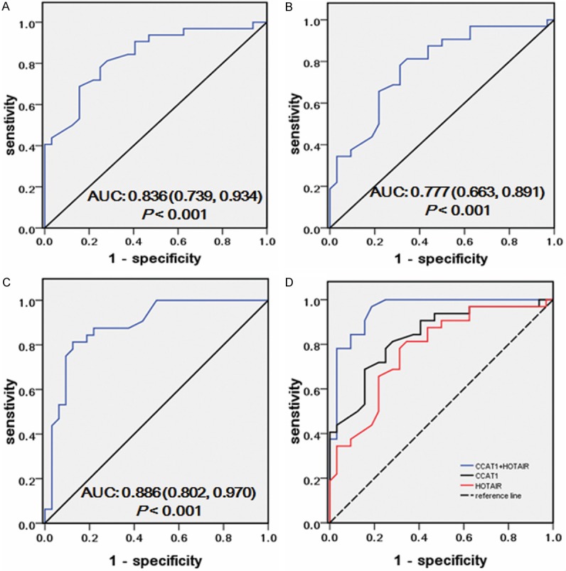
Evaluation of HOTAIR, CCAT1 or lincRNA-p21 in plasma as a predictive CRC-related biomarker. Receiver operating characteristics (ROC) curves are drawn with the data of plasma lncRNAs, CCAT1 (A), HOTAIR (B) and lincRNA-p21 (C), from 32 CRC patients and 32 healthy controls. ROC corves of a combination of HOTAIR and CCAT1 to discriminate CRC from healthy controls (D).
Table 3.
Performance of CCAT1, HOTAIR and both CCAT1 and HOTAIR in the detection of different clinical stage CRC
| Clinical stage | |||
|---|---|---|---|
|
|
|||
| I/II | III/IV | Total | |
| CCAT1 | 40.0% (8/20) | 66.7.0% (8/12) | 50.0% (16/32) |
| HOTAIR | 25.0% (5/20) | 58.3% (7/12) | 37.5% (12/32) |
| CCAT1 + HOTAIR | 85.0% (17/20) | 91.7% (11/12) | 87.5% (28/32) |
| P value | |||
| Combination vs. CCAT1 | < 0.001 | 0.042 | < 0.001 |
| Combination vs. HOTAIR | < 0.001 | 0.018 | < 0.001 |
Discussion
Recent genome-wide studies have indicated that the mammalian genome is abundantly transcribed and that at least 80% of this transcription is exclusively associated with lncRNAs [31]. So far, numerous studies have indicated that lncRNAs play an important role in tumor occurrence, invasion and metastasis by regulating gene expression as well as signaling pathways [32]. Emerging data strongly implicates that lncRNAs in plasma or serum may be utilized as a tool for cancer diagnosis [7]. However, we found that the effect of lncRNAs in the early diagnosis and prediction of CRC had not been investigated thoroughly. Therefore, we tried to investigate the role of CCAT1 and HOTAIR in the screening of CRC.
In our work, the initial CRC-related lncRNAs screening was performed based on different expression profiling between tumor tissues and corresponding non-tumourous specimens, and all lncRNAs of interest were then subjected to real-time PCR validation. The results demonstrated that the levels of CCAT1 and HOTAIR were significantly higher in plasma from CRC patients compared with healthy controls. We used the ROC curve to analyze the diagnostic value of plasma CCAT1 and HOTAIR. The results showed that the individual AUC of CCAT1 and HOTAIR for the diagnosis of CRC were about 0.836 and 0.777, respectively, Moreover, the combination of CCAT1 and HOTAIR could provide a more effective screening for CRC. The results indicated that combination of CCAT1 and HOTAIR yielded an AUC of 0.954, which was significantly improved as compared to CCAT1 (AUC = 0.836) or HOTAIR (AUC = 0.777) alone. These results indicated that CRC-related lncRNAs could be released into the circulation and that their different expression profiles in plasma could be used as diagnostic markers for ESCC. Intriguingly, CCAT1 combining with HOTAIR could provide a more powerful differential diagnosis between CRC patients and healthy controls than use of POU3F3 or SCCA alone. Some classic tumor biomarkers have limited utility in the early detection of CRC [4-6], however, the levels of lncRNA in plasma appear to play an important role in the early screening of CRC. In our study, the results indicated that plasma CCAT1 was more effective than HOTAIR for early detection of CRC (40% vs. 25%), and the combination of CCAT1 and HOTAIR could significantly provide the positivity rate (85%) for CRC screening.
As we know, early discovery, early diagnosis, and early treatment could greatly increase the survival rate of cancer patients. Biomarkers in body fluid have a potential capacity to detect cancers in early stage [32]. Measurements obtained from plasma and serum were strongly correlated for CCAT1 and HOTAIR. The results suggested that circulating lncRNAs were acceptable for CRC screening. HOTAIR as a candidate lncRNA has been shown to play an important modulatory role in the development and progression of cancer, and as a biomarker has been applied to screen gastric cancer [33] and breast cancer [34]. The circulating HOTAIR shows a 2.15-fold change in breast cancer patients compared with healthy controls (P < 0.0001, AUC = 0.786), and the optimal cutoff value for diagnosis was 0.30 with sensitivity of 80.0% and specificity of 68.3% [34]. In our study, the levels of HOTAIR showed a 4-fold change in plasma from CRC patients compared with healthy controls (P < 0.001, AUC = 0.777). Moreover, plasma level of HOTAIR could discriminate CRC from normal controls with 67.5% sensitivity and 89.9% specificity, and the sensitivity and specificity of CCAT1 for CRC diagnosis were 75.7% and 85.3% respectively. The sensitivity and specificity of combination (SCCA + POU3F3) group for distinguishing CRC from healthy controls were 84.3% and 80.2% respectively.
In conclusion, our results demonstrated that increased plasma HOTAIR and CCAT1 could be used as a predictive biomarker for CRC screening, and that combination of HOTAIR and CCAT1 had a higher positive diagnostic rate of CRC than HOTAIR or CCAT1 alone.
Disclosure of conflict of interest
None.
References
- 1.Liang WC, Fu WM, Wong CW, Wang Y, Wang WM, Hu GX, Zhang L, Xiao LJ, Wan DC, Zhang JF, Waye MM. The LncRNA H19 promotes epithelial to mesenchymal transition by functioning as MiRNA sponges in colorectal cancer. Oncotarget. 2015;6:22513–25. doi: 10.18632/oncotarget.4154. [DOI] [PMC free article] [PubMed] [Google Scholar]
- 2.Qiu JJ, Yan JB. Long non-coding RNA LINC01296 is a potential prognostic biomarker in patients with colorectal cancer. Tumour Biol. 2015;36:7175–83. doi: 10.1007/s13277-015-3448-5. [DOI] [PubMed] [Google Scholar]
- 3.Zheng ZX, Zheng RS, Zhang SW, Chen WQ. Colorectal cancer incidence and mortality in China, 2010. Asian Pac J Cancer Prev. 2014;15:8455–8460. doi: 10.7314/apjcp.2014.15.19.8455. [DOI] [PubMed] [Google Scholar]
- 4.Ye HM, Lu YZ, Liang XM, Lin YZ, Li Y, Zhang ZY, Tzeng CM. Clinical significance of combined testing of YKL-40 with CEA in Chinese colorectal cancer patients. Clin Lab. 2014;60:397–405. doi: 10.7754/clin.lab.2013.121027. [DOI] [PubMed] [Google Scholar]
- 5.Ayude D, Rodriguez-Berrocal FJ, Ayude J, Blanco-Prieto S, Vazquez-Iglesias L, Vazquez-Cedeira M, Paez de la Cadena M. Preoperative serum CA 72.4 as prognostic factor of recurrence and death, especially at TNM stage II, for colorectal cancer. BMC Cancer. 2013;13:543. doi: 10.1186/1471-2407-13-543. [DOI] [PMC free article] [PubMed] [Google Scholar]
- 6.Wang YR, Yan JX, Wang LN. The diagnostic value of serum carcino-embryonic antigen, alpha fetoprotein and carbohydrate antigen 19-9 for colorectal cancer. J Cancer Res Ther. 2014;10(Suppl):307–309. doi: 10.4103/0973-1482.151538. [DOI] [PubMed] [Google Scholar]
- 7.Tong YS, Wang XW, Zhou XL, Liu ZH, Yang TX, Shi WH, Xie HW, Lv J, Wu QQ, Cao XF. Identification of the long non-coding RNA POU3F3 in plasma as a novel biomarker for diagnosis of esophageal squamous cell carcinoma. Mol Cancer. 2015;14:3. doi: 10.1186/1476-4598-14-3. [DOI] [PMC free article] [PubMed] [Google Scholar]
- 8.Gibb EA, Brown CJ, Lam WL. The functional role of long non-coding RNA in human carcinomas. Mol Cancer. 2011;10:38. doi: 10.1186/1476-4598-10-38. [DOI] [PMC free article] [PubMed] [Google Scholar]
- 9.Martens-Uzunova ES, Bottcher R, Croce CM, Jenster G, Visakorpi T, Calin GA. Long noncoding RNA in prostate, bladder, and kidney cancer. Eur Urol. 2014;65:1140–1151. doi: 10.1016/j.eururo.2013.12.003. [DOI] [PubMed] [Google Scholar]
- 10.Xiang JF, Yin QF, Chen T, Zhang Y, Zhang XO, Wu Z, Zhang S, Wang HB, Ge J, Lu X, Yang L, Chen LL. Human colorectal cancer-specific CCAT1-L lncRNA regulates long-range chromatin interactions at the MYC locus. Cell Res. 2014;24:513–531. doi: 10.1038/cr.2014.35. [DOI] [PMC free article] [PubMed] [Google Scholar]
- 11.Wang F, Ren S, Chen R, Lu J, Shi X, Zhu Y, Zhang W, Jing T, Zhang C, Shen J, Xu C, Wang H, Wang H, Wang Y, Liu B, Li Y, Fang Z, Guo F, Qiao M, Wu C, Wei Q, Xu D, Shen D, Lu X, Gao X, Hou J, Sun Y. Development and prospective multicenter evaluation of the long noncoding RNA MALAT-1 as a diagnostic urinary biomarker for prostate cancer. Oncotarget. 2014;5:11091–11102. doi: 10.18632/oncotarget.2691. [DOI] [PMC free article] [PubMed] [Google Scholar]
- 12.Ren S, Wang F, Shen J, Sun Y, Xu W, Lu J, Wei M, Xu C, Wu C, Zhang Z, Gao X, Liu Z, Hou J, Huang J, Sun Y. Long non-coding RNA metastasis associated in lung adenocarcinoma transcript 1 derived miniRNA as a novel plasma-based biomarker for diagnosing prostate cancer. Eur J Cancer. 2013;49:2949–2959. doi: 10.1016/j.ejca.2013.04.026. [DOI] [PubMed] [Google Scholar]
- 13.Shao Y, Ye M, Jiang X, Sun W, Ding X, Liu Z, Ye G, Zhang X, Xiao B, Guo J. Gastric juice long noncoding RNA used as a tumor marker for screening gastric cancer. Cancer. 2014;120:3320–3328. doi: 10.1002/cncr.28882. [DOI] [PubMed] [Google Scholar]
- 14.Zhou X, Yin C, Dang Y, Ye F, Zhang G. Identification of the long non-coding RNA H19 in plasma as a novel biomarker for diagnosis of gastric cancer. Sci Rep. 2015;5:11516. doi: 10.1038/srep11516. [DOI] [PMC free article] [PubMed] [Google Scholar]
- 15.Zanke BW, Greenwood CM, Rangrej J, Kustra R, Tenesa A, Farrington SM, Prendergast J, Olschwang S, Chiang T, Crowdy E, Ferretti V, Laflamme P, Sundararajan S, Roumy S, Olivier JF, Robidoux F, Sladek R, Montpetit A, Campbell P, Bezieau S, O'Shea AM, Zogopoulos G, Cotterchio M, Newcomb P, McLaughlin J, Younghusband B, Green R, Green J, Porteous ME, Campbell H, Blanche H, Sahbatou M, Tubacher E, Bonaiti-Pellie C, Buecher B, Riboli E, Kury S, Chanock SJ, Potter J, Thomas G, Gallinger S, Hudson TJ, Dunlop MG. Genome-wide association scan identifies a colorectal cancer susceptibility locus on chromosome 8q24. Nat Genet. 2007;39:989–994. doi: 10.1038/ng2089. [DOI] [PubMed] [Google Scholar]
- 16.He X, Tan X, Wang X, Jin H, Liu L, Ma L, Yu H, Fan Z. C-Myc-activated long noncoding RNA CCAT1 promotes colon cancer cell proliferation and invasion. Tumour Biol. 2014;35:12181–12188. doi: 10.1007/s13277-014-2526-4. [DOI] [PubMed] [Google Scholar]
- 17.Nissan A, Stojadinovic A, Mitrani-Rosenbaum S, Halle D, Grinbaum R, Roistacher M, Bochem A, Dayanc BE, Ritter G, Gomceli I, Bostanci EB, Akoglu M, Chen YT, Old LJ, Gure AO. Colon cancer associated transcript-1: a novel RNA expressed in malignant and pre-malignant human tissues. Int J Cancer. 2012;130:1598–1606. doi: 10.1002/ijc.26170. [DOI] [PubMed] [Google Scholar]
- 18.Ohana P, Schachter P, Ayesh B, Mizrahi A, Birman T, Schneider T, Matouk I, Ayesh S, Kuppen PJ, de Groot N, Czerniak A, Hochberg A. Regulatory sequences of H19 and IGF2 genes in DNA-based therapy of colorectal rat liver metastases. J Gene Med. 2005;7:366–374. doi: 10.1002/jgm.670. [DOI] [PubMed] [Google Scholar]
- 19.Hrdlickova B, de Almeida RC, Borek Z, Withoff S. Genetic variation in the non-coding genome: Involvement of micro-RNAs and long non-coding RNAs in disease. Biochim Biophys Acta. 2014;1842:1910–1922. doi: 10.1016/j.bbadis.2014.03.011. [DOI] [PubMed] [Google Scholar]
- 20.Zhang X, Zhou Y, Mehta KR, Danila DC, Scolavino S, Johnson SR, Klibanski A. A pituitary-derived MEG3 isoform functions as a growth suppressor in tumor cells. J Clin Endocrinol Metab. 2003;88:5119–5126. doi: 10.1210/jc.2003-030222. [DOI] [PubMed] [Google Scholar]
- 21.Qi P, Du X. The long non-coding RNAs, a new cancer diagnostic and therapeutic gold mine. Mod Pathol. 2013;26:155–165. doi: 10.1038/modpathol.2012.160. [DOI] [PubMed] [Google Scholar]
- 22.Graham LD, Pedersen SK, Brown GS, Ho T, Kassir Z, Moynihan AT, Vizgoft EK, Dunne R, Pimlott L, Young GP, Lapointe LC, Molloy PL. Colorectal Neoplasia Differentially Expressed (CRNDE), a Novel Gene with Elevated Expression in Colorectal Adenomas and Adenocarcinomas. Genes Cancer. 2011;2:829–840. doi: 10.1177/1947601911431081. [DOI] [PMC free article] [PubMed] [Google Scholar]
- 23.Matouk IJ, Abbasi I, Hochberg A, Galun E, Dweik H, Akkawi M. Highly upregulated in liver cancer noncoding RNA is overexpressed in hepatic colorectal metastasis. Eur J Gastroenterol Hepatol. 2009;21:688–692. doi: 10.1097/meg.0b013e328306a3a2. [DOI] [PubMed] [Google Scholar]
- 24.Cheetham SW, Gruhl F, Mattick JS, Dinger ME. Long noncoding RNAs and the genetics of cancer. Br J Cancer. 2013;108:2419–2425. doi: 10.1038/bjc.2013.233. [DOI] [PMC free article] [PubMed] [Google Scholar]
- 25.Zhai H, Fesler A, Schee K, Fodstad O, Flatmark K, Ju J. Clinical significance of long intergenic noncoding RNA-p21 in colorectal cancer. Clin Colorectal Cancer. 2013;12:261–266. doi: 10.1016/j.clcc.2013.06.003. [DOI] [PubMed] [Google Scholar]
- 26.Yang F, Huo XS, Yuan SX, Zhang L, Zhou WP, Wang F, Sun SH. Repression of the long noncoding RNA-LET by histone deacetylase 3 contributes to hypoxia-mediated metastasis. Mol Cell. 2013;49:1083–1096. doi: 10.1016/j.molcel.2013.01.010. [DOI] [PubMed] [Google Scholar]
- 27.Qi P, Xu MD, Ni SJ, Shen XH, Wei P, Huang D, Tan C, Sheng WQ, Zhou XY, Du X. Downregulation of ncRAN, a long non-coding RNA, contributes to colorectal cancer cell migration and invasion and predicts poor overall survival for colorectal cancer patients. Mol Carcinog. 2015;54:742–50. doi: 10.1002/mc.22137. [DOI] [PubMed] [Google Scholar]
- 28.Wang G, Li Z, Zhao Q, Zhu Y, Zhao C, Li X, Ma Z, Li X, Zhang Y. LincRNA-p21 enhances the sensitivity of radiotherapy for human colorectal cancer by targeting the Wnt/beta-catenin signaling pathway. Oncol Rep. 2014;31:1839–1845. doi: 10.3892/or.2014.3047. [DOI] [PubMed] [Google Scholar]
- 29.Iguchi T, Uchi R, Nambara S, Saito T, Komatsu H, Hirata H, Ueda M, Sakimura S, Takano Y, Kurashige J, Shinden Y, Eguchi H, Sugimachi K, Maehara Y, Mimori K. A long noncoding RNA, lncRNA-ATB, is involved in the progression and prognosis of colorectal cancer. Anticancer Res. 2015;35:1385–1388. [PubMed] [Google Scholar]
- 30.Xu MD, Qi P, Weng WW, Shen XH, Ni SJ, Dong L, Huang D, Tan C, Sheng WQ, Zhou XY, Du X. Long non-coding RNA LSINCT5 predicts negative prognosis and exhibits oncogenic activity in gastric cancer. Medicine (Baltimore) 2014;93:e303. doi: 10.1097/MD.0000000000000303. [DOI] [PMC free article] [PubMed] [Google Scholar]
- 31.Wang Y, Chen W, Yang C, Wu W, Wu S, Qin X, Li X. Long non-coding RNA UCA1a(CUDR) promotes proliferation and tumorigenesis of bladder cancer. Int J Oncol. 2012;41:276–284. doi: 10.3892/ijo.2012.1443. [DOI] [PubMed] [Google Scholar]
- 32.Li Q, Shao Y, Zhang X, Zheng T, Miao M, Qin L, Wang B, Ye G, Xiao B, Guo J. Plasma long noncoding RNA protected by exosomes as a potential stable biomarker for gastric cancer. Tumour Biol. 2015;36:2007–2012. doi: 10.1007/s13277-014-2807-y. [DOI] [PubMed] [Google Scholar]
- 33.Arita T, Ichikawa D, Konishi H, Komatsu S, Shiozaki A, Shoda K, Kawaguchi T, Hirajima S, Nagata H, Kubota T, Fujiwara H, Okamoto K, Otsuji E. Circulating long non-coding RNAs in plasma of patients with gastric cancer. Anticancer Res. 2013;33:3185–3193. [PubMed] [Google Scholar]
- 34.Zhang L, Song X, Wang X, Xie Y, Wang Z, Xu Y, You X, Liang Z, Cao H. Circulating DNA of HOTAIR in serum is a novel biomarker for breast cancer. Breast Cancer Res Treat. 2015;152:199–208. doi: 10.1007/s10549-015-3431-2. [DOI] [PubMed] [Google Scholar]