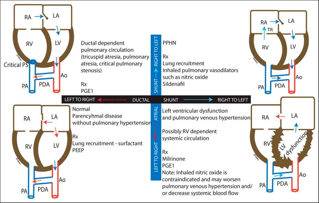
Figure 4.
Echocardiographic evaluation of neonatal hypoxemia based on ductal (black bar) and atrial (blue bar) shunts. Left-to-right shunt at the ductal and atrial level is considered normal but can also be seen in the presence of parenchymal lung disease, resulting in hypoxemia in the absence of persistent pulmonary hypertension of the newborn (PPHN) (lower left quadrant). The presence of right-to-left shunt at the atrial and ductal levels is associated with PPHN (upper right quadrant). Right-to-left shunt at the ductal level but a left-to-right shunt at the atrial level is associated with left ventricular dysfunction, pulmonary venous hypertension, and ductal-dependent systemic circulation (lower right quadrant) and is a contraindication for inhaled pulmonary vasodilators, such as inhaled nitric oxide. In patients with right-sided obstruction (such as critical pulmonary stenosis [PS]), right atrial blood flows to the left atrium through the PFO. Pulmonary circulation is dependent on a left-to-right shunt at the patent ductus arteriosus (PDA) (upper left quadrant).
Ao = aorta; LA = left atrium; LV = left ventricle; PA = pulmonary artery; PGE1 = prostaglandin E1; RA = right atrium; RV = right ventricle; Rx = treatment; TR = tricuspid regurgitation.
Modified from Nair and Lakshminrusimha. (15) Copyright Satyan Lakshminrusimha.

