

Abstract
AIM: To investigate the cellular effects of hybrid polar compound hexamethylene bisacetamide (HMBA) on the growth and apoptosis of human hepatocellular carcinoma cells and to provide the molecular mechanism for potential application of HMBA in the treatment of liver cancer.
METHODS: Effects of HMBA on the growth of human hepatocellular carcinoma SMMC-7721 cells were assayed by MTT chronometry. Apoptosis induced by HMBA was detected by phase-contrast microscopy, flow cytometry, propidium iodide staining and immunocytochemical analysis.
RESULTS: The growth of SMMC-7721 cells was significantly inhibited by HMBA, and the growth inhibitory rate was 51.1%, 62.6%, 68.7% and 73.9% respectively after treatment with 5.0, 7.5, 10.0 and 12.5 mmol/L of HMBA. In the cells treated with 10 mmol/L of HMBA for 72 h, the population of cells at sub-G1 phase significantly increased, and the apoptotic bodies and condensed nuclei were detected. Moreover, treatment of SMMC-7721 cells with 10 mmol/L of HMBA down-regulated the expression of Bcl-2 anti-apoptotic protein, while slightly up-regulated the level of pro-apoptotic protein Bax.
CONCLUSION: Treatment with 10.0 mmol/L of HMBA can significantly inhibit the growth and induce apoptosis of human hepatocellular carcinoma SMMC-7721 cells by decreasing the ratio of Bcl-2 to Bax.
INTRODUCTION
Hepatocellular carcinoma (HCC) is one of the most common malignancies worldwide and accounts for as many as one million deaths annually[1]. HCC is a leading cause for cancer-related deaths of adults in Asia and sub-Saharan[2,3]. In China, the mortality rate of HCC ranks first in rural areas and second in cities[4,5].The main environmental risk factors identified to be closely associated with hepatocellular carcinoma incidences are hepatitis B virus (HBV) and hepatitis C virus (HCV) infections, which account for more than 80% of HCC cases worldwide. Other agents that also play an important role in HCC development include aflatoxin B1 (AFB1) exposure, heavy alcohol consumption and cigarette smoking[3,6]. However, the cellular and molecular mechanism underlying HCC development remains poorly understood. HCC is still one of the worst malignant diseases without an effective treatment. Therefore, it is critical to search for novel chemotheraputic agents that can inhibit the growth or induce the apoptosis of hepatocellular carcinoma cells.
Hybrid polar compounds are potent inducers of cell differentiation for a wide variety of tumor cells[7]. Hexamethylene bisacetamide (HMBA), a prototype of hybrid polar compounds, has been used to induce terminal differentiation in a number of leukemic and solid tumor cell lines[8-12]. In the previous reports[13,14], we have shown that HMBA at a low concentration induced differentiation of human hepatocellular carcinoma SMMC-7721 cells, a cell line that has been previously used as an appropriate cell model in vitro to study the cellular mechanism of HCC development[15-22]. However, whether HMBA at a higher concentration can induce apoptosis of hepatocellular carcinoma cells has not been determined yet. Here we reported the effects of HMBA on the growth and apoptosis of human hepatocellular carcinoma SMMC-7721 cells. We revealed that treatment with 10.0 mmol/L of HMBA significantly inhibited the growth and induced apoptosis of SMMC-7721 cells by down-regulating the Bcl-2/Bax ratio.
MATERIALS AND METHODS
Materials
Hexamethylene bisacetamide (HMBA), [3-(4,5)-dimethylthiazol-2-yl]-2,5-diphenyltetrazolium bromide (MTT), dimethyl sulphoxide (DMSO) and propidium iodide (PI) were purchased from Sigma. DMEM was obtained from Invitrogen Inc. Fetal bovine serum was supplied by Si-Ji-Qing Biotechnology Co. (Hangzhou, China). Mouse anti-human Bcl-2 and Bax monoclonal antibodies were obtained from Santa Cruz Biotechnology. SP detection kit and DAB kit were purchased from Beijing Zhongshan Biotechnology Co.
Cell culture
SMMC-7721 cell line was obtained from the Institute of Biochemistry and Cell Biology, Shanghai Institute of Biological Sciences, Chinese Academy of Sciences. Cells were maintained in DMEM supplemented with 100 mL/L heat-inactivated fetal bovine serum, 100 units/mL of penicillin and 100 mg/L of streptomycin at 37 °C with 50 mL/L CO2 in atmosphere.
MTT assay
SMMC-7721 cells were seeded in 96-well plates at a density of 7 × 10³ cells/well. After 24 h, the cells were treated with different concentrations of HMBA for different times. One hundred µL MTT (250 mg/mL) was added to the cells per well. The plate was incubated for 4 h at 37 °C until purple formazan crystal developed. Then MTT-containing medium was removed and 200 µL DMSO solution (containing 900 mL/L DMSO and 100 mL/L 0.1 mol/L of glycine-NaOH) was added to each well and incubated at room temperature for 30 min. The absorbance at 570 nm was read and four wells were examined with an ELISA plate reader (Bio-Rad) for each treatment.
Phase-contrast microscopy
SMMC-7721 cells and the cells treated with 10 mmol/L of HMBA for 72 h on 6-well plates were examined under phase-contrast microscopy (Leica DM IRB).
Flow cytometry assay and fluorescence microscopy
SMMC-7721 cells untreated and treated with 10 mmol/L of HMBA for 72 h were assayed for DNA content using the propidium iodide staining method and subsequent flow cytometry analysis. Briefly, the cells (generally 2 × 10⁶) were collected, rinsed in PBS, resuspended and fixed in 70% ethanol at 4 °C overnight. The fixed cells were pelleted, resuspended in PBS, and incubated in 100 mg/L of RNase A at 37 °C for 30 min and in 50 mg/L of propidium iodide at 4 °C for 30 min in the dark. Cell cycle distribution at different phase was analyzed with FACScan flow cytometry (Becton Dickinson). More than 10 000 events were acquired for analysis. The nuclear morphology of the stained cells was also observed under fluorescence microscope (Leica DM IRB).
Immunocytochemical analysis
SMMC-7721 cells untreated and treated with 10 mmol/L of HMBA were cultured on coverslips in a 6-well plate. After 72 h, the cells growing on coverslips were fixed with cold acetone for 10 min, rinsed twice in PBS for 10 min. Endogenous peroxidases were inactivated by immersing the sections in 3% hydrogen peroxide for 10 min, washed with distilled water and PBS for 15 min, blocked with 100 mL/L normal goat serum for 10 min to reduce the non-specific binding, and incubated with monoclonal mouse anti-human Bcl-2, Bax antibodies (1:200) at 4 °C overnight. After incubation, the cells on coverslips were incubated with biotinylated anti-mouse IgG at 37 °C for 10 min, rinsed twice in PBS for 15 min, and then incubated in streptavidin-peroxidase at 37 °C for 10 min. The chromogenic reaction was developed with diaminobenzidine (DAB). Negative controls were incubated in the absence of primary antibodies.
Statistical analysis
Data of MTT assay were expressed as mean ± SD. Difference between means was analyzed using Student’s t –test, P < 0.05 was considered statistically significant.
RESULTS
Effects of HMBA on growth of SMMC-7721 cells
To address whether HMBA affected the growth of hepatocellular carcinoma cells, SMMC-7721 cells were treated with different concentrations of HMBA and then examined by MTT dye reduction assay. As is shown in Table 1, HMBA significantly inhibited the growth of hepatocellular carcinoma cells at concentrations of 5.0, 7.5, 10.0 and 12.5 mmol/L, and the growth inhibitory rate on SMMC-7721 cells at the 72 h was 51.1%, 62.6%, 68.7% and 73.9%, respectively (P < 0.05).
Table 1.
Growth inhibition of SMMC-7721 cells by HMBA
| Concentrition (mmol/L) |
24h treatment |
48h treatment |
72h treatment |
|||
| A570 | I R (%) | A570 | I R (%) | A570 | I R (%) | |
| 0 | 0.201 ± 0.029 | / | 0.409 ± 0.064 | / | 0.788 ± 0.037 | / |
| 5.0 | 0.155 ± 0.009 | 22.9 | 0.238 ± 0.031 | 47.2 | 0.385 ± 0.033 | 51.1 |
| 7.5 | 0.154 ± 0.011 | 23.4 | 0.216 ± 0.046 | 47.2 | 0.295 ± 0.031 | 62.6 |
| 10.0 | 0.146 ± 0.013 | 27.4 | 0.209 ± 0.020 | 48.9 | 0.247 ± 0.019 | 68.7 |
| 12.5 | 0.133 ± 0.021 | 33.8 | 0.195 ± 0.024 | 52.3 | 0.206 ± 0.021 | 73.9 |
I R: Inhibitory rate.
Apoptosis of SMMC-7721 cells indueed by HMBA
In the previous reports, we have revealed that treatment with 5 mmol/L of HMBA induced differentiation of SMMC-7721. In this study, we found that treatment with 10 mmol/L of HMBA caused more dramatic changes of cellular morphology of SMMC-7721 cells (Figure 1). To test whether HMBA-mediated growth inhibition and morphological alteration of hepatocellular carcinoma cells were associated with the induction of apoptosis, SMMC-7721 cells were cultured in the presence of 10 mmol/L of HMBA for 72 h, stained with propidium iodide and then cellular morphology was examined. A significant population of cells treated with 10 mmol/L of HMBA displayed the features of apoptosis, such as fragmented nuclei and apoptotic bodies (Figure 2). In addition, in the untreated control cells, only 2.74% cells were found in sub-G1 phase, but in the cells treated with 10 mmol/L of HMBA for 24, 48 and 72 h, 5.49%, 7.31% and 21.6% cells were found in sub-G1 phase respectively, indicating that HMBA treatment at a high concentration could significantly induce apoptosis of SMMC-7721 hepatocellular carcinoma cells.
Figure 1.
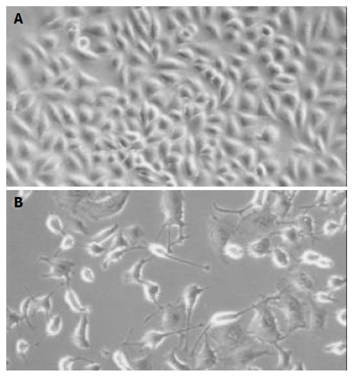
Detection of cell morphology by phase-contrast microscopy ( × 200). A: SMMC-7721 cells, B: SMMC-7721 cells treated with 10 mmol/L of HMBA for 72 h.
Figure 2.
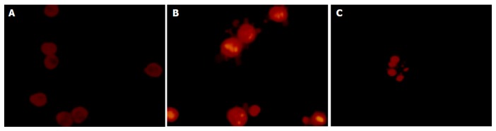
Detection of apoptotic morphology in SMMC-7721 cells treated with 10 mmol/L of HMBA for 72 h ( × 400). SMMC-7721 cells displayed fragmented nuclei and apoptotic bodies (B, C) after treatment with 10 mmol/L of HMBA for 72 h, while the untreated cells did not show these apoptotic characteristics (A).
Effects of HMBA on expression of Bcl-2 and Bax in SMMC-7721 cells
To elucidate the mechanism by which HMBA induces apoptotic cell death of SMMC-7721 cells, we analyzed the expression of two critical apoptosis-associated proteins, Bcl-2 and Bax, which are known to regulate the cell death/survival in opposite manners. As is shown in Figure 3, Bcl-2 protein level was significantly decreased while Bax protein level was slightly increased in the cells treated with 10 mmol/L of HMBA for 72 h. This result suggested that HMBA could induce apoptosis of SMMC-7721 cells by down-regulating the ratio of Bcl-2 to Bax.
Figure 3.
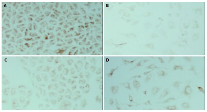
Immunocytochemical analysis of Bcl-2 and Bax expression in SMMC-7721 cells treated with 10 mmol/L of HMBA for 72 h (×200). A: The high level of Bcl-2 protein in SMMC-7721 cells, B: The decreased level of Bcl-2 protein in the cells treated with 10 mmol/L of HMBA, C: The low level of Bax protein in SMMC-7721 cells, D: The slightly increased level of Bax protein in the cells treated with 10 mmol/L of HMBA.
DISCUSSION
Dysregulations of cell proliferation, differentiation and apoptosis are hallmarks of cancer cells[23]. It is possible that these malignant features could be altered by many differentiation inducers such as hybrid polar compound HMBA. Considerable progress has been made toward elucidating the mechanism by which HMBA induces terminal differentiation of cancer cells. In recent year, some reports showed that HMBA induced apoptosis on a number of cancer cells[24-28], which appeared characteristics of cell shrinkage, chromatin condensation, DNA fragmentation and membrane blebbing[29,30].
In the present study, we investigated the cellular effects of HMBA on the growth and apoptosis of human hepatocellular carcinoma SMMC-7721 cells. Our data demonstrated that HMBA could effectively inhibit the growth of SMMC-7721 cells in a dose- and time-dependent manner. A significant portion of the cells treated with 10 mmol/L of HMBA for 72 h displayed the classical hallmarks of apoptosis with typical condensed nuclei and apoptotic bodies. In addition, as was shown in the previous reports[13,14], HMBA at 5 mmol/L concentration could arrest most of SMMC-7721 cells in G0/G1 phase and induce differentiation but not apoptosis. Our present results indicated that HMBA at 10 mmol/L concentration could not only inhibit cell proliferation and induce cell terminal differentiation, but also significantly induce apoptosis in human hepatocellular carcinoma SMMC-7721 cells.
To investigate the mechanism by which HMBA induces cellular apoptosis, we studied the effects of HMBA on apoptotic regulatory proteins in SMMC-7721 cells. Numerous molecular entities have been shown to regulate the apoptosis. Among these, the Bcl-2 family of proteins is well known for its regulatory role during apoptosis, and the interaction between anti-apoptotic and pro-apoptotic members of the family, such as between Bcl-2 and Bax, seemed to be crucial for regulation of this process[31-33]. Bcl-2, originally described as a proto-oncogene, is known as a cell death inhibitor that is regulated by its interaction with pro-apoptotic factors such as Bax family. Bax, one of the pro-apoptotic proteins, could counteract the anti-apoptotic effects of Bcl-2 by forming a heterodimer with Bcl-2[34]. In fact, the ratio of Bcl-2 to Bax, rather than the levels of individual proteins, has been considered to be critical in determining the survival or death of cells[35-38]. A number of studies have revealed that apoptosis induced by many agents is mediated through a decrease of the Bcl-2/Bax ratio[39-43]. Our results also showed that HMBA could reduce the ratio of Bcl-2 to Bax, indicating that the decreased ratio of Bcl-2 to Bax caused by HMBA treatment might trigger apoptosis of SMMC-7721 cells.
In conclusion, HMBA as a potent inducer can effectively inhibit the proliferation of human hepatocellular carcinoma cells at a low concentration and induce cellular apoptosis at a higher concentration. HMBA induces cell apoptosis of human hepatocellular carcinoma cells by decreasing the ratio of Bcl-2 to Bax.
Footnotes
Supported by the National Natural Science Foundation of China, No. 30170463 and Science Research Foundation of Xiamen University and Natural Science Foundation of Fujian Province, No. C0210005
Edited by Zhang JZ and Wang XL Proofread by Xu FM
References
- 1.Varela M, Sala M, Llovet JM, Bruix J. Treatment of hepatocellular carcinoma: is there an optimal strategy? Cancer Treat Rev. 2003;29:99–104. doi: 10.1016/s0305-7372(02)00123-8. [DOI] [PubMed] [Google Scholar]
- 2.Kew MC. Epidemiology of hepatocellular carcinoma. Toxicology. 2002;181-182:35–38. doi: 10.1016/s0300-483x(02)00251-2. [DOI] [PubMed] [Google Scholar]
- 3.Chen CJ, Yu MW, Liaw YF. Epidemiological characteristics and risk factors of hepatocellular carcinoma. J Gastroenterol Hepatol. 1997;12:S294–S308. doi: 10.1111/j.1440-1746.1997.tb00513.x. [DOI] [PubMed] [Google Scholar]
- 4.Wu MC. Clinical research advances in primary liver cancer. World J Gastroenterol. 1998;4:471–474. doi: 10.3748/wjg.v4.i6.471. [DOI] [PMC free article] [PubMed] [Google Scholar]
- 5.Zeng ZC, Jiang GL, Wang GM, Tang ZY, Curran WJ, Iliakis G. DNA-PKcs subunits in radiosensitization by hyperthermia on hepatocellular carcinoma hepG2 cell line. World J Gastroenterol. 2002;8:797–803. doi: 10.3748/wjg.v8.i5.797. [DOI] [PMC free article] [PubMed] [Google Scholar]
- 6.Yu MC, Yuan JM, Govindarajan S, Ross RK. Epidemiology of hepatocellular carcinoma. Can J Gastroenterol. 2000;14:703–709. doi: 10.1155/2000/371801. [DOI] [PubMed] [Google Scholar]
- 7.Herrero R, Moncelli MR, Guidelli R, Carla M, Arcangeli A, Olivotto M. Hybrid polar compounds produce a positive shift in the surface dipole potential of self-assembled phospholipid monolayers. Biochim Biophys Acta. 2000;1466:278–288. doi: 10.1016/s0005-2736(00)00181-4. [DOI] [PubMed] [Google Scholar]
- 8.Rifkind RA, Richon VM, Marks PA. Induced differentiation, the cell cycle, and the treatment of cancer. Pharmacol Ther. 1996;69:97–102. doi: 10.1016/0163-7258(95)02044-6. [DOI] [PubMed] [Google Scholar]
- 9.Leszczyniecka M, Roberts T, Dent P, Grant S, Fisher PB. Differentiation therapy of human cancer: basic science and clinical applications. Pharmacol Ther. 2001;90:105–156. doi: 10.1016/s0163-7258(01)00132-2. [DOI] [PubMed] [Google Scholar]
- 10.Guilbaud NF, Gas N, Dupont MA, Valette A. Effects of differentiation-inducing agents on maturation of human MCF-7 breast cancer cells. J Cell Physiol. 1990;145:162–172. doi: 10.1002/jcp.1041450122. [DOI] [PubMed] [Google Scholar]
- 11.Marks PA, Richon VM, Kiyokawa H, Rifkind RA. Inducing differentiation of transformed cells with hybrid polar compounds: a cell cycle-dependent process. Proc Natl Acad Sci U S A. 1994;91:10251–10254. doi: 10.1073/pnas.91.22.10251. [DOI] [PMC free article] [PubMed] [Google Scholar]
- 12.Li XN, Du ZW, Huang Q. Modulation effects of hexamethylene bisacetamide on growth and differentiation of cultured human malignant glioma cells. J Neurosurg. 1996;84:831–838. doi: 10.3171/jns.1996.84.5.0831. [DOI] [PubMed] [Google Scholar]
- 13.Ouyang GL, Li QF, Peng XX, Hong SG. [Differentiation of human hepatocarcinoma SMMC-7721 cells induced by HMBA] Shiyan Shengwu Xuebao. 2001;34:269–273. [PubMed] [Google Scholar]
- 14.Ouyang GL, Li QF, Peng XX, Hong SG. [Effects of HMBA on the expression of cell-cycle-associated genes in human hepatocarcinoma SMMC-7721 cells] Shi Yan Sheng Wu Xue Bao. 2002;35:173–178. [PubMed] [Google Scholar]
- 15.Ren JG, Zheng RL, Shi YM, Gong B, Li JF. Apoptosis, redifferentiation and arresting proliferation simultaneously triggered by oxidative stress in human hepatoma cells. Cell Biol Int. 1998;22:41–49. doi: 10.1006/cbir.1998.0226. [DOI] [PubMed] [Google Scholar]
- 16.Li J, Huang CY, Zheng RL, Cui KR, Li JF. Hydrogen peroxide induces apoptosis in human hepatoma cells and alters cell redox status. Cell Biol Int. 2000;24:9–23. doi: 10.1006/cbir.1999.0438. [DOI] [PubMed] [Google Scholar]
- 17.Yuan JH, Wang XW, Luo D, Xie Y, Xie H. Anti-hepatoma activity of taxol in vitro. Acta Pharmacol Sin. 2000;21:450–454. [PubMed] [Google Scholar]
- 18.Zhang SW, Lin WS, Ying XL, Zhu D, Guo MY, Gu JX. Effect of suppression of TGF-beta1 expression on cell-cycle and gene expression of β-1,4-galactosyltransferase 1 in human hepatocarcinoma cells. Biochem Biophys Res Commun. 2000;273:833–838. doi: 10.1006/bbrc.2000.3028. [DOI] [PubMed] [Google Scholar]
- 19.Chan EW, Cheng SC, Sin FW, Xie Y. Triptolide induced cytotoxic effects on human promyelocytic leukemia, T cell lymphoma and human hepatocellular carcinoma cell lines. Toxicol Lett. 2001;122:81–87. doi: 10.1016/s0378-4274(01)00353-8. [DOI] [PubMed] [Google Scholar]
- 20.Ouyang GL, Li QF, Peng XX, Liu QR, Hong SG. Effects of tachyplesin on proliferation and differentiation of human hepatocellular carcinoma SMMC-7721 cells. World J Gastroenterol. 2002;8:1053–1058. doi: 10.3748/wjg.v8.i6.1053. [DOI] [PMC free article] [PubMed] [Google Scholar]
- 21.Rui-Chuan C, Jin-Hua S, Gao-Liang O, Ke-Xia C, Jin-Quan L, Xiao-Guang X. Induction of differentiation in human hepatocarcinoma cells by isoverbascoside. Planta Med. 2002;68:370–372. doi: 10.1055/s-2002-26759. [DOI] [PubMed] [Google Scholar]
- 22.Geng CX, Zeng ZC, Wang JY. Docetaxel inhibits SMMC-7721 human hepatocellular carcinoma cells growth and induces apoptosis. World J Gastroenterol. 2003;9:696–700. doi: 10.3748/wjg.v9.i4.696. [DOI] [PMC free article] [PubMed] [Google Scholar]
- 23.Hanahan D, Weinberg RA. The hallmarks of cancer. Cell. 2000;100:57–70. doi: 10.1016/s0092-8674(00)81683-9. [DOI] [PubMed] [Google Scholar]
- 24.Siegel DS, Zhang X, Feinman R, Teitz T, Zelenetz A, Richon VM, Rifkind RA, Marks PA, Michaeli J. Hexamethylene bisacetamide induces programmed cell death (apoptosis) and down-regulates BCL-2 expression in human myeloma cells. Proc Natl Acad Sci U S A. 1998;95:162–166. doi: 10.1073/pnas.95.1.162. [DOI] [PMC free article] [PubMed] [Google Scholar]
- 25.Hyman T, Rothmann C, Heller A, Malik Z, Salzberg S. Structural characterization of erythroid and megakaryocytic differentiation in Friend erythroleukemia cells. Exp Hematol. 2001;29:563–571. doi: 10.1016/s0301-472x(01)00616-6. [DOI] [PubMed] [Google Scholar]
- 26.Purtell DJ. Drug/alcohol abuse patients and the confidentiality of medical records. Hosp Med Staff. 1976;5:24–27. [PubMed] [Google Scholar]
- 27.Ruefli AA, Smyth MJ, Johnstone RW. HMBA induces activation of a caspase-independent cell death pathway to overcome P-glycoprotein-mediated multidrug resistance. Blood. 2000;95:2378–2385. [PubMed] [Google Scholar]
- 28.Zhang Z, Liong EC, Lau TY, Leung KM, Fung PC, Tipoe GL. Induction of apoptosis by hexamethylene bisacetamide is p53-dependent associated with telomerase activity but not with terminal differentiation. Int J Oncol. 2000;16:887–892. doi: 10.3892/ijo.16.5.887. [DOI] [PubMed] [Google Scholar]
- 29.Liu JR, Chen BQ, Yang YM, Wang XL, Xue YB, Zheng YM, Liu RH. Effect of apoptosis on gastric adenocarcinoma cell line SGC-7901 induced by cis-9, trans-11-conjugated linoleic acid. World J Gastroenterol. 2002;8:999–1004. doi: 10.3748/wjg.v8.i6.999. [DOI] [PMC free article] [PubMed] [Google Scholar]
- 30.Kroemer G, Dallaporta B, Resche-Rigon M. The mitochondrial death/life regulator in apoptosis and necrosis. Annu Rev Physiol. 1998;60:619–642. doi: 10.1146/annurev.physiol.60.1.619. [DOI] [PubMed] [Google Scholar]
- 31.Chao DT, Korsmeyer SJ. BCL-2 family: regulators of cell death. Annu Rev Immunol. 1998;16:395–419. doi: 10.1146/annurev.immunol.16.1.395. [DOI] [PubMed] [Google Scholar]
- 32.Kluck RM, Bossy-Wetzel E, Green DR, Newmeyer DD. The release of cytochrome c from mitochondria: a primary site for Bcl-2 regulation of apoptosis. Science. 1997;275:1132–1136. doi: 10.1126/science.275.5303.1132. [DOI] [PubMed] [Google Scholar]
- 33.Evan G, Littlewood T. A matter of life and cell death. Science. 1998;281:1317–1322. doi: 10.1126/science.281.5381.1317. [DOI] [PubMed] [Google Scholar]
- 34.Kobayashi T, Ruan S, Clodi K, Kliche KO, Shiku H, Andreeff M, Zhang W. Overexpression of Bax gene sensitizes K562 erythroleukemia cells to apoptosis induced by selective chemotherapeutic agents. Oncogene. 1998;16:1587–1591. doi: 10.1038/sj.onc.1201681. [DOI] [PubMed] [Google Scholar]
- 35.Oltvai ZN, Milliman CL, Korsmeyer SJ. Bcl-2 heterodimerizes in vivo with a conserved homolog, Bax, that accelerates programmed cell death. Cell. 1993;74:609–619. doi: 10.1016/0092-8674(93)90509-o. [DOI] [PubMed] [Google Scholar]
- 36.Oltvai ZN, Korsmeyer SJ. Checkpoints of dueling dimers foil death wishes. Cell. 1994;79:189–192. doi: 10.1016/0092-8674(94)90188-0. [DOI] [PubMed] [Google Scholar]
- 37.Reed JC. Bcl-2 family proteins. Oncogene. 1998;17:3225–3236. doi: 10.1038/sj.onc.1202591. [DOI] [PubMed] [Google Scholar]
- 38.Fukamachi Y, Karasaki Y, Sugiura T, Itoh H, Abe T, Yamamura K, Higashi K. Zinc suppresses apoptosis of U937 cells induced by hydrogen peroxide through an increase of the Bcl-2/Bax ratio. Biochem Biophys Res Commun. 1998;246:364–369. doi: 10.1006/bbrc.1998.8621. [DOI] [PubMed] [Google Scholar]
- 39.Panaretakis T, Pokrovskaja K, Shoshan MC, Grandér D. Interferon-alpha-induced apoptosis in U266 cells is associated with activation of the proapoptotic Bcl-2 family members Bak and Bax. Oncogene. 2003;22:4543–4556. doi: 10.1038/sj.onc.1206503. [DOI] [PubMed] [Google Scholar]
- 40.Yan J, Xu YH. Tributyrin inhibits human gastric cancer SGC-7901 cell growth by inducing apoptosis and DNA synthesis arrest. World J Gastroenterol. 2003;9:660–664. doi: 10.3748/wjg.v9.i4.660. [DOI] [PMC free article] [PubMed] [Google Scholar]
- 41.Zhou HB, Zhu JR. Paclitaxel induces apoptosis in human gastric carcinoma cells. World J Gastroenterol. 2003;9:442–445. doi: 10.3748/wjg.v9.i3.442. [DOI] [PMC free article] [PubMed] [Google Scholar]
- 42.Aranha O, Grignon R, Fernandes N, McDonnell TJ, Wood DP, Sarkar FH. Suppression of human prostate cancer cell growth by ciprofloxacin is associated with cell cycle arrest and apoptosis. Int J Oncol. 2003;22:787–794. [PubMed] [Google Scholar]
- 43.Pettersson F, Dalgleish AG, Bissonnette RP, Colston KW. Retinoids cause apoptosis in pancreatic cancer cells via activation of RAR-gamma and altered expression of Bcl-2/Bax. Br J Cancer. 2002;87:555–561. doi: 10.1038/sj.bjc.6600496. [DOI] [PMC free article] [PubMed] [Google Scholar]