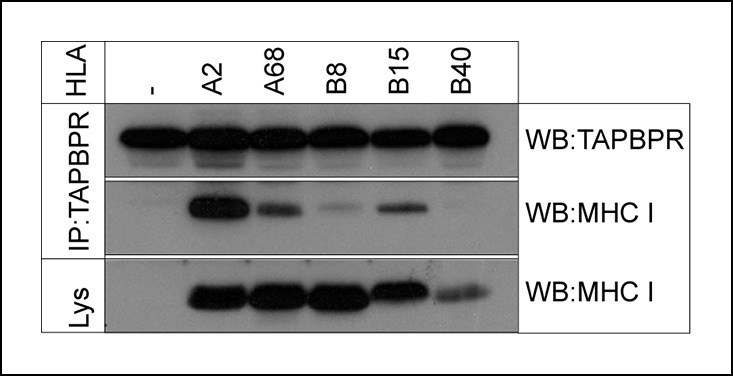
Figure 3. TAPBPR associates with HLA-A and HLA-B molecules.

TAPBPR was isolated by immunoprecipitation (using R014) from the MHC class I negative cell line 721.221 and 721.221 stably transduced with HLA-A2, A68, B8, B15 or B40. Western blot analysis was performed for TAPBPR and the MHC class I heavy chain (using HCA2 and HC10) on lysates (labelled lys) and TAPBPR immunoprecipitates as indicated. The data is representative of three independent experiments.
