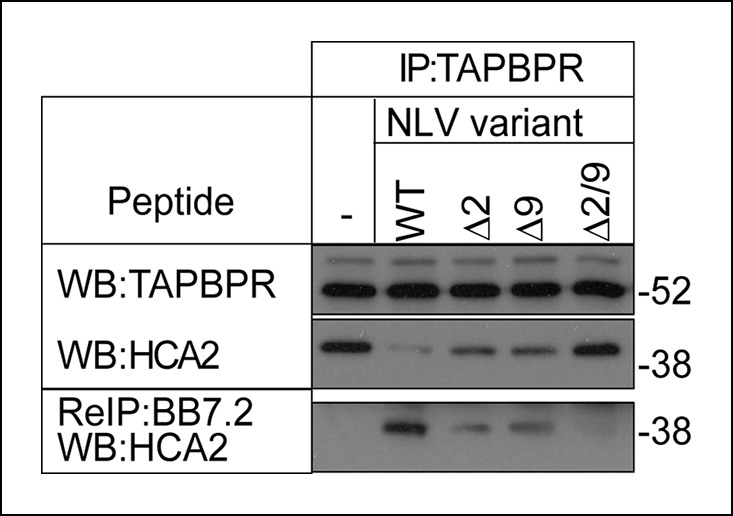
Figure 7. The MHC I bound to TAPBPR is peptide receptive.

The TAPBPR:MHC I complex was immunoprecipitated from IFN-γ induced KBM-7 cells. Equal aliquots were divided, then incubated -/+ 100 μM (20 μg) of the indicated peptide (WT:NLVPMVATV, Δ2: NAVPMVATV, Δ9: NLVPMVATM, Δ2/9:NAVPMVATM) for 30 min at 4°C. Subsequently all eluates (- or + peptide) were re-immunoprecipitated with BB7.2. Extensive washing was performed to remove any released MHC I before denaturation. Western blot analysis was performed for TAPBPR, and HLA-A2 (using HCA2) under reducing conditions. Data shown are representative of three independent experiments.
