Abstract
Introduction:
Myositis ossificans [MO] is a benign heterotropic bone forming (often- self resolving) pathology of bone and soft tissue. Here we are reporting the first time in literature for osteomyelitis of myositis ossificans in arm of a male due to trauma as a perusal of rare entity.
Case Report:
It is a case report of a 25 years old male presented to us in out-patient department with chief complaint of discharging wounds over mid part of left arm since six months. Clinically provisional diagnosis of chronic osteomylitis of left humerus made and his x-ray sought. X- Ray showed geographic appearance of myositis ossificans around the upper two third of left arm. Sinuses curetted and infected bone (part of myositis ossificans) removed and sent for biopsy. Now the patient is discharge and sinus free, and has resumed his work.
Conclusion:
Osteomyleitis of myositis ossificans should be recognized as a possible differential diagnosis chronic discharging sinus. This type of presentation of myositis ossificans is rarest.
Keywords: Osteomyelitis, myositis ossificans, arm
Introduction
Myositis ossificans [MO] is a benign heterotropic bone forming (often- self resolving) pathology of bone and soft tissue. It has also been reported to occur due to strain or overuse of muscles[1]. Here we are reporting the first time in literature for osteomyelitis of myositis ossificans in the arm of a male due to trauma as a perusal of a rare entity.
Case Report
It is a case report of a 25 years old male presented to us in outpatient departments with chief complaint of discharging wounds over mid part of his left arm since six months. On the complete history taking he revealed that he had an episode of trauma one year back over his left arm due to fall on the ground and swelling develops at that time. For it he did not take any medical advice and he took few pain killers and repeated massage over his left arm. Subsequently he noticed that the swelling over his whole left arm is permanent. After six months of this incident he felt that now mid of his arm is pain full and after 2 to 3 episodes of fever pus discharge came out. Since then, on and off the pus (sometimes with chunk of bone) is coming out.
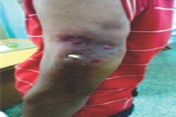
On physical examination there were multiple discharging sinuses (with the protruding bony chunk) over his lateral aspect of his mid part of the left arm with puckered and hyper pigmented skin [Fig 1]. On palpation his left arm was thickened and tenderness was found around the arm. Range of movements over left shoulder and elbow were terminally restricted and pain full. Vital signs of patient were stable and distal neurovascular status was intact. Clinical provisional diagnosis of chronic osteomyelitis of left humerus made and his hematological examination and X-ray sought. His hematological study was near normal except the erythrocyte sedimentation rate (ESR) which was raised (Westergren’s method=35 mm/hour).
Figure 1.

Image showing multiple discharging sinuses and protruding bony chunk.
X- Ray was astonishing, entire humerus bone was normal, and there was no any osteomyelitic change in the humerus. There was a geographic appearance of myositis ossificans around the upper two third of left arm [Fig 2].
Figure 2.

Anteroposterior and lateral X-Ray of left arm showing oseomyelitis ossificans.
After consent taking, the patient got operated. Sinuses curetted out and sequestrum (infected part of myositis mass, because it was not feasible to remove the all myositis mass) removed and sent for biopsy and culture sensitivity. Now after the one year of follow-up, there is no recurrence of the myositis ossificans. And the patient is free from discharging sinuses and has resumed his work.
Discussion
The exact pathogenesis of MO is not clear but one theory stated that due to injury there are fibroblastic proliferation or osteoblastic migration (from injured periosteum) into haematoma, have been blamed for causing the pathology of myositis ossificans [2]. Neither is it found exclusively in skeletal muscle nor there is muscle inflammation so its name is misnomer, even the name myositis ossificans is most commonly used clinical term. So the term heterotrophic ossification seems clinically worth [3,4]. Myositis ossificans have three variants, myositis ossificans circumscripta (MOC), neurogenic myositis ossificans and fibrodysplasia (myositis) ossificans progressiva.
Myositis ossificans circumscripta (or traumatica) is a pseudosarcomatous pathology with restricted growth tendency. If its excision is done in matured phase (well demarcated from surrounding tissue) and if repeated injury is avoided then it may prevent the recurrence of it [5].
Neurogenic form of myositis involves the large joints of the body (hip, knee) if they have been immobilized due to traumatic neurological damage, burn or arthroplasty. But on the contrary to circumscripta, it involves the connective tissue between the muscle plane and around the joint rather than skeletal muscle [6, 7].
Fibrodysplasia (myositis) ossificans progressiva (FOP) have the preponderance for young age while the other two forms do so rarely. FOP is an autosomal dominating inherited disabling disease (a disorder of bone morphogenic protein 4) have a predilection to paraspinal, scalp and temporomandibular joints (but never involves facial muscles, tongue, diaphragm and viscera). FOP has the characteristic feature of (potentially recognizable at birth) short big toe, hallux valgus (also thumb and cervical spine sometimes involved) and broad femoral neck. There is spontaneous (also by trauma) endochondral ectopic ossification in tendons, ligaments, joint capsule and progressively in a specific manner and give a feature of “second skeleton” [8, 9, 10].
MOC has peculiar clinico-radiological and cytological feature and clinically present as painful soft tissue swelling with restricted range of movement in a joint following a trauma. In the early stage it appears as irregular, hazy, flocculent structure, but after 3 weeks zonal calcified area is evident. So for the early diagnosis of MOC, sonography and three phase bone scan is needed [11, 12]. This characteristic radiological feature of “zoning,” is due to distinct mature ossification at the periphery and centrally situated radiolucent (which is due to immature osteoid and primitive mesenchymal tissue) nidus [5]. This characteristic feature of “zoning” differentiate it from extra skeletal osteosarcoma in which there is centrally situated radio opaque matured osteoid cell. Zonal architecture is obvious after 6 weeks of trauma and Computer tomography is needed to demonstrate this zonal mineralization pattern [13].
For the management of MOC initially conservative management (i.e. - rest, immobilization and physiotherapy) should be given and surgical excision can be used as a last resort for the matured stage (6-12 months of trauma) to avoid the recurrence. Even after the diligent search we did not get any article on osteomyelitis in MO. Etiopathogenesis of osteomyelitis in myositis mass can be conceded same, as it occurs in other bone (i.e. direct inoculation due to trauma or hematogenous spread). In our case the osteomyelitis would have been occurred via hematogenous spread. Our case had the feature of chronic osteomyelitis (chronic discharging sinuses and protruded sequestrum) so the treatment protocol was the same as usual of the chronic osteomyelitis of normal bone.
In summary, as a rare case in literature we are presenting the case of osteomyelitis of MOC due to trauma. In this case MO was in matured stage and infected so the surgical excision done. As a perusal of rare presentation, clinicians should be aware of this unusual presentation of MO.
Conclusion
Osteomyelitis of myositis ossificans can be conceded as a possible differential diagnosis of chronic discharging sinus in a few feasible conditions (following trauma around joints). This type of presentation of myositis ossificans is rare, and we are reporting to it as a first time in literature.
Clinical Message.
This unusual presentation of myositis ossificans is not mentioned in the literature till now. So we are bringing it to horizon of knowledge to disclose the unusual presentation of myositis ossificans.
Biography


Footnotes
Conflict of Interest: Nil
Source of Support: None
References
- 1.Defoorts, Arnout NA, Debeer PD. Myositis ossificans circumscripta of the triceps due to overuse in a female swimmer. Int J Shoulder Surg. 2012 Jan;6(1):19–22. doi: 10.4103/0973-6042.94315. doi: 10.4103/0973-6042.94315. [DOI] [PMC free article] [PubMed] [Google Scholar]
- 2.Meffert O, Weber HG. Beitrag zur Myositis ossificans localisata. Dtsch med Wschr. 1973;98:653–656. doi: 10.1055/s-0028-1106878. [DOI] [PubMed] [Google Scholar]
- 3.Nuovo MA, Norman A, Chumas J, Ackerman LV. Myositis ossificans with atypical clinical, radiographic, or pathologic findings: a review of 23 cases. Skeletal Radiol. 1992;21:87. doi: 10.1007/BF00241831. [DOI] [PubMed] [Google Scholar]
- 4.Fuselier CO, Tlapek TA, Sowell RD. Heterotrophic ossification (myositis ossificans) in the foot. A case report. J Am Podiatr Med Assoc. 1986;76:524. doi: 10.7547/87507315-76-9-524. [DOI] [PubMed] [Google Scholar]
- 5.Dorfman HD, Czerniak B. Reactive and metabolic conditions simulating neoplasms of bone. In: Dorfman HD, Czerniak B, editors. Bone Tumors. St Louis: Mosby; 1998. p. 1139. [Google Scholar]
- 6.Carlier RY, Safa DM, Parva P, et al. Ankylosing neurogenic myositis ossificans of the hip. An enhanced volumetric CT study. J Bone Joint Surg Br. 2005;87:301. doi: 10.1302/0301-620x.87b3.14737. [DOI] [PubMed] [Google Scholar]
- 7.Gunduz B, Erhan B, Demir Y. Subcutaneous injections as a risk factor of myositis ossificans traumatica in spinal cord injury. Int J Rehabil Res. 2007;30:87. doi: 10.1097/MRR.0b013e3280146f85. [DOI] [PubMed] [Google Scholar]
- 8.Smith R, Athanasou NA, Vipond SE. Fibrodysplasia (myositis) ossificans progressiva: clinicopathological features and natural history. QJM. 1996;89:445. doi: 10.1093/qjmed/89.6.445. [DOI] [PubMed] [Google Scholar]
- 9.Connor JM. Fibrodysplasia ossificans progressiva lessons from rare maladies. N Engl J Med. 1996;335:591. doi: 10.1056/NEJM199608223350812. [DOI] [PubMed] [Google Scholar]
- 10.Mahboubi S, Glaser DL, Shore EM, Kaplan FS. Fibrodysplasia ossificans progressiva. Pediatr Radiol. 2001;31:307. doi: 10.1007/s002470100447. [DOI] [PubMed] [Google Scholar]
- 11.Okayama A, Futani H, Kyo F, et al. Usefulness of ultrasonography for early recurrent myositis ossificans. J Orthop Sci. 2003;8:239. doi: 10.1007/s007760300041. [DOI] [PubMed] [Google Scholar]
- 12.Drane WE. Myositis ossificans and the three-phase bone scan. American Journal of Roentgenology. 1984;142:179. doi: 10.2214/ajr.142.1.179. [DOI] [PubMed] [Google Scholar]
- 13.Amendola MA, Glazer GM, Agha FP, et al. Myositis ossificans circumscripta: computed tomographic diagnosis. Radiology. 1983;149:775. doi: 10.1148/radiology.149.3.6647854. [DOI] [PubMed] [Google Scholar]


