Abstract
Introduction:
Malignant pigmented villonodular synovitis (PVNS) or Malignant giant cell tumour tendon sheath (MGCTTS) is a controversial and debatable lesion. Few case reports have indicated the potential for metastasis.1These aggressive cases are designated malignant giant cell tumour tendon sheath or malignant PVNS. Less than 20 cases are described in literature. We report a case of 65 year old lady who was diagnosed eight years back as pigmented villonodular synovitis. She had multiple local recurrences and now presented with lymphnodal metastases, which is extremely rare.
Case Report:
Sixty five year old lady presented with swelling in left inguinal region of six months duration. She gave a past history of swelling around medial condyle of left femur eight years back. Swelling was excised three times. At the time of third recurrence, swelling was extensive, infiltrating surrounding tissues and underlying bone, encasing femoral and popliteal vessels for which she underwent an above knee amputation. She now presented with inguinal swelling measuring 5.7×3.0 cms. Positron Emission Tomography Scan (PET-CT) revealed multiple enlarged left common iliac, internal and external iliac nodes, largest measuring 7.0 cms. Both the inguinal and pelvic nodes were excised. Lesion was diagnosed as metastatic deposits of Malignant pigmented villonodular synovitis based on morphological and Immunohistochemical findings.
Conclusion:
It is important to have a high index of clinical suspicion because these lesions can have an aggressive behaviour even with bland cytological features. Our experience suggests that in a recurrent lesion for GCTTS. A wide surgical excision with safe surgical margins and close follow up with radiological evaluation might help to diagnose these lesions early and be amenable to limb salvage surgeries.
Keywords: Lymphnode, Metastasis, Pigmented villonodular synovitis, Malignant
Introduction
Malignant pigmented villonodular synovitis (PVNS) or Malignant giant cell tumour tendon sheath (MGCTTS) is a controversial and debatable lesion. They are heterogenous lesions and sarcomatous areas can have features of malignant fibrous histiocytomas, fibrosarcoma or myxofibrosarcoma. [1] Three cases are described with metastasis to lung and lymphnodes. [1] The case under discussion had cytological features of malignant lesion with extensive local recurrences and lymph nodal metastases which warrant the diagnosis of malignant PVNS, eight years after the initial diagnosis.
Case Report
Sixty five year old lady presented with swelling in left inguinal region of six months duration. Swelling was gradually increasing in size and was not painful. There were no associated systemic symptoms. There was restriction of movement in the hip joint.
She gave a past history of swelling around medial condyle of left femur eight years back. Swelling was excised three times. After the second recurrence she received telecobalt therapy for 15 days. At the time of third recurrence, swelling was extensive, infiltrating surrounding tissues and underlying bone, encasing femoral and popliteal vessels for which she underwent an above knee amputation. The swelling recurred after one and half years above the amputation site and debulking of the tumour was done. She presented after one year with swelling in inguinal region for which a disarticulation was suggested. First biopsy was reported as Giant cell tumour tendon sheath/PVNS and subsequent biopsies were diagnosed as pigmented villonodular synovitis, recurrent. She now presented to our hospital for further management. There was no significant family history or personal history.
On examination, she was a thin frail lady with an above knee amputation stump.
Swelling in the inguinal region was irregular measuring 5.7×3.0 cms. Margins were ill defined, swelling was adherent to surrounding structures. Her hematological investigations revealed microcytic hypochromic anemia. Leukocytes and platelets were normal. Her biochemical parameters were within normal limits. She was HIV non reactive, HBsAg and HCV negative. Chest x-ray was normal. Positron Emission Tomography Scan (PET-CT) revealed multiple enlarged left common iliac, internal and external iliac nodes, largest measuring 7.0 cms. Large enhancing deposits were seen in inguinal region measuring 5.7 cms. No lesions were seen in lungs or other organs. Both the inguinal and pelvic nodes were excised.
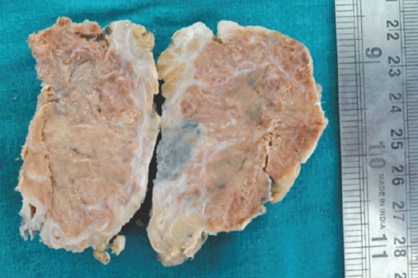
Pathology: Grossly pelvic lesion was an irregular mass measuring 6.5×4.5 cms. Inguinal mass was a nodular mass measuring 5×5 cms with adherent skin. Surface was smooth. Cut section was yellowish brown (fig 1)
Figure 1.

Gross specimen showing a well circumscribed lesion, surface is yellow brown.
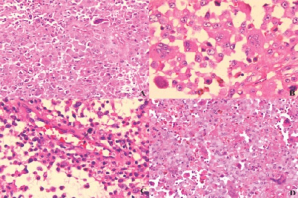
Microscopic examination revealed compressed lymphoid tissue with a cellular lesion composed of cells arranged in sheets. Cell showed moderate eosinophilic cytoplasm, nuclei were vesicular, there was mild anisonucleosis with prominent nucleoli. Mitoses were 3-4/10 high power fields. Interspersed through out the lesion were osteoclast type giant cells. (Fig 2 a-d) Groups of pigment laden macrophages were seen. There was no necrosis. A panel of immunostains was done. S-100, HMB-45, EMA were negative. Vimentin and CD-68 were positive. A targeted therapy was planned, so an immunostain with anti-platelet derived growth factor receptor (PDGFR) was done which was diffusely and strongly positive. With these features, lesion was diagnosed as malignant giant cell tumour tendon sheath/malignant PVNS metastatic to lymphnodes. She was put on Imatinib with a dosage of 400 mg per day. An ultrasound abdomen done after six months revealed recurrence of pelvic mass, which measured 6×5 cms. She also complained of weakness and bone pains. Investigations revealed anemia of 6.7 gms and an ESR of 140 mm/hr. Immuno-electrophoresis revealed M band. Bone marrow revealed 22% plasma cells. She is presently on treatment for myeloma with good clinical response.
Figure 2.

a) Cells with eosinophilic cytoplasm. Hematoxylin & Eosin(H&EX200); b) Nuclei with prominent nucleoli. (H&E×400); c) Discohesive cells, mitotic figure (arrow) (H&E×400); d) Hemosiderin laden cells. (H&E×200).
Discussion
In 1941 Jaffe et al termed fibrohistiocytic tumours in large joints as pigmented villonodular synovitis. They are considered benign, which are capable of local recurrence but not capable of distant metastasis. Recently a few case reports have indicated the potential for metastasis. [2] These aggressive cases are designated malignant giant cell tumour tendon sheath or malignant PVNS. This is a rare entity and can occur de novo or arise in recurrent disease. The incidence of malignant transformation of PVNS is 3% in a study by Bertoni. [3] Carstens and Howell4 first reported malignant PVNS in 1979. There is a controversy in literature about the diagnosis of malignant change in PVNS. Lesions now designated as malignant PVNS have multiple local recurrences as well as metastasis to lymph nodes or lungs. Bertoni et al defined the criteria which include 1) nodular lesions, infiltrative growth pattern 2) large plump or round to oval cells 3) cells with large nuclei with eosinophilic cytoplasm and prominent nucleoli 4) fewer giant cells, xanthoma cells and inflammatory cells.5) lack of normal zonal patterns 6) areas of necrosis.
The criteria according to AFIP to designate a lesion as malignant PVNS should have five of the eight criteria.1) diffuse pleomorphism, 2) prominent nucleoli, 3) high nuclear cytoplasmic ratio, 4) mitoses of 10/10hpfs, 5) necrosis, 6) discohesion of cells, 7) paucity of giant cells and 8) diffuse growth pattern..
Our case had diffuse growth pattern, discohesive cells, cells with moderate eosinophilic cytoplasm, prominent nucleoli, few xanthoma cells and 3-4 mitoses/10hpfs. There were no areas of necrosis. Hence the lesion is clinically malignant in the form of local recurrences and lymphnodal metastasis and also had cytological criteria to classify it as malignant GCTTS.
Common sites of occurrence are knee (47%), foot (20%), ankle (13%), hip (7%) and thigh (7%).5Mean age at diagnosis is 48 yrs. Despite aggressive therapy it is associated with guarded prognosis. Local recurrence is reported in 54-70% of cases and metastasis to lungs or lymph nodes is observed in 38-70% and death occurs in 50% of cases. It is a rare tumour with only 20 cases reported in literature. [6] Peculiar findings in this case was lack of necrosis and mitotic rate was in agreement with the rates reported by Bertoni et al. [3] The lesion recurred in pelvis six months after excision. There were no clinical or radiological signs of pulmonary metastasis.
She is presently on treatment for myeloma with clinical remission.
Conclusion
It is important to have a high index of clinical suspicion because these lesions can have an aggressive behaviour even with bland cytological features. Our experience suggests that in a recurrent lesion for GCTTS. A wide surgical excision with safe surgical margins and close follow up with radiological evaluation might help to diagnose these lesions early and be amenable to limb salvage surgeries.
Clinical Message.
Malignant Pigmented Villonodular Synovitis is extremely rare. They arise de novo or in recurrent lesions. They have various histological features and can metastasise to lungs or lymphnodes.
Biography


Footnotes
Conflict of Interest: Nil
Source of Support: None
References
- 1.Yoon HJ, Cho YA, Lee JI, Hong SP, Hong SD. Malignant pigmented villonodular synovitis of the temporomandibular joint with lung metastasis: a case report and review of the literature. Oral Surg Oral Med Oral Pathol Oral Radiol Endod. 2011;111(5):30–6. doi: 10.1016/j.tripleo.2010.11.031. [DOI] [PubMed] [Google Scholar]
- 2.Nielsen AL, Kiaer T. Malignant giant cell tumor of synovium and locally destructive pigmented villonodular synovitis: ultrastructural and immunohistochemical study and review of literature. Hum Pathol. 1989;20:765–71. doi: 10.1016/0046-8177(89)90070-1. [DOI] [PubMed] [Google Scholar]
- 3.Bertoni F, Unni KK, Beabout JW, Sim FH. Malignant giant cell tumor of tendon sheath and joints (malignant pigmented villonodular synovitis) Am J Surg Pathol. 1997;21:153–63. doi: 10.1097/00000478-199702000-00004. [DOI] [PubMed] [Google Scholar]
- 4.Carstens PH, Howell RS. Malignant giant cell tumour of tendon sheath. Virchows Arch. 1979;382:237–43. doi: 10.1007/BF01102878. [DOI] [PubMed] [Google Scholar]
- 5.Fanburg-Smith JC, Miettinen M. Scientific expansions. Nice France: International Academy of Pathology; 1998. Malignant tenosynovial giant cell tumors (MGCTTS) [Google Scholar]
- 6.Naoaki Imakiire, Takashi Fujino, Takeshi Morii, Keita Honya, Kazuo Mochizuki, Kazuhiko Satomi, et al. Malignant pigmented villonodular synovitis in the knee-Report of a case with Rapid Clinical Progression. The Open Orthopaedics Journal. 2011;5:13–6. doi: 10.2174/1874325001105010013. [DOI] [PMC free article] [PubMed] [Google Scholar]


