Abstract
Introduction:
Fractures involving bones containing a component of a prosthetic joint are becoming more common. The causation is multifactorial but most of these injuries are associated with trivial trauma. The options available for operative management of these fractures include internal fixation of the fracture alone, fixation of the fracture with revision of the prosthesis, and reconstruction of proximal femur with either modified impaction bone grafting or proximal femoral replacement.
Case Report:
We present here a case of periprosthetic fracture Vancouver type B1 with a broken cemented bipolar prosthesis insitu, in which the broken implant was firmly fixed in the proximal fragment and could not be removed following which the whole of the proximal fragment along with the broken implant was removed and replaced by a customized steel long stem cemented mega prosthesis.
Conclusion:
This case is being presented on account of its unusual presentation and fracture pattern. A broken prosthesis along with a periprosthetic fracture is not a common incident. Thus the treatment had to be individualized. Since the prosthesis was well fixed, its broken stem could not be removed from the proximal fragment and so the whole of the proximal fragment along with stem was removed and replaced with a long stem custom made bipolar prosthesis.
Keywords: Periprosthetic fracture, mega prosthesis, Proximal femoral bone loss, early mobilization
Introduction
Periprosthetic femoral fracture is a devastating complication after total hip arthroplasty and is associated with a high rate of postoperative complications and often a poor clinical result [1,2]. The rate of intra-operative fracture (with cemented or uncemented stems) has been reported as ranging from 1% and 3%-20% respectively [3,4,5]. The exact prevalence of postoperative periprosthetic fracture is more difficult to determine but is estimated to be approximately 1% over the life of the prosthesis [6]. Moreover, the mortality rate after periprosthetic femoral fracture is alarmingly high [7]. The causation is multifactorial but most of these injuries are associated with trivial trauma. Conditions which result in distorted anatomy or diminished bone quality are responsible for intraoperative periprosthetic fracture such as osteopenia, rheumatoid arthritis, osteomalacia, Paget's disease, osteopetrosis, poliomyelitis and parkinsonism [4,8]. On the other hand the various risk factors for post operative periprosthetic fractures are loosening of the femoral component, osteolysis due to wear debris and most importantly cortical stress risers [4,9,10]. The underlying cause in almost all cases is a decrease in mechanical strength of the host bone either due to osteoporosis, stress shielding or osteolytic lesions [6,11]. The common classification systems include those of Johansson et al.,[12] Parrish and Jones,[13] Bethea et al., [10] however, the Vancouver Classification [14] currently seems to be the most widely used.
Figure 1.

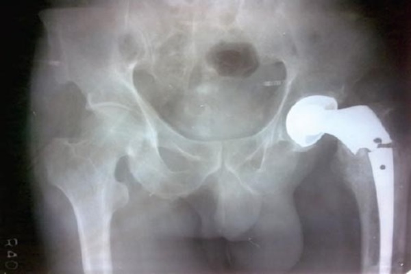
Preoperative radiograph of the patient - Plain radiograph of the left hip and thigh region showing a Vancouver type B1 periprosthetic fracture with a broken cemented bipolar prosthesis insitu.
Treatment recommendations have varied from non-operative [15] to more complicated algorithms based upon the site of the fracture [6,16]. The options available for operative management of these fractures include internal fixation of the fracture alone, fixation of the fracture with revision of the prosthesis, and reconstruction of proximal femur with either modified impaction bone grafting or proximal femoral replacement. We present here a case of periprosthetic fracture Vancouver type B1 with a broken Austin Moore prosthesis insitu, in which the broken implant was firmly fixed in the proximal fragment and could not be removed following which the whole of the proximal fragment along with the broken implant was removed and replaced by a customized steel long stem cemented mega prosthesis.
Case Report
A 60 years old male presented in June 2010, to our department with complaints of severe pain and swelling in left hip and upper thigh region since last 2 days, following a history of trauma. He was unable to walk and bear weight on the left lower limb following the trauma. Pain was present on the anterior and lateral aspect of left hip and upper thigh region. It was constant in nature, was present even at rest, dull aching type, severe in intensity and aggravated by hip movements. It was accompanied with difficulty in walking due to pain and the patient was unable to bear weight on the left lower limb. It was also associated with diffuse swelling over the upper part of thigh.
The patient had a history of hip hemireplacement operation on the left side 5 years back. There was no other significant past history. Patient was a sedentary worker with no history of tobacco or drug intake. No sensitivity or allergy to any drug. There was no family history of tuberculosis or malignancy. On examination the left lower limb was externally rotated. There was a diffuse swelling over antero-lateral aspect of upper part of left thigh. The skin overlying the swelling was neither warm nor inflamed. There was deep tenderness over the swelling. Bony crepitus was present and there was loss of transmitted movements. There was no apparent shortening or true shortening. Straight leg raising test was negative. Distal neurovascular status of the limb was intact.
Routine blood investigations were under normal limits. Plain radiographs of the left hip and thigh showed a Vancouver type B1 periprosthetic fracture with a broken cemented bipolar prosthesis insitu [14]. There was sufficient bone stock and there was no osteopenia and osteolysis.
The patient was planned for surgery with options for internal fixation of the fracture with revision of the prosthesis, and reconstruction of proximal femur with proximal femoral mega prosthesis.
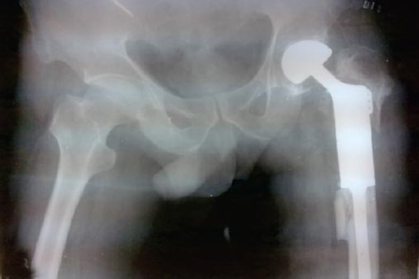
The hip was opened by posterior approach. The head of the broken prosthesis was removed easily but the stem was well fixed in the bone stock and could not be removed despite of all the efforts. So we opted for proximal femoral replacement with customized hip mega prosthesis. An osteotomy of the greater trochanter was performed and it was raised separately along with the attached abductors. The rest of the proximal part of the femur up to the fracture site was resected and removed along with the broken prosthesis. It was replaced by long stem steel cemented proximal femoral mega prosthesis. The remaining portion of the greater trochanter along with the abductors was attached to the ports provided at the lateral side of the prosthesis. Wound was closed over a suction drain. The patient was allowed to bear weight after removal of the stitches on the 12th post operative day with the help of four post walker. He has completed 2 years of follow up and is totally asymptomatic, pain free and walks independently without support.
Discussion
Fractures of the ipsilateral femur in patients with previous hip replacements have long been recognized as a significant problem associated with the procedure, but these are becoming more common, as the number of patients with a hip implant increases. Treatment options that have been described over the years include non-operative methods including protection of the fracture as described by Dysart SH et.al [17]. Various studies like Beals RK et. al [6], Johansson JE et. al [12], Adolphson P et.al [15], have advised traction as a method of non-operative treatment of these fractures specially in patients with high risk for surgery. Casts and braces can also be used to treat these fractures as shown in studies by Mont MA et. al [16], McElfresh EC et. al [18] and Missakian ML et. al [19].
Figure 2.

Postoperative radiograph of the patient - Plain radiograph of the left hip and thigh region showing the fracture treated by a custom made long stem cemented bipolar prosthesis.
Figure 3.

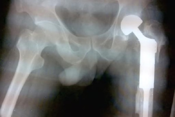
One year follow up radiograph of the patient - Plain radiograph of the left hip and thigh region after one year of follow up showing a stable prosthesis with no signs of loosening or any other complication.
Surgical options include either internal fixation using circlage wires or cables as shown by Beals RK et. al [6] and Mont MA et. al [16] in their study. Serocki JH et. al [20] described the use of screws with and without plates along with the use of circlage wires and cables. Special plates that have claws, bands or circlage wires to allow fixation in the region of the femoral stem have been used by Missakian ML et. al [19], Dave DJ et. al [21], Jensen TT et. al [22], Partridge AJ [23], Radcliffe SN et. al [24] and Zenni Jr EJ [25] in their studies. Other commonly used mode of treatment is revision of the femoral component. Options for revision include cemented and uncemented stems, long stems with proximal or extensive porous coating and stems with distal interlocking screws. In rare instances the whole of the proximal femur can also be removed and replaced by proximal femoral mega prosthesis. Patients with a failed total hip arthroplasty and massive proximal femoral bone loss can be salvaged with a proximal femoral megaprosthesis if there is no other alternative as shown by Shu-Tai Shih et. al [26] and Parvizi J et. al [27] in their studies. However, this procedure is technically demanding and has a high rate of complications.
If the prosthesis is loose, the bone is undergoing resorption and is at increased risk of failure. It therefore follows that treatment of a fracture secondary to a loose prosthesis requires revision of the prosthesis. On the other hand, a fracture in a bone with a well-fixed prosthesis following significant trauma requires treatment along the same principles as any other fracture, the only extra consideration is that of restrictions on choice of trauma implant due to the presence of an intramedullary prosthesis, unless the security of fixation is compromised by the fracture configuration. The choice of treatment is therefore determined primarily by whether or not the prosthesis is well fixed, and only secondarily by the site of the fracture. In our case the prosthesis itself was broken along with a periprosthetic fracture and the stem of the prosthesis was well fixed in the bone. Every attempt to remove the stem of the prosthesis failed and so the whole proximal part of the femur had to be sacrificed and replaced by a long stem customized cemented bipolar prosthesis.
Conclusion
The variable results of treatment for late periprosthetic femoral fracture, makes it necessary for undertaking every means to prevent this complication. The surgeon must keep in mind patient factors that increase the chance of fracture, including age, gender and index diagnosis. The quality of bone in both complex primary and revision surgery must be assessed. The surgical management of periprosthetic fractures is complex and can have potential complications. The treatment for each case must be individualized. While there is no set of rules that can be applied to all cases, the Vancouver classification combines the important factors in the management of these fractures; fracture location, implant stability and bone quality and can be useful in guiding treatment. The goal is to obtain near-anatomic alignment, stable fracture fixation, and a secure and well-fixed femoral component in proper alignment which allows for early mobilization of the patient to prevent any complications associated with prolonged recumbency in old age. Periprosthetic fractures are likely to increase even further in the coming years as the survival rate of the prostheses increases and the life expectancy of the population in general increases.
Clinical Message.
The treatment of periprosthetic fractures is very complex and the results are very variable. The treatment for each case must be individualized. Custom made prosthesis may be used in places where the prosthesis is well fixed in the femur. The goal is to obtain stable fracture fixation, and a secure and well-fixed femoral component in proper alignment which allows for early mobilization of the patient to prevent any complications associated with prolonged recumbency in old age.
Biography


Footnotes
Conflict of Interest: Nil
Source of Support: None
References
- 1.Lewallen DG, Berry DJ. Periprosthetic fracture of the femur after total hip arthroplasty: treatment and results to date. Instr Course Lect. 1998;47:243–9. [PubMed] [Google Scholar]
- 2.Tower SS, Beals RK. Fractures of the femur after hip replacement: the Oregon experience. Orthop Clin North Am. 1999;30:235–47. doi: 10.1016/s0030-5898(05)70078-x. [DOI] [PubMed] [Google Scholar]
- 3.Garcia-Cimbrelo E, Munuera L, Gil-Garay E. Femoral shaft fractures after cemented total hip arthroplasty. Int Orthop. 1992;16-1:97–100. doi: 10.1007/BF00182995. [DOI] [PubMed] [Google Scholar]
- 4.Kelley SS. Periprosthetic Femoral Fractures. J Am Acad Orthop Surg. 1994;2-3:164–72. doi: 10.5435/00124635-199405000-00005. [DOI] [PubMed] [Google Scholar]
- 5.Scott RD, Turner RH, Leitzes SM, Aufranc OE. Femoral fractures in conjunction with total hip replacement. J Bone Joint Surg Am. 1975;57-4:494–501. [PubMed] [Google Scholar]
- 6.Beals RK, Tower SS. Periprosthetic fractures of the femur. An analysis of 93 fractures. Clin Orthop Relat Res. 1996;327:238–46. doi: 10.1097/00003086-199606000-00029. [DOI] [PubMed] [Google Scholar]
- 7.Bhattacharyya T. Mortality after periprosthetic fracture of the femur (unpublished data) doi: 10.2106/JBJS.F.01538. [DOI] [PubMed] [Google Scholar]
- 8.Haddad FS, Masri BA, Garbuz DS, Duncan CP. The prevention of periprosthetic fractures in total hip and knee arthroplasty. Orthop Clin North Am. 1999;30-2:191–207. doi: 10.1016/s0030-5898(05)70074-2. [DOI] [PubMed] [Google Scholar]
- 9.Pazzaglia U, Byers PD. Fractured femoral shaft through an osteolytic lesion resulting from the reaction to prosthesis. A case report. J Bone Joint Surg Br. 1984;66-3:337–9. doi: 10.1302/0301-620X.66B3.6725341. [DOI] [PubMed] [Google Scholar]
- 10.Bethea JS, 3rd, DeAndrade JR, Fleming LL, Lindenbaum SD, Welch RB. Proximal femoral fractures following total hip arthroplasty. Clin Orthop Relat Res. 1982;170:95–106. [PubMed] [Google Scholar]
- 11.Schmidt AH, Kyle RF. Periprosthetic fractures of the femur. Orthop Clin North Am. 2002;33-1:p143–52. doi: 10.1016/s0030-5898(03)00077-4. ix. [DOI] [PubMed] [Google Scholar]
- 12.Johansson JE, McBroom R, Barrington TW, Hunter GA. Fracture of the ipsilateral femur in patients with total hip replacement. J Bone Joint Surg [Am] 1981;63:1435–42. [PubMed] [Google Scholar]
- 13.Parrish TF, Jones JR. Fracture of the femur following prosthetic arthroplasty of the hip. J Bone Joint Surg [Am] 1964;46:241–8. [PubMed] [Google Scholar]
- 14.Brady OH, Kerry R, Masri BA, et al. The Vancouver Classification of periprosthetic fractures of the hip: a rational approach to treatment techniques in orthopaedics. 1999;14(2):107–14. [Google Scholar]
- 15.Adolphson P, Jonsson U, Kalen R. Fractures of the ipsilateral femur after total hip arthroplasty. Arch Orthop Trauma Surg. 1987;106:353–7. doi: 10.1007/BF00456869. [DOI] [PubMed] [Google Scholar]
- 16.Mont MA, Marr DC. Fractures of the ipsilateral femur after hip arthroplasty: a statistical analysis of outcome based on 487 patients. J Arthroplasty. 1994;9:511–9. doi: 10.1016/0883-5403(94)90098-1. [DOI] [PubMed] [Google Scholar]
- 17.Dysart SH, Savoy CG, Callaghan JJ. Nonoperative treatment of a postoperative fracture around an uncemented porous coated femoral component. J Arthroplasty. 1989;4:187–90. doi: 10.1016/s0883-5403(89)80074-9. [DOI] [PubMed] [Google Scholar]
- 18.McElfresh EC, Coventry MB. Femoral and pelvic fractures after total hip arthroplasty. J Bone Joint Surg. 1974;56A:483–92. [PubMed] [Google Scholar]
- 19.Missakian ML, Rand JA. Fractures of the femoral shaft adjacent to long stem femoral components of total hip arthroplasty: report of seven cases. Orthopedics. 1993;16:149–52. doi: 10.3928/0147-7447-19930201-08. [DOI] [PubMed] [Google Scholar]
- 20.Serocki JH, Chandler RW, Dorr LD. Treatment of fractures about hip prostheses with compression plating. J Arthroplasty. 1992;7:129–35. doi: 10.1016/0883-5403(92)90005-b. [DOI] [PubMed] [Google Scholar]
- 21.Dave DJ, Koka SR, James SE. Mennen plate fixation for fracture of the femoral shaft with ipsilateral total hip and knee arthroplasties. J Arthroplasty. 1995;10:113–5. doi: 10.1016/s0883-5403(05)80111-1. [DOI] [PubMed] [Google Scholar]
- 22.Jensen TT, Overgaard S, Mossing NB. Partridge cerclene system for femoral fractures in osteoporotic bones with ipsilateral hemi/total arthroplasty. J Arthroplasty. 1990;5:123–6. doi: 10.1016/s0883-5403(06)80230-5. [DOI] [PubMed] [Google Scholar]
- 23.Partridge AJ, Evans PEL. The treatment of fractures of the shaft of femur using nylon circlage. J Bone Joint Surg. 1982;64-B(2):210–4. doi: 10.1302/0301-620X.64B2.7068743. [DOI] [PubMed] [Google Scholar]
- 24.Radcliffe SN, Smith DN. The Mennen plate in periprosthetic hip fractures. Injury. 1996;27:27–30. doi: 10.1016/0020-1383(95)00163-8. [DOI] [PubMed] [Google Scholar]
- 25.Zenni EJ, Jr, Pomeroy DL, Claude RJ. Ogden plate and other fixations for fractures complicating femoral endoprostheses. Clin Orthop. 1988;231:83–90. [PubMed] [Google Scholar]
- 26.Shu-Tai Shih MD, Jun-Wen Wang MD, Chia-Chen Hsu MD. Proximal Femoral Megaprosthesis for Failed Total Hip Arthroplasty. Chang Gung Med J. 2007 Jan-Feb;30(No. 1) [PubMed] [Google Scholar]
- 27.Parvizi J, Sim FH. Proximal femoral replacements with megaprostheses. Clin Orthop Relat Res. 2004;420:169–75. doi: 10.1097/00003086-200403000-00023. [DOI] [PubMed] [Google Scholar]


