Abstract
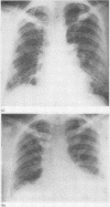
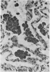
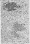
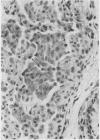
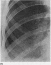
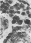
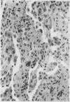
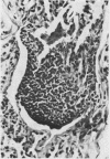
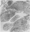
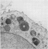
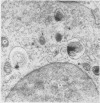
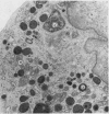
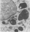
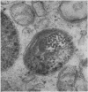
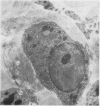
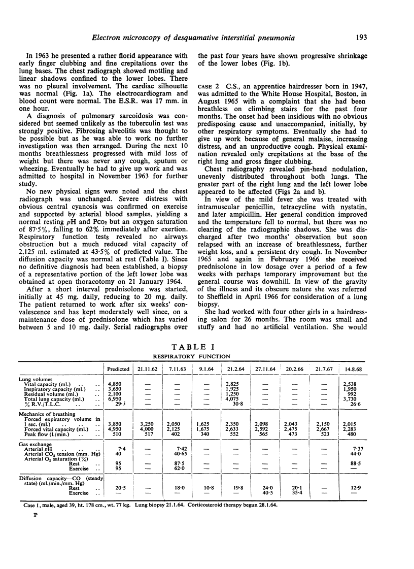
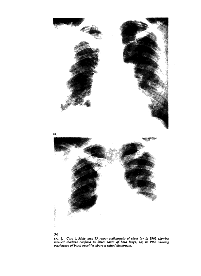
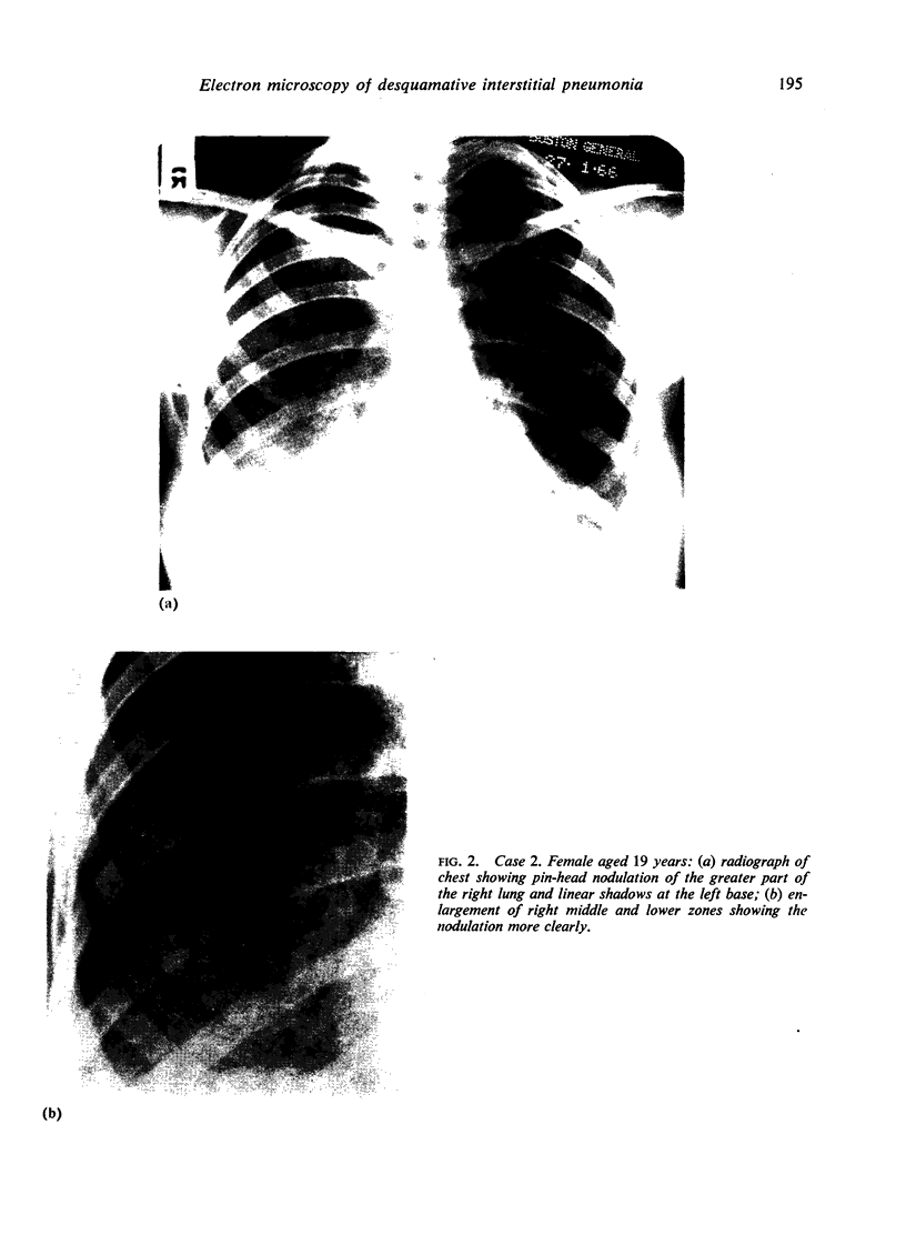
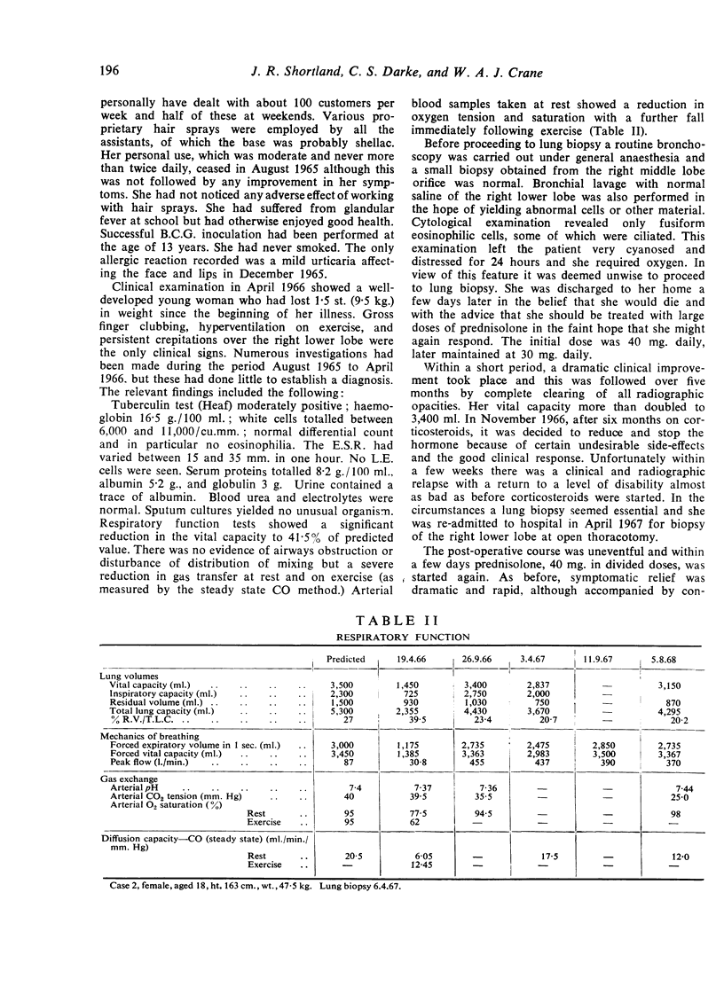
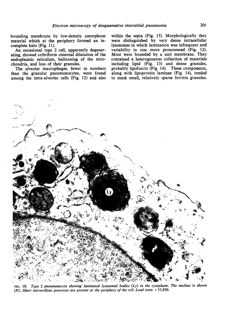
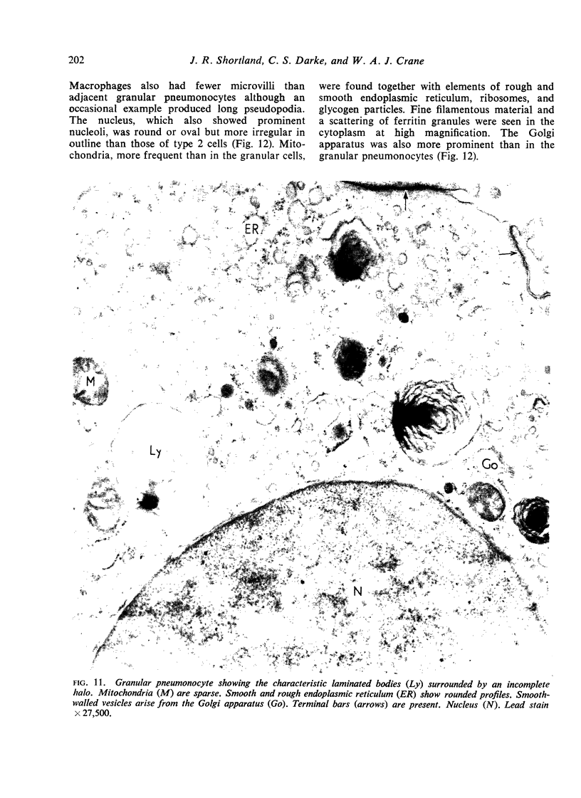
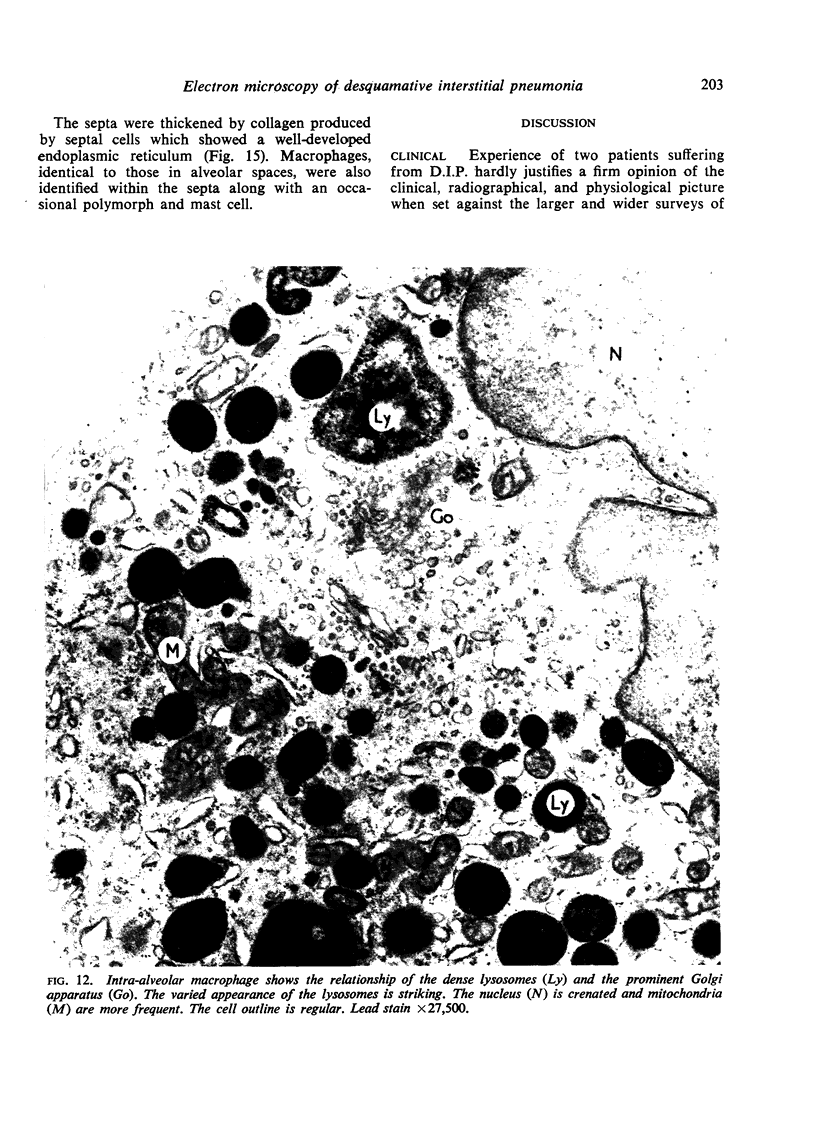
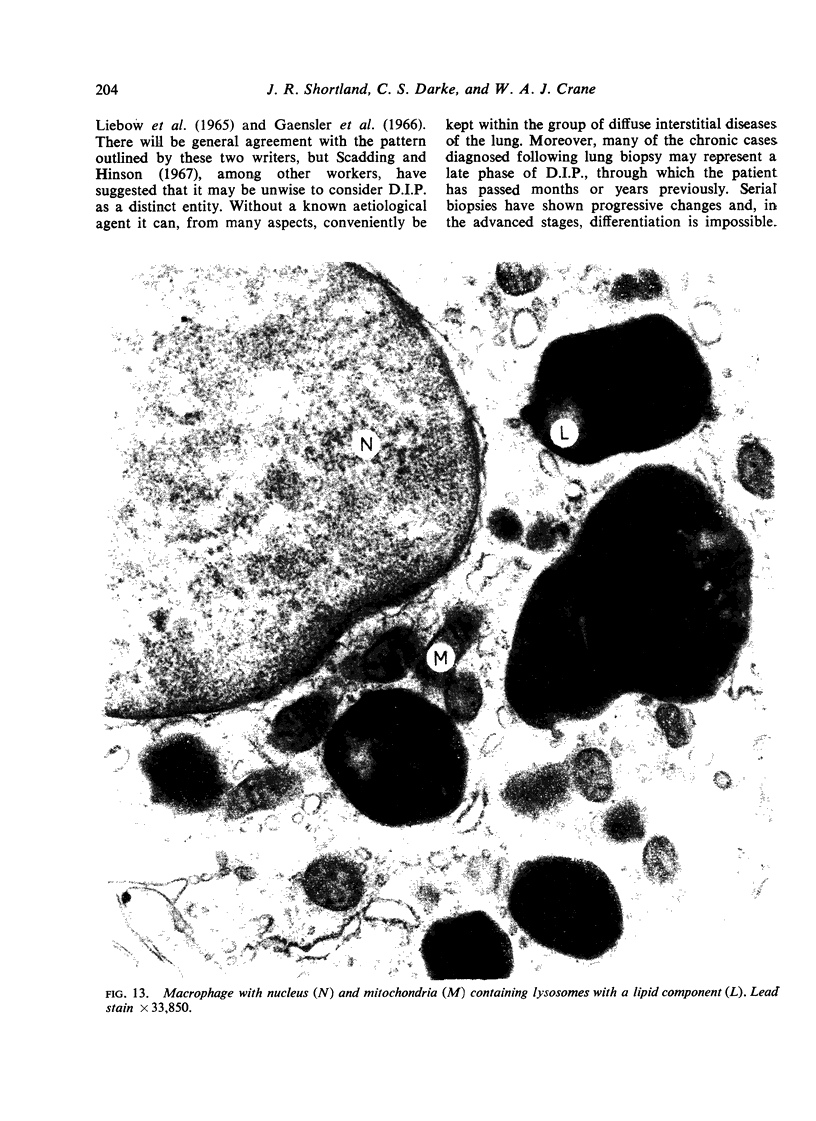
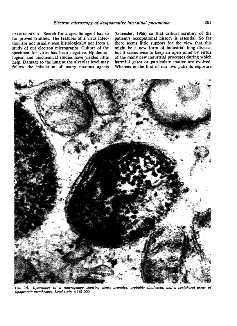
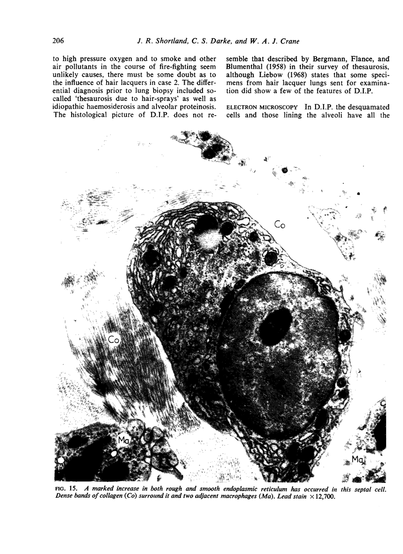
The clinical, radiographical, and physiological picture of two patients suffering from desquamative interstitial pneumonia is described. The diagnosis was established by lung biopsy when the characteristic histological features were found on light microscopy. The dramatic response to adequate corticosteroid therapy is recorded, and attention is directed to the danger of serious relapse on early withdrawal of this treatment and the subsequent satisfactory response to a second course. Electron microscopical studies of the tissue from one patient add materially to the understanding of the clinical course and the nature of the tissue response. At the ultrastructural level the attenuated membranous (type 1) pneumonocytes which normally line the alveoli were replaced by granular (type 2) pneumonocytes. The desquamated intra-alveolar cells comprised two main groups. These were granular pneumonocytes, similar to those lining the alveoli, and smaller numbers of macrophages. The cytopathic effects of virus infection were not detected by light or electron microscopy.
Full text
PDF
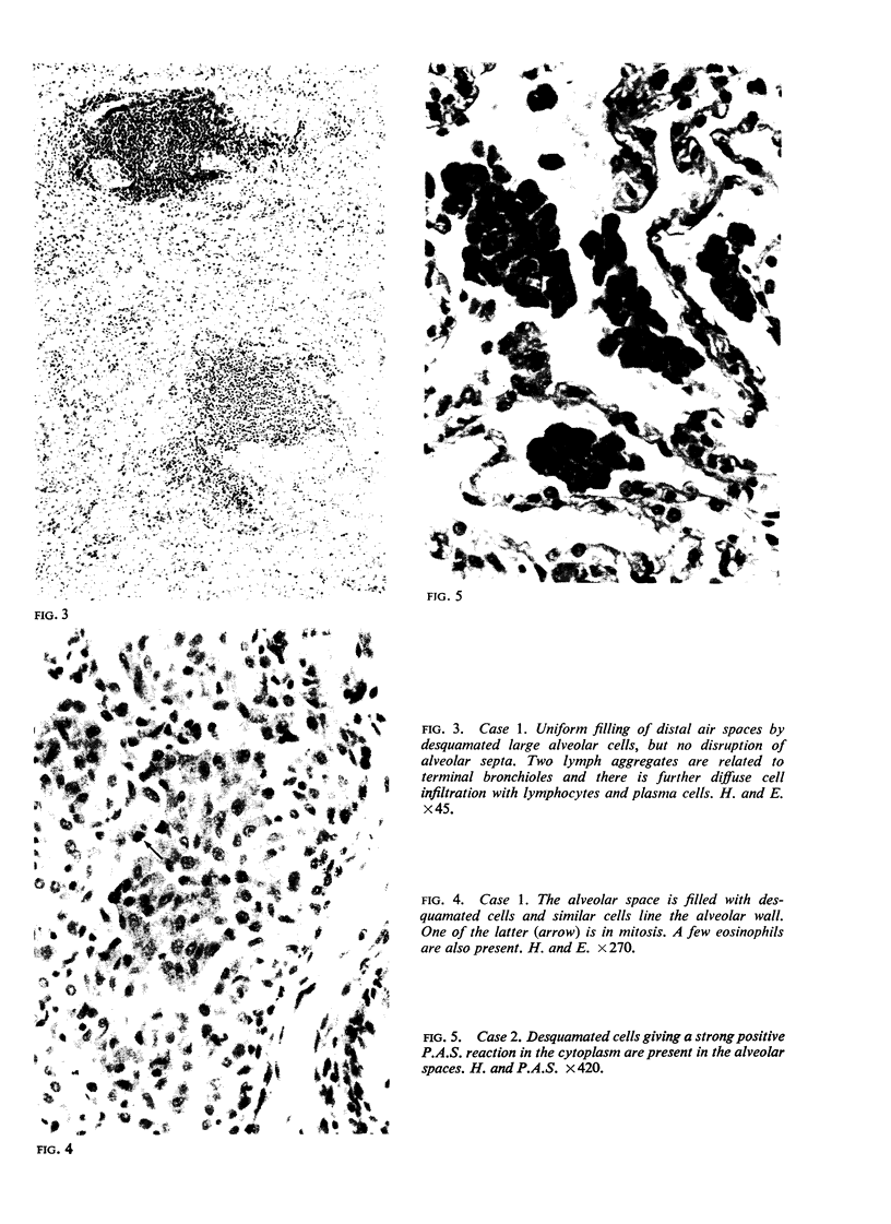
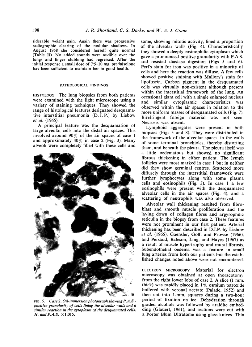
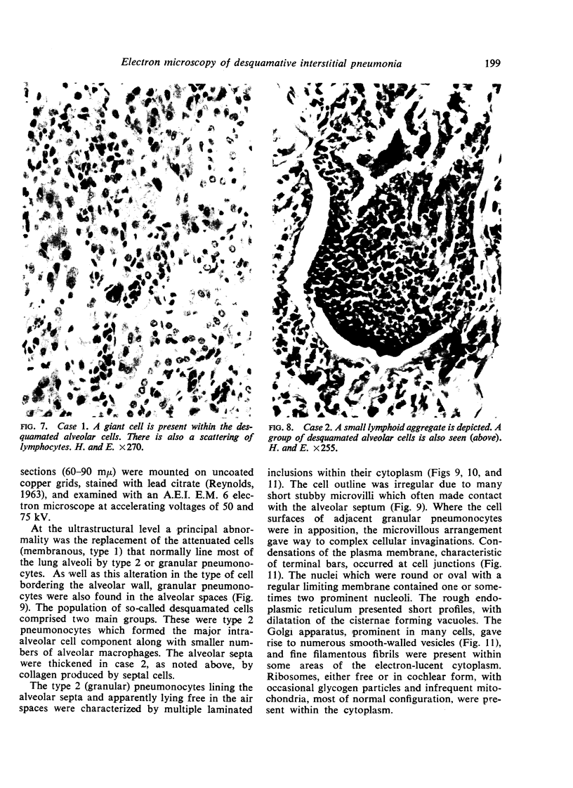
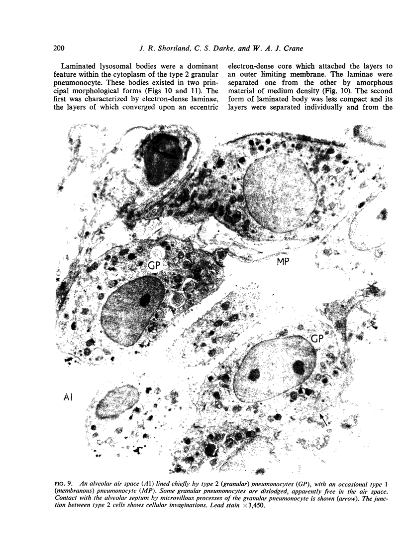
Images in this article
Selected References
These references are in PubMed. This may not be the complete list of references from this article.
- BERGMANN M., FLANCE I. J., BLUMENTHAL H. T. Thesaurosis following inhalation of hair spray; a clinical and experimental study. N Engl J Med. 1958 Mar 6;258(10):471–476. doi: 10.1056/NEJM195803062581004. [DOI] [PubMed] [Google Scholar]
- BUCKINGHAM S., AVERY M. E. Time of appearance of lung surfactant in the foetal mouse. Nature. 1962 Feb 17;193:688–689. doi: 10.1038/193688a0. [DOI] [PubMed] [Google Scholar]
- BUCKINGHAM S., MCNARY W. F., Jr, SOMMERS S. C. PULMONARY ALVEOLAR CELL INCLUSIONS: THEIR DEVELOPMENT IN THE RAT. Science. 1964 Sep 11;145(3637):1192–1193. doi: 10.1126/science.145.3637.1192. [DOI] [PubMed] [Google Scholar]
- Gaensler E. A., Goff A. M., Prowse C. M. Desquamative interstitial pneumonia. N Engl J Med. 1966 Jan 20;274(3):113–128. doi: 10.1056/NEJM196601202740301. [DOI] [PubMed] [Google Scholar]
- Goff A. M., McNary W. F., Jr, Gaensler E. A. Desquamative interstitial pneumonia. Med Thorac. 1967;24(5):317–329. doi: 10.1159/000192536. [DOI] [PubMed] [Google Scholar]
- Hatasa K., Nakamura T. Electron microscopic observations of lung alveolar epithelial cells of normal young mice, with special reference to formation and secretion of osmiophilic lamellar bodies. Z Zellforsch Mikrosk Anat. 1965 Oct 12;68(2):266–277. doi: 10.1007/BF00342433. [DOI] [PubMed] [Google Scholar]
- KARRER H. E. The ultrastructure of mouse lung; general architecture of capillary and alveolar walls. J Biophys Biochem Cytol. 1956 May 25;2(3):241–252. doi: 10.1083/jcb.2.3.241. [DOI] [PMC free article] [PubMed] [Google Scholar]
- Kikkawa Y., Motoyama E. K., Cook C. D. The ultrastructure of the lungs of lambs. The relation of osmiophilic inclusions and alveolar lining layer to fetal maturation and experimentally produced respiratory distress. Am J Pathol. 1965 Nov;47(5):877–903. [PMC free article] [PubMed] [Google Scholar]
- LEESON T. S., LEESON C. R. A LIGHT AND ELECTRON MICROSCOPE STUDY OF DEVELOPING RESPIRATORY TISSUE IN THE RAT. J Anat. 1964 Apr;98:183–193. [PMC free article] [PubMed] [Google Scholar]
- LIEBOW A. A., STEER A., BILLINGSLEY J. G. DESQUAMATIVE INTERSTITIAL PNEUMONIA. Am J Med. 1965 Sep;39:369–404. doi: 10.1016/0002-9343(65)90206-8. [DOI] [PubMed] [Google Scholar]
- LOW F. N. The electron microscopy of sectioned lung tissue after varied duration of fixation in buffered osmium tetroxide. Anat Rec. 1954 Dec;120(4):827–851. doi: 10.1002/ar.1091200402. [DOI] [PubMed] [Google Scholar]
- MOE H., ROSTGAARD J., BEHNKE O. ON THE MORPHOLOGY AND ORIGIN OF VIRGIN LYSOSOMES IN THE INTESTINAL EPITHELIUM OF THE RAT. J Ultrastruct Res. 1965 Apr;12:396–403. doi: 10.1016/s0022-5320(65)80107-1. [DOI] [PubMed] [Google Scholar]
- NOVIKOFF A. B., BEAUFAY H., DE DUVE C. Electron microscopy of lysosomerich fractions from rat liver. J Biophys Biochem Cytol. 1956 Jul 25;2(4 Suppl):179–184. [PMC free article] [PubMed] [Google Scholar]
- ORZALESI M. M., MOTOYAMA E. K., JACOBSON H. N., KIKKAWA Y., REYNOLDS E. O., COOK C. D. THE DEVELOPMENT OF THE LUNGS OF LAMBS. Pediatrics. 1965 Mar;35:373–381. [PubMed] [Google Scholar]
- PALADE G. E. A study of fixation for electron microscopy. J Exp Med. 1952 Mar;95(3):285–298. doi: 10.1084/jem.95.3.285. [DOI] [PMC free article] [PubMed] [Google Scholar]
- Persaud V., Bateson E. M., Ling J. A., Hayes J. A. Desquamative interstitial pneumonia. Br J Dis Chest. 1967 Jul;61(3):159–162. doi: 10.1016/s0007-0971(67)80045-7. [DOI] [PubMed] [Google Scholar]
- REYNOLDS E. S. The use of lead citrate at high pH as an electron-opaque stain in electron microscopy. J Cell Biol. 1963 Apr;17:208–212. doi: 10.1083/jcb.17.1.208. [DOI] [PMC free article] [PubMed] [Google Scholar]
- SCADDING J. G. FIBROSING ALVEOLITIS. Br Med J. 1964 Sep 12;2(5410):686–686. doi: 10.1136/bmj.2.5410.686. [DOI] [PMC free article] [PubMed] [Google Scholar]
- SCHLIPKOTER H. W. Elektronenoptische Untersuchungen ultradünner Lungenschnitte. Dtsch Med Wochenschr. 1954 Nov 5;79(45):1658–1659. doi: 10.1055/s-0028-1119938. [DOI] [PubMed] [Google Scholar]
- Scadding J. G., Hinson K. F. Diffuse fibrosing alveolitis (diffuse interstitial fibrosis of the lungs). Correlation of histology at biopsy with prognosis. Thorax. 1967 Jul;22(4):291–304. doi: 10.1136/thx.22.4.291. [DOI] [PMC free article] [PubMed] [Google Scholar]
- WOODSIDE G. L., DALTON A. J. The ultrastructure of lung tissue from newborn and embryo mice. J Ultrastruct Res. 1958 Nov;2(1):28–54. doi: 10.1016/s0022-5320(58)90046-7. [DOI] [PubMed] [Google Scholar]


