Abstract
Introduction:
Osteochondromas usually arise from the metaphyseal region of the growing skeleton but extraskeletal cartilaginous tumors are rarest.
Case Report:
A 65 year old woman presented with anterior knee pain and inability to flex her knee more than 90° since 1 year. Clinical examination and imaging studies revealed a nodular calcific mass in the anterior portion of the knee i.e. patella. Following excision, histopathology confirmed the diagnosis of extra-osseous osteochondroma-like soft tissue mass, with no recurrence in 36 weeks.
Conclusion:
An integrated clinical-pathologic diagnosis helps to clarify the nature of extraskeletal cartilaginous tumors that can arise at unusual anatomic site viz. patella. Complete local surgical excision is the management of choice.
Keywords: osteochondroma, clinical, patella, pathological, excision, extraskeletal cartilaginous
Introduction
Osteochondromas usually develop in relation to the periosteum, and occur around the growth plate of long bones, especially the knee. The tumor usually stops to grow with closure of the growth plate [1] BUT, some of these tumors continue to grow after skeletal maturity [2] we report a patient with an extra-osseous osteochondroma-like soft tissue mass in the anterior portion of the knee joint.
Case report
65 Years old women presented with one-year history of anterior knee pain and unable to flex knee more than 90 degree. There was no history of trauma, constitutional symptoms, fever, loss of wait, morning stiffness and involvement of other joints.
Clinical examination shows Firm nodular mass was present in the anterior part of the knee, extending superiorly, inferiorly, medially and laterally as well. There was no joint line tenderness present Range of motion was Flexion was restricted to 90 degree and full extension was possible. Neurovascular examination was normal Serology was normal. Surgical excision of mass was done. Intraoperatively Mass involves complete patella but femoral condyle, tibial condyle and patellar tendon was not involved. Post operatively Recovery was uneventful and patient returned to activity of daily living after 2 weeks. She regained 110 degree of flexion Histopathological examination reveals Osteochondroma features with no evidence of malignancy After 6 months patient was asymptomatic and there was no clinical and radiological evidence of reoccurrence.
Figure 1.
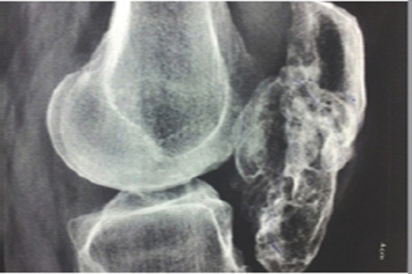
Pre Operative X-Rav
Figure 2.
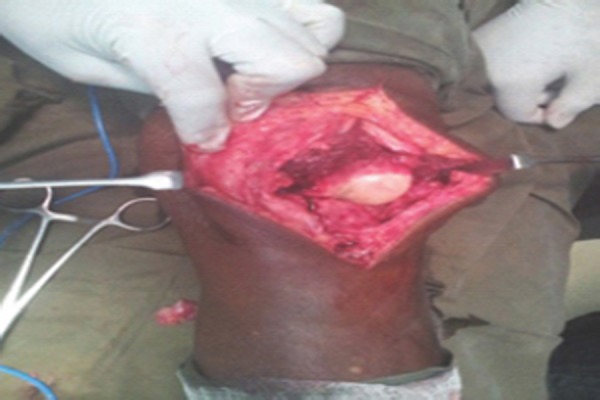
Intraoperative
Figure 3.
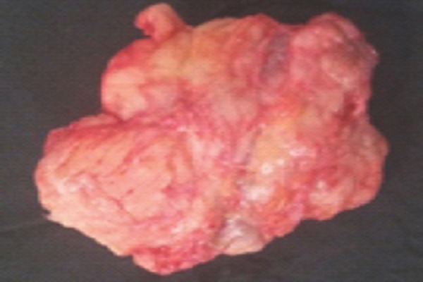
Mass excised
Figure 4.
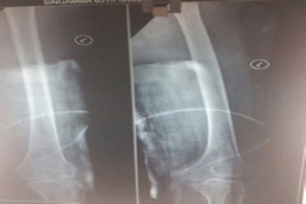
Post Op X-ray
Figure 5.
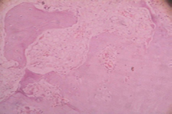
Histopathologial picture
Discussion
Large osteochondromas involving the joint capsule or paraarticular soft tissues adjacent to a joint are rare osteocartilaginous tumors, first described by Jaffe in 1958 [3]. Subsequently, these lesions have been reported using various terms such as paraarticular osteochondroma [4, 5,6], intracapsular chondroma [7], para-articular chondroma [8,9], giant extrasynovial osteochondroma [10], Hoffa’s disease [11] and giant intra-articular osteochondroma [12].
But in our case this lesion involve whole of patella that patella cannot be distinguished and at histological examination the mass was characterized by trabecular bone tissue surrounded by hyaline cartilage. The pathogenesis and classification of this lesion are still controversial. Rizzello et al. [6] reported that this tumor seems to originate from a cartilaginous metaplasia of the articular and para-articular connective tissue. Rodriguez- Peralto et al., [7] emphasize a possible metaplastic origin of the tumor, due to a traumatic event of Hoffa’s fat pad. Other authors [10,13] reported a relation between this type of osteochondroma of the knee and a chronic impingement of the infrapatellar fat pad; they conclude that this lesion is the end-stage of Hoffa’s disease. But in our case there was no history of trauma or symptomatic history of infrapatellar fat pad Usually after fusion of epiphysis chances of osteosarcoma is more but in this case at the age of 65yrs no signs of malignancy were seen.
Conclusion
An integrated clinical-pathologic diagnosis helps to clarify the nature of extra skeletal cartilaginous tumors that can arise at unusual anatomic site. Complete local surgical excision is the management of choice with proper follow up for reoccurrence.
Clinical Messege
Unusual site of any tumor should be discussed clinically, radiological and pathologically before deciding treatment.
Biography


Footnotes
Conflict of Interest: Nil
Source of Support: None
Reference
- 1.Kienb.ck R. Über die Gelenkskapsel-Osteome. Kniegelenk Fortschr Röntgenstr. 1924;32:527–546. [Google Scholar]
- 2.Reith JD, Bauer TW, Joyce MJ. Paraarticular osteochondroma of the knee. Report of 2 cases and review of the literature. ClinOrthop. 1997;334:225–232. [PubMed] [Google Scholar]
- 3.Jaffe HL. Philadelphia: Lea and Febiger; 1958. Tumors and tumorous conditions of the bones and joints; pp. 558–567. [Google Scholar]
- 4.Milgram JW, Dunn EJ. Para-articular chondromas and osteochondromas: a report of three cases. Clin Orthop Relat Res. 1980;(148):147–51. [PubMed] [Google Scholar]
- 5.Reith JD, Bauer TW, Joyce MJ. Paraarticular osteochondroma of the knee: report of 2 cases and review of the literature. Clin Orthop Relat Res. 1997;(334):225–32. [PubMed] [Google Scholar]
- 6.Rizzello G, Franceschi F, Meloni MC, et al. Para-articular osteochondroma of the knee. Arthroscopy. 2007;23(8):910, e1–4. doi: 10.1016/j.arthro.2006.05.030. [DOI] [PubMed] [Google Scholar]
- 7.Rodriguez-Peralto JL, Lopez-Barrea F, Gonzalez-Lopez J. Intracapsular chondroma of the knee: an unusual neoplasm. Int J Surg Pathol. 1997;5(1&2):49–54. [Google Scholar]
- 8.Sakai H, Tamai K, Iwamoto A, Saotome K. Para-articular chondroma and osteochondroma of the infrapatellar fat pad: a report of three cases. Int Orthop. 1999;23(2):114–7. doi: 10.1007/s002640050322. [DOI] [PMC free article] [PubMed] [Google Scholar]
- 9.Schmidt-Rohlfing B, Staatz G, Tietze L, Ihme N, Siebert CH, Niethard FU. Diagnosis and differential diagnosis of extraskeletal, para-articular chondroma of the knee. Z Orthop Ihre Grenzgeb. 2002;140(5):544–7. doi: 10.1055/s-2002-33998. [DOI] [PubMed] [Google Scholar]
- 10.Turhan E, Doral MN, Atay AO, Demirel M. A giant extrasynovial osteochondroma in the infrapatellar fat pad: end stage Hoffa’s disease. Arch Orthop Trauma Surg. 2008;128(5):515–9. doi: 10.1007/s00402-007-0397-5. [DOI] [PubMed] [Google Scholar]
- 11.Krebs VE, Parker RD. Arthroscopic resection of an extrasynovial ossifying chondroma of the infrapatellar fat pad: end-stage Hoffa’s. doi: 10.1016/s0749-8063(05)80117-3. [DOI] [PubMed] [Google Scholar]
- 12.Sarmiento A, Elkins RW. Giant intra-articular osteochondroma of the knee. J Bone Joint Surg Am. 1975;57(4):560–1. [PubMed] [Google Scholar]
- 13.Nouri H, Ben Hmida F, Ouertatani M, Bouaziz M, Abid L, Jaafoura H, Zehi K, Mestiri M. Tumour-like lesions of the infrapatellar fat pad. Knee Surg Sports Traumatol Arthrosc. 2010;18(10):1391–4. doi: 10.1007/s00167-009-1034-3. [DOI] [PubMed] [Google Scholar]


