

Abstract
So far it has been difficult to repair and reconstruct the composite tissue defects in knee. Saphenous artery flap has been widely used to repair complex wounds, but the design and clinical application of composite tissue flap at perforating branches of saphenous artery were not reported. In this research, we design a new composite tissue flap by carrying fascial flap in the medial gastrocnemius muscle with perforators flap in saphenous artery to repair and reconstruct the composite tissue defects in knee. By anatomic observation and analysis, we find that there exists blood-supply in netty form among saphenous arteries, medial artery below the knee, intermuscular branch in high-order position of posterior tibial artery and perforating branch in medial artery of calf. We chose saphenous artery as blood-supplying artery; utilized the netty blood-supplying mode in middle-up and medial part of shank; cut the composite tissue flap at perforating branches of saphenous artery with fascial flap carried in the medial gastrocnemius muscle; reconstructed the ligamentum patellae using medial head of gastrocnemius muscle and Achilles’s tendon; and covered the wounds at front side of knee with flap. Composite tissues were survived completely, free from infection at wounds and exosmosis of joint fluid. Motion function of knee-joint proved satisfactory, and ambulatory function was recovered. There was no complication in donor site. Composite tissue flap at perforating branches of saphenous artery with fascial flap carried in the medial gastrocnemius muscle is one of the most ideal solutions for repairing the composite tissue defects at front side of knee joint.
Keywords: Composite tissue flap, saphenous artery, perforators flap, the design of the flap, the knee joint reconstruction
Introduction
The skin defects at front side of knee joint caused by traffic accident injuries, burn or ulcer are commonly seen may bring about the defect of ligamentum patellae at front side of knee joint that can affect the function of knee joint, so local flaps transfer repair is often used to treat simple skin defects at front side of knee joint. But the repair of complex wounds at front side of knee joint, especially of those accompanied with defects in deep subcutaneous tissue at front side of knee joint, is still a challenging task. Anterior wounds of knee joint are often accompanied with skin defects, ligamentum patellae and joint capsule, as well as by potential lacunas. In order for repair and reconstruction of composite tissue at front side of knee joint, it is also necessary to seal the articular cavity and to reconstruct the ligamentum patellae apart from covering the wounds with flap.
Normally, there is enough blood supply in muscular flaps and myocutaneous flaps that can be used to packing lacuna and reconstructing composite tissue defects. Gastrocnemius myocutaneous flap has been reportedly widely used in the repair and reconstruction of composite tissue defects at front side of knee joint caused by trauma, and often, good operation results can be expected. This method has however, certain disadvantages, including swollen pedicle, comparatively long pedicle with no effect, unavailable pedicle tissue and more sacrificed tissue. Free composite tissue flap can also be used to repair composite tissue defects at front side of knee joint, but microsyrgical technique is needed in this method with longer operation time and higher risks. Using saphenous arterial blood supply, we designed a new composite tissue flap by carrying fascial flap in the medial gastrocnemius muscle with perforators flap in saphenous artery; chose the emerging point of saphenous artery at medial side of knee joint as rotation point of pedicle, and chain-like anastomosis line of blood vessel in high-order position of posterior tibial artery and saphenous artery as the axis for cutting off flap; cut off the flap at middle-up part and medial side of shank; designed fascial flap in the medial gastrocnemius muscle of appropriate volume according to the defect size of ligamentum patellaea and joint capsule at front side of knee joint; reconstructed the ligamentum patellae at front side of knee joint; closed the joint capsule; designed the flap according to the size and shape of wounds; and covered the wound with flap at the same time, in order to repair and reconstruct the defects of composite tissue at front side of knee joint with less sacrifice of pedicle and less damage of donor site. The author proposed a case of applying a new composite tissue flap by carrying fascial flap in the medial gastrocnemius muscle with perforators flap in saphenous artery to repair and reconstruct the composite tissue defects at front side of knee joint.
Case report
A patient with 38 years of age, male, was hospitalized for skin soft-tissue defects and necrosis in left knee joint caused by accidental burning by brazier due to acute onset of seizures in April. Physical examination results showed that the sign of life was stable, with no abnormalities in heart, lung and abdomen. Specialized examination results showed that the skin at front side of left knee joint was blackened and necrotic, with a defect area of about 13 cm×8 cm; the defects were found in the exposed part of patella and ligamentum patellae, with partial defects in joint capsule, exposed knee joint and local joint fluid exudation, making the knee joint incapable of flexion and blood supply in limb not good enough. The operation was conducted after successful drug control of seizure by repair and reconstruction of ligamentum patellae using composite tissue flap of saphenous artery and wounds repair + skin grafting
Preoperative design
We will measure the size of wounds before operation, design a composite tissue flap at perforating branches of saphenous artery that carrying with fascial flap in the medial gastrocnemius muscle and partial Achilles’s tendon; detect the direction of the perforating branches in intermuscular space of saphenous artery and high-order position of posterior tibial artery at medial side of knee joint using a portable ultrasonic Doppler with the route marked; detect the direction of perforating branches at medial side of calf at the position of middle part of the medial head of gastrocremius muscle with the route marked; choose the emerging point of saphenous artery at medial side of knee joint as rotation point of pedicle, and chain-like anastomosis line of blood vessel in high-order position of posterior tibial artery and saphenous artery as the axis for cutting off flap; design a flap at medial side and middle-up part of shank according to the size of wound, with both the width and length of flap being 1 cm longer than that of the wound; and design a fascial flap that need to be carried with the center point chosen at arterial perforating branch point of medial part of the calf. The size of the flap is 15 cm×10 cm.
Operating procedures
The detailed description of the operation is shown in Figure 1. After the patient took supine position after successful anesthesia with conventional sterilization and debridement to have cleared away necrotic infection tissue, it was found that the properly trimmed broken ends of ligamentum patellae were prepared for repair and reconstruction. We cut off composite tissue flap according to the preoperative design, open the flap at posterior side; cut open the fascial flap in the medial gastrocnemius muscle at intermuscular space of medial and lateral heads of sural muscle; chose arterial perforating point at medial side of calf as center point; cut off myolemma flap of gastrocnemius muscle in upward and downward directions; carried some Achille’s tendon tissue at medial side of gastrocnemius muscle, sutured myolemma flap with flap and cutting off them both, so as to prevent the separation of myolemma flap with flap; tied the bleeder and opened the front part of the flap and pedicle of the flap; sutured the tendinous tissue of myolemma flap to reconstruct the ligamentum patellae; seamed the fascia tissue of muscle to close the joint capsule; covered the wound with flap; provided skin-grafting repair to flap-donating site and provided aseptic dressing with petrolatum gauze to skin-donating site at a proper dressing pressure; and fixed the knee joint to the extended position with plaster after aseptic dressing.
Figure 1.
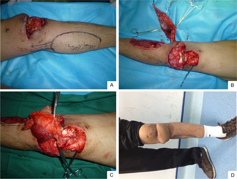
A. Design the composite tissue flap before operation. B. Cut off the composite tissue flap that carries myolamma flap of gastrocnemius muscle during the operation. C. Reconstruct the ligamentum patellae using myolemma flap of gastrocnemius muscle during the operation. D. Stepping ability recovers after 6 months.
Postoperative management
We provided lamp-baking to knee joint for seven consecutive days in 1 week after operation; conducted conventional “three resistance” treatment for 1 week; employed plaster immobilization for 4 weeks; changed the dressing in a timely manner; and bound loosely to protect the blood vessel pedicle from being pressed. We removed stitches at the position of skin-grafting in flap-donating site after 12 days; and in suture site, 14 days. We enhanced the functional training of knee joint gradually after tearing down the external fixation in the 4th week.
Results
The flap survived and no vessel crisis happened. After a continuous follow-up of 6 months, we found that color, character and appearance of the flap were all close to that of the peripheral tissue; no contracture happened in knee joint. The reconstructed ligamentum patellae was powerful enough to extend the knee joint in resistance of the gravity of shank and the knee joint could extend to 0 degree and flex to 90 degrees. Therefore, the range of flexion and extension of knee joint was satisfactory. The skin grafts in flap-donating site were all survived in phase I, and flap-donating site was a little sunken. There was no obvious weakening in the strength of Achilles’s tendon of injured limb compared with the normal one. The area of numbness at the dominant region of saphenous nerve became smaller and smaller during the follow-up visit. Peripheral nerves could be compensated with no serious complication.
Discussion
To human beings, knee joint is one of the most important joints which are locally exposed and having larger range of motion, so the peripheral skin soft-tissue thereof is vulnerable to injury. The skin soft-tissue injury of knee joint is often caused by burn, local blunt trauma, direct heat injury, traffic accidents. Skin injury at front side of the knee joint, such as accompanied infection, can also lead to the necrosis and defects of a large area of skin. Knee joint would suffer from dysfunction at varying degrees if left untreated. A simple way to repair it is to provide partial skin-grafting to cover the granulation wound and partial arbitrary flap transfer to cover the wound, but after skin-grafting, the resulting scar contracture would affect the knee-joint function. Furthermore, skin-grafting can’t repair the composite tissue defects, so composite tissue flap that carries blood vessel pedicle is often adopted at present. There are lots of methods for repairing flap transferring for comparatively larger area of soft tissue defects at knee joint. Flaps that are merely used for covering wounds include: sural neurouascular flap, arteria peronea and perforating branches flap [1], gastrocnemius myocutaneous flap [2,3], arterial perforators flap at medial side of calf [4], retrograde anterolateral thigh flap, saphenous artery flap [5], cross-leg flap and so on. Free semitendinosus tendon [6], allogenic ligamentum patellae, overlapped muscle tendon of gracilis, artificial muscle tendon are often used to repair and reconstruct the ligamentum patellae for simple ligamentum patellae defects. Composite tissue flap repair with blood supply is most suitable for treating wounds that accompanied with ligament defects for myocutaneous flap in gastrocnemius muscle; because of its good blood supply, this method is appropriate for repairing infected wounds. Myocutaneous flap in gastrocnemius muscle can be used to repair and reconstruct the ligamentum patellae and wounds [7,8], but there are some drawbacks, such as much more serious trauma, bloated pedicle after transfer and sunken donor site that would affect the physical strength of lower limbs restricted by length of blood vessel pedicle. Myocutaneous flaps in gastrocnemius muscle are not suitable for the repair of soft tissue defects at near-end and front side of soft tissue in knee [9-12]. Along with the development of vascular anatomy of skin-muscle perforators, myolemma flap of muscle has gradually replaced some muscle flaps and myocutaneous flaps because of its conformance with the reconstruction principle of “supplying the shortage” throughout the repair and reconstruction of soft tissue. In recent years, myolemma flap of muscle has been gaining popularities and has achieved desirable results [13-15]. A recombination flap of saphenous artery and myolemma is designed to repair the composite tissue defects at front side of knee joint, for fear of the drawbacks from use of myocutaneous flap. The blood supply of saphenous artery flap mainly comes from branched saphenous artery in the descending genicular artery. And the area that can be cut off is about 10 cm above knee and 20 cm below the knee [16-20], which can meet the demand of repairing the wound at front side of knee joint. The blood supply of recombination flap of saphenous artery and myolemma mainly come from saphenous artery [21]. The reticulated vascular anastomosis among saphenous artery, posterior tibial artery perforators, medial genicular artery and medial sural artery perforators enable the transfer of flap that carries myolemma flap at medial side of gastrocnemius muscle with blood supply [22]. In that case, the pedicle can be much closer to the wound, and also the carrying of a small amount of muscle can bring lesser damage, avoiding the bloated pedicle and the sunken donor site [23-25].
We have obtained the satisfactory effect in the application of composite tissue flap of saphenous artery to repair the soft tissue defects at front side of knee joint. The said operation treatment has the following advantages: 1) the larger cutting area of saphenous artery flap can meet the demand of wound repair of skin defects at front side of knee joint; 2) the blood supply of composite tissue flap at saphenous artery is sufficient with a anterograde blood supply and anterograde venous return; 3) no edema of the tissue flap happens after operation; and 4) the anti-infection ability is strong enough, all of which can meet the demand of wound repair under bad blood-supply condition. The direction of saphenous artery is relatively constant, and the pedicle of tissue flap is long enough to transfer neatly with no need for anastomosing with blood vessel, contributing to a higher rate of success. The flap carries some saphenous nerve which can bring a protective feel. There are reticular vascular anastomosis among saphenous artery, arteria tibialis perforator and medial sural artery perforator, so it is sensible to use a composite tissue flap of appropriate size to repair the wound at front side of knee joint that accompanies with ligamentum patellae defects. The donor site of the flap, as well as the skin texture, is close to the recipient site. But the skin tension at skin-donating site is strong, so a skin-grafting operation is often needed for the wound after the skin is made available. There is still a disadvantage by this method: the cutaneous sensation of the flap-cutting area, the area below it and inferior side of arcrotarsium, is often lost after the saphenous nerve is cut off, but the area of numbness can become smaller and smaller during the long-term follow-up, which can be compensated for by peripheral nerves [26].
Because myocutaneous flaps in local or vicinal parts of knee joint can repair almost all of the soft tissue defects and bone exposures at front side of knee. It is however difficult to repair the soft tissue defects using free flap because of the deeper position of the blood vessel at recipient site around the knee, especially at front side of knee. As a result, free flap is seldom used to repair the soft tissue defects at front side of knee in clinics. But when large-area defects happened or when local and vicinal flaps are unavailable, free flap would be the last choice [27]. In clinical practice, appropriate flap should be chosen to repair the skin defects according to the different depths and scopes of defects. Local saphenous artery vascular anatomy is relatively constant in reliable blood supply, and does not have to include use of special microsurgery technique [28]; it is characterized by simple cutting, high success rate, fine reconstruction appearance, less donor site defects and no effects on lower limbs’ motional function; it can carry the myolemma flap at medial head of gastrocnemius muscle which has enough blood supply to form a composite tissue flap to repair the composite tissue defects at front side of knee joint [29]; and it also can meet the demand of ligamentum patellae repairment, becoming one of the best methods for repairing composite tissue defects at front side of knee.
Acknowledgements
This study is supported by National clinical key college fund.
Disclosure of conflict of interest
None.
References
- 1.Ruan HJ, Cai PH, Fan CY. Flap transfer of peroneal and perforators vessel pedicle to repair soft tissue defects around the knee joint. Zhongguo Xiu Fu Chong Jian Wai Ke Za Zhi. 2009;23:303–305. [PubMed] [Google Scholar]
- 2.Zhang B, Du G, Huang K. Using island shaped myocutaneous flap of medial gastrocnemius muscle to repair the soft tissue defects at knee joint. Chinese Journal of Clinical Medicine. 2010;3:365–367. [Google Scholar]
- 3.Tang F, Wang HJ, X J. The observation of using myocutaneous flap at medial gastrocnemius muscle to repair the deep wound of knee joint. Chinese Journal of Clinical Physicians (electronic edition) 2012;6:5338–5339. [Google Scholar]
- 4.Zhang GL, Ge BF, Wang Y. The clinical application analysis of pedicle-carried islandshaped flap in arterial perforators at medial side of calf. J Pract Orthopaedics. 2009;15:673–675. [Google Scholar]
- 5.Acland RD, Schusterman M, Godina M. The saphenous neurovascular free flap. Plast Reconstr Surg. 1981;67:763–774. doi: 10.1097/00006534-198106000-00009. [DOI] [PubMed] [Google Scholar]
- 6.Li NS. The repair and reconstruction of oboslete patellar tendon fracture. Chinese J Orthopeadic. 1984;4:380. [Google Scholar]
- 7.Wang ZQ, Wang Q, Jin LG. Using Achilles’s tendon-carried myocutaneous flap at medial gastrocnemius muscle to repair the soft tissue and patellar tendon defects at front side of knee. Orthopedic Journal of China. 2002;9:662–663. [Google Scholar]
- 8.Wang XW. The transposition of flap at medial gastrocnemius muscle to repair skin defects at ligamentum patellae and anteromedial side of knee. China Practical Medical. 2011;6:64–65. [Google Scholar]
- 9.Ries MD, Bozic KJ. Medial gastrocnemius fl ap coverage for treatment of skin necrosis after total knee arthroplasty. Clin Orthop Relat Res. 2006;446:186–192. doi: 10.1097/01.blo.0000218723.21720.51. [DOI] [PubMed] [Google Scholar]
- 10.Wei ZR, Xiao EC, Tan J. Using composite tissue flap of perforators pedicle on ankle in posterior tibial artery with reticular blood supply to repair composite tissue defects behind the heel. Chinese Journal of Reconstruction Surgery. 2010;24:635–636. [Google Scholar]
- 11.Wei ZR, Sun GF, Tang XJ. Using composite tissue flap of perforators pedicle at posterior tibial artery with chain typed blood supply from medial and rear side of shank to repair the infected wound at ankle. Third Military Medical University Journals. 2009;31:2161–2162. [Google Scholar]
- 12.Hou CL, Gu YD. Chirurgery of flap. Shanghai: Shanghai Science and Technology Press; 2006. pp. 636–639. [Google Scholar]
- 13.Ju JH, Jin GZ, Zhao Q. Using free saphenous arterial flap to repair skin soft tissue defects of hand and foot. Chinese Journal of Clinical Anatomy. 2010;28:690–693. [Google Scholar]
- 14.Gupta A, Kumer S, Kaplan B. Novel immunosuppressive strategies for composite tissue allografts. Curr Opin Organ Transplant. 2014;19:552–557. doi: 10.1097/MOT.0000000000000135. [DOI] [PubMed] [Google Scholar]
- 15.Wang ZT, Sun WH. Cosmetic Reconstruction of the Digits in the Hand by Composite Tissue Grafting. Clin Plast Surg. 2014;41:407–427. doi: 10.1016/j.cps.2014.03.001. [DOI] [PubMed] [Google Scholar]
- 16.Diaz-Siso JR, Bueno EM, Sisk GC. Vascularized composite tissue allotransplantationstate of the art. Clin Transplant. 2013;27:330–337. doi: 10.1111/ctr.12117. [DOI] [PMC free article] [PubMed] [Google Scholar]
- 17.Ren X, Laugel MC. The next frontier in composite tissue allotransplantation. CNS Neurosci Ther. 2013;19:1–4. doi: 10.1111/cns.12029. [DOI] [PMC free article] [PubMed] [Google Scholar]
- 18.Glaus SW, Johnson PJ, Mackinnon SE. Clinical strategies to enhance nerve regeneration in composite tissue allotransplantation. Hand Clin. 2011;27:495–509. doi: 10.1016/j.hcl.2011.07.002. [DOI] [PMC free article] [PubMed] [Google Scholar]
- 19.Schneeberger S, Landin L, Jableki J. Achievements and challenges in composite tissue allotransplantation. Transpl Int. 2011;24:760–769. doi: 10.1111/j.1432-2277.2011.01261.x. [DOI] [PubMed] [Google Scholar]
- 20.Murphy BD, Zuker RM, Borschel GH. Vascularized composite allotransplantation: an update on medical and surgical progress and remaining challenges. J Plast Reconstr Aesthet Surg. 2013;66:1449–1455. doi: 10.1016/j.bjps.2013.06.037. [DOI] [PubMed] [Google Scholar]
- 21.Hui-Chou HG, Rodriguez ED. Clinical Facial Composite Tissue Allotransplantation: A Review of the Global Experience and Future Implications. The Know-How of Face Transplantation. London: Springer; 2011. pp. 315–330. [DOI] [PubMed] [Google Scholar]
- 22.Li Z, Shang X, Cao X. Surgical Reconstruction of a Severe Crush Injury of the Lateral Part of the Forefoot with Use of a Cross-Leg Osteocutaneous Pedicled Fibular Graft. JBJS Case Connector. 2013;3:e125. doi: 10.2106/JBJS.CC.M.00112. [DOI] [PubMed] [Google Scholar]
- 23.Siemionow M, Klimczak A. Advances in the development of experimental composite tissue transplantation models. Transpl Int. 2010;23:2–13. doi: 10.1111/j.1432-2277.2009.00948.x. [DOI] [PubMed] [Google Scholar]
- 24.Yu L, Tan JH, Cai L. Repair of severe composite tissue defects in the lower leg using two different cross-leg free composite tissue flaps. Ann Plas Surg. 2012;68:83–87. doi: 10.1097/SAP.0b013e3181fe9351. [DOI] [PubMed] [Google Scholar]
- 25.Turgut G, Kayalı MU, Köse Ö. Repair of a wide lower extremity defect with cross-leg free transfer of latissimus dorsi and serratus anterior combined flap: a case report. Strategies Trauma Limb Reconstr. 2010;5:155–158. doi: 10.1007/s11751-010-0094-8. [DOI] [PMC free article] [PubMed] [Google Scholar]
- 26.Zhang G, Ge B, Hu Y. Reconstruction of degloved thumb with prefabricated flap. Chin J Traumatol. 2009;12:184–186. [PubMed] [Google Scholar]
- 27.Takagi S, Oyama T, Yamazumi K. Vascular augmentation of an extended latissimus dorsi myocutaneous flap through an intercostal vessel: A preliminary report. J Plast Surg Hand Surg. 2013;47:123–125. doi: 10.3109/2000656X.2012.738608. [DOI] [PubMed] [Google Scholar]
- 28.Elsaftawy A, Jabłecki J, Domanasiewicz A. Treatment possibilities of reverse-flow sural flap in covering the defects of lower extremities. Pol Przegl Chir. 2013;85:192–197. doi: 10.2478/pjs-2013-0029. [DOI] [PubMed] [Google Scholar]
- 29.Zhang G, Chen K, Zhang J. Repair of a large soft tissue defect in the leg with cross-leg bridge free transfer of a latissimus dorsi myocutaneous flap: a case report. Chin J Traumatol. 2012;15:373–375. [PubMed] [Google Scholar]