Abstract
Background:
Human cystic echinococcosis (hydatidosis) continues to be an essential cause of morbidity and mortality in many parts of the world.
Methods:
We studied hydatid cyst pattern in hospitalized adult patients from 2003 to 2012 in Mashhad and Neyshabour, northeast of Iran.
Results:
Overall, 1342 patients, 711 females (53%) and 631 males (47%) diagnosed as infected with hydatid cyst were evaluated. Their age was between 1 and 91 yr (mean age 37.75). The most affected age group was 20–30 yr old. Totally, 953 cases (71%) were urban and 375 cases (27.8%) were rural residents. The most common localization of cysts was the liver and lung. The housewives were the most frequently infected occupations.
Conclusion:
The rate of infection with hydatid cyst is high in Mashhad, northeast of Iran, and the incidence of human hydatidosis tends to increase in recent years so control and prevention programs are recommended.
Keywords: Hydatid cyst, Echinococcus granulosus, Hydatidosis, Iran
Introduction
Cystic echinococcosis (CE) is a near-cosmopolitan zoonosis caused by larval stages of tapeworms belonging to the genus Echinococcus (Family Taeniidae). Six species of Echinococcus have been recognized, but the most important members of the genus in respect of their public health importance and their geographical distribution is E. granulosus, causes cystic echinococcosis) (1–7). Hydatidosis is a very serious problem of man in the world (5, 8–10).
The greatest prevalence of hydatidosis in human and animal hosts is found in sheep-raising areas, including southern South America, the entire Mediterranean littoral, southern and central parts of the former Soviet Union, central Asia, China, Australia, and north and east Africa (1, 2, 5, 11, 12). Hydatidosis is one of the most important diseases (12, 13). Iran is an endemic region for hydatidosis (5, 11, 14, 15). Hydatid cyst is reported in 1% of Iranian patients admitted for surgery (15).
Definitive hosts for E. granulosus are carnivores particularly dogs and other caniners and many mammals may serve as intermediate hosts, but herbivorous species such as sheep, goat, cattle, swine, deer and human are most likely to become infected by contact with dog, eating vegetable, geophagy and contact with sheep, pastoral occupation and poor education(2, 3, 15–18). After ingestion of the egg excreted from dog fecal material, the larva reach the blood and lymphatic circulation and transport to the liver, lungs, kidney and other organs (15, 19).
Hydatid cyst can occur in any part of body, but mostly in liver and lungs. It is rarely seen in the kidney, spleen, heart, brain, bone and muscle (11, 20). Symptoms of hydatidosis are related with size, location, rupture and infection for cysts. Identification of cyst is confirmed by ultrasonography and CT scan (11). By a very slow process of growth, the asymptomatic period is too long and hydatidosis might be diagnosed after 20–25 yr post infection (8).
Khorasan Province, northeast part of Iran had the highest incidence rate for hydatidosis in Iran (15). In the present retrospective study we reviewed the gender, age, jobs, location of hydatid cysts during ten year in three main hospitals of Mashhad and Neyshaboor, Iran.
Materials and Methods
This retrospective and descriptive study was performed in hospitalized patients in Qaem and Emam Reza and 22 Bahman hospitals in Mashhad and Neyshabur, Iran from March 2003, Dec2012. Data were collected by searching through patient’s files in hospitals archives considering different factors such as age, sex, occupation, organ involvement and geographical distribution of patients. After surgery in all cases, hydatidosis had been approved by parasitological and histopathological examinations.
Statistical analysis was carried out using the SPSS ver. 16 software (Chicago, IL, USA).
Results
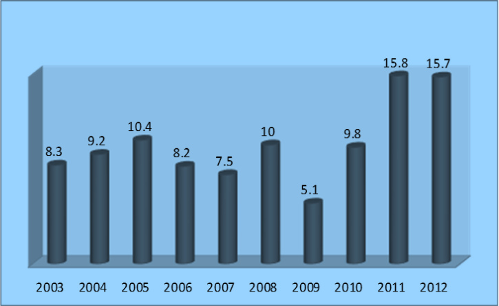
In this study, we evaluated 1342 patients, 711 females (53%) and 631 males (47%) who had hydatid cysts, during ten years. Their age was between 1 and 91 yr, (mean age 37.75). The most affected age group was 20–30 yr old. The homemakers had the highest rate of infection. The distribution of residency showed 953 cases (71%) of urban origin. The highest annually operation rate (15.8%) was seen in 2011 and the lowest (5.1%) in 2009 (Fig. 1).
Fig. 1: Distribution of the rate of hydatidosis (%) during 10 yr examination from 2003–2012.
The highest prevalence of infection was in Mashhad (449 cases, 33.5%), 124 cases (9.35%) in Neyshabur, 108 cases (8%) in Torbat Heydarieh, 77cases (5.8%) in Torbat Jam, 50 cases (3.7%) in Chenaran, and in other cities was 39.65%. The liver was the most frequently infected organ followed by the lungs, kidney, brain, spleen, diaphragm, heart, subcutaneous, pancreas, ovary, spine, pelvic, spinal cord, bladder (Table 1).
Table 1: Distribution of infected organs by sex.
| Organ | Sex | |||
|---|---|---|---|---|
| Male | Female | |||
| Number | Percent | Number | Percent | |
| Liver | 321 | 50.9 | 442 | 62.2 |
| Lung | 252 | 39.9 | 210 | 29.5 |
| Diaphragm | 1 | 0.2 | 2 | 0.3 |
| Subcutaneous | 1 | 0.2 | 0 | 0 |
| Kidney | 10 | 1.6 | 6 | 0.8 |
| Brain | 2 | 0.3 | 5 | 0.7 |
| Pancreas | 1 | 0.2 | 0 | 0 |
| Ovary | 0 | 0 | 1 | 0.1 |
| Spleen | 3 | 0.5 | 2 | 0.3 |
| Spinal Cord | 1 | 0.2 | 1 | 0.1 |
| Heart | 1 | 0.2 | 1 | 0.1 |
| Pelvic | 0 | 0 | 1 | 0.1 |
| Bladder | 1 | 0.2 | 0 | 0 |
| Total | 631 | 100 | 711 | 100 |
Discussion
In the present study from March 2003– Dec 2012, 1342 cases of hydatid cyst were operated; the average number of operated cysts per year was 134.2.
Hydatidosis is a serious public health problem in Iran. The existence of very young children with hydatidosis and the new cases registered every year showed that the disease is being actively transmitted in Iran (21).
In Iran, there are several reports of human hydatidosis. Mousavi et al. reported 202 cases in Urmia City during 1991–2001 (11). Mohammadzadeh Hajipirloo et al. reported 294 cases in West Azerbaijan, Northwest Iran from 2000–2009 (21). Comparing to these reports the rate of hydatidosis in Mashhad (Khorasan Razavi) is higher than other parts of Iran. Dopchiz et al. reported 120 cases in one of Mar del Plata City Hospital Buenos Aires Argentina between 1992 and 2002 (22). Neghina et al. reported 81 cases in Romania from 1994–2001 (23). These reports also showed that north east of Iran is an endemic area for hydatidosis.
In Iran, the most common localization of hydatid cyst was the liver like our study (2, 11, 14–16, 24, 25). Mousavi et al. in his study in Iran reported the most affected organs were liver, lung, brain, spinal cord, pelvis, spleen, kidney and heart (11). Neghina et al. in Romania reported the most affected organs were liver, lung, kidney, ovary and peritoneal (23). However in Mauritania, the most affected organs were lung (50%), and then 33% in the liver and 17% elsewhere (26).
The present study shows that women underwent surgery more often than men did, and that homemakers had the highest rate of surgery (53%). This rate has been reported as 53.3% in west Azerbaijan (21), 56% in Shohada Tajrish Hospital in Tehran (11), and 56.4% in report of Nourjah from Iran(8).
In rural areas, the highest incidence of female hydatidosis might be due to swallowing the sweeping dust containing eggs of Echinococcus from dog’s feces, and in urban area cleaning and eating raw vegetables can be the cause of higher incidence of hydatidosis in women. This finding is similar to other reports of hydatidosis in Iran like (8, 11, 12, 15, 21).
In our study, patients’ age ranged from 1–91 yr old. The peak incidence of the disease was between 20 and 30 yr old. This result is consistent with previous studies in Iran (8, 11, 15, 21). Dopchiz et al. reported the mean age of the patients was 42.2+16.8 years (22). Caremani et al. in one study in Italy reported the mean age of the patients was 45.38 (27). Probably most human hydatidosis are acquired in childhood and it may be undetected until adolescence. Hydatidosis is a disease of long incubation period (might be 20 to 30 yr) and a wide range of different ages is obvious in patients. But the age group of 20 to 40 yr has more contact with livestock in farms and this can be the reason of the higher prevalence of hydatid cyst among them (11, 15).
Our study showed that 28% patients were from rural areas. In one study in Iran, the rate of infection of urban areas was greater than rural areas (12).
Conclusion
The rate of infection with hydatid cyst is high in Mashhad, northeast of Iran, and the incidence of human hydatidosis tends to increase in recent years so control and prevention programs are recommended. Infected animals can act as reservoirs of human hydatidosis, finally treatment and vaccination of sheep and dogs are recommended. Personal hygiene must be noticed in order to prevent ingestion of infective eggs from soil contaminated with dog’s feces.
Acknowledgments
The authors greatly acknowledge the Research Council of Mashhad University of Medical Sciences (MUMS), Mashhad, Iran, for their financial grant. The results presented in this work have been taken from Zohreh Andalib Aliabady thesis, with the ID number “921380.” The authors declare that there is no conflict of interests.
References
- 1. Grosso G, Gruttadauria S, Biondi A, Marventano S, Mistretta A. Worldwide epidemiology of liver hydatidosis including the mediterranean area. World J Gasteroenterol. 2012; 18 (13): 1425– 37. [DOI] [PMC free article] [PubMed] [Google Scholar]
- 2. Moro P, Schantz PM. Echinococcosis: A review. Int J Infect Dis. 2009; 13 (2): 125– 133. [DOI] [PubMed] [Google Scholar]
- 3. Roberts L, Janovy J. Foundation of parasitology, 5th. WCB Company, UK: 2000: 347– 410. [Google Scholar]
- 4. Rojo-Vazquez FA, Pardo-Lledias J, Francos-Von Hunefeld M, Cordero-Sanchez M, Alamo-Sanz R, Hernandez-Gonzalez A, Brunetti E, Siles-Lucas M. Cystic echinococcosis in Spain: Current situation and relevance for other endemic areas in europe. PLoS Negl Trop Dis. 2011; 5 (1): e893. [DOI] [PMC free article] [PubMed] [Google Scholar]
- 5. Jahangir A, Taherikalani M, Asadolahi K, Emaneini M. Echinococcosis/hydatidosis in Ilam province, western Iran. Iran J Parasitol. 2013; 8 (3): 417– 22 [PMC free article] [PubMed] [Google Scholar]
- 6. Mandal S, Deb Mandal M. Human cystic echinococcosis: Epidemiologic, zoonotic, clinical, diagnostic and therapeutic aspects. Asian Pac J Trop Med. 2012; 5 (4): 253– 260. [DOI] [PubMed] [Google Scholar]
- 7. Karimi A, Asadi K, Mohseni F, Hossein Akbar M. Hydatid cyst of the biceps femoris muscle (a rare case in orthopedic surgery). Shiraz E-Med J. 2011; 12 (3): 150– 4. [Google Scholar]
- 8. Nourjah N, Sahba G, Baniardalani M, Chavshin A. Study of 4850 operated hydatidosis cases in Iran. Southeast Asian J Trop Med Public Health. 2004; 35 (Supp1): 218– 222. [Google Scholar]
- 9. Reyes MM, Taramona CP, Saire-Mendoza M, Gavidia CM, Barron E, Boufana B, Craig PS, Tello L, Garcia HH, Santivañez SJ. Human and canine echinococcosis infection in informal, unlicensed abattoirs in Lima, Peru. PLoS Negl Trop Dis. 2012; 6 (4): e1462. [DOI] [PMC free article] [PubMed] [Google Scholar]
- 10. Akhlaghi L, Massoud J, Housaini A. Observation on hydatid cyst infection in Kordestan province (west of Iran) using epidemiological and seroepidemiological criteria. Iran J Public Health. 2005; 34 (4): 73– 75. [Google Scholar]
- 11. Mousavi SR, Samsami M, Fallah M, Zirakzadeh H. A retrospective survey of human hydatidosis based on hospital records during the period of 10 years. J Parasit Dis. 2012; 36 (1): 7– 9. [DOI] [PMC free article] [PubMed] [Google Scholar]
- 12. Vejdani M, Vejdani S, Lotfi S, Najafi F, Nazari N, Hamzavi Y. Study of operated primary and secondary (recurrence) hydatidosis in hospitals of Kermanshah, west of islamic republic of Iran. East Mediterr Health J. 2013; 19 (7): 671– 5. [PubMed] [Google Scholar]
- 13. Hajizadeh M, Ahmadpour E, Sadat A, Spotin A. Hydatidosis as a cause of acute appendicitis: A case report. Asian Pac J Trop Dis. 2013; 3 (1): 71– 73. [Google Scholar]
- 14. Geramizadeh B. Unusual locations of the hydatid cyst: A review from Iran. Iran J Med Sci. 2013; 38 (1): 2– 14. [PMC free article] [PubMed] [Google Scholar]
- 15. Rokni M. Echinococcosis/hydatidosis in Iran. Iran J Parasitol. 2009; 4 (2): 1– 16. [Google Scholar]
- 16. Craig PS, McManus DP, Lightowlers MW, Chabalgoity JA, Garcia HH, Gavidia CM, Gilman RH, Gonzalez AE, Lorca M, Naquira C, Nieto A, Schantz PM. Prevention and control of cystic echinococcosis. Lancet Infect Dis. 2007; 7 (6): 385– 394. [DOI] [PubMed] [Google Scholar]
- 17. Parsa F, Haghpanah B, Pestechian N, Salehi M. Molecular epidemiology of Echinococcus granulosus strains in domestic herbivores of lorestan, Iran. Jundishapur J Microbiol. 2011; 4 (2): 123– 130. [Google Scholar]
- 18. Ziaei H, Fakhar M, Armat S. Epidemiological aspects of cystic echinococcosis in slaughtered herbivores in Sari abattoir, north of Iran. J Parasit Dis. 2011; 35 (2): 215– 218 [DOI] [PMC free article] [PubMed] [Google Scholar]
- 19. Berenji F, Mirsadraei S, Asaadi L, Maroufi A, Fata A., Shahi M. A case report of alveolar echinococcosis. Med J Mashad Univ Med Sci. 2007; 50 (97): 354– 357. [Google Scholar]
- 20. Rokni Yazdi H, Sotoudeh H, Shargh H, Yazdabadi A. A case of primary adductor muscle hydatidosis: “Water-lily sign” on magnetic resonance imaging. Iran J Radiol. 2007; 4 (4): 223– 226. [Google Scholar]
- 21. Hajipirloo HM, Bozorgomid A, Alinia T, Tappeh KH, Mahmodlou R. Human cystic echinococcosis in west Azerbaijan, northwest Iran: A retrospective hospital based survey from 2000 to 2009. Iran J Parasitol. 2013; 8 (2): 323– 326. [PMC free article] [PubMed] [Google Scholar]
- 22. Dopchiz MC, Elissondo MC, Rossin MA, Denegri G. Hydatidosis cases in one of mar Del plata city hospitals, Buenos Aires, Argentina. Rev Soc Bras Med Trop. 2007; 40 (6): 635– 639. [DOI] [PubMed] [Google Scholar]
- 23. Neghina R, Neghina AM, Marincu I, Iacobiciu I. Epidemiology and epizootology of cystic echinococcosis in Romania 1862–2007. Foodborne Pathog Dis. 2010; 7 (6): 613– 618. [DOI] [PubMed] [Google Scholar]
- 24. McManus DP, Zhang W, Li J, Bartley PB. Echinococcosis. Lancet. 2003; 362 (9392): 1295– 1304. [DOI] [PubMed] [Google Scholar]
- 25. Uraiqat AA, Al-Awamleh A. Hydatid cyst in the muscles: A case report. JRMS. 2010;17(Supp1)72–74. [Google Scholar]
- 26. Salem COA, Schneegans F, Chollet J, Et Jemli M. Epidemiological studies on echinococcosis and characterization of human and livestock hydatid cysts in mauritania. Iran J Parasitol. 2011; 6 (1): 49– 57. [PMC free article] [PubMed] [Google Scholar]
- 27. Caremani M, Maestrini R, Occhini U, Sassoli S, Accorsi A, Giorgio A, Filice C. Echographic epidemiology of cystic hydatid disease in Italy. Eur J Epidemiol. 1993; 9 (4): 401– 404. [DOI] [PubMed] [Google Scholar]


