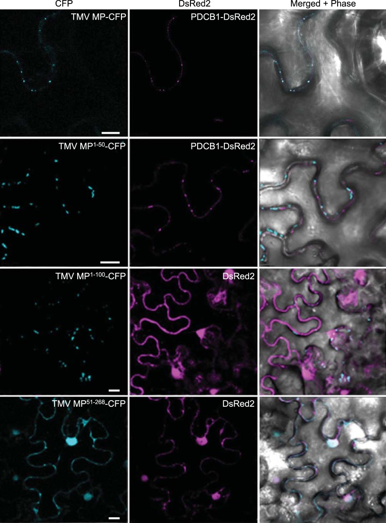
FIG 2 .
Subcellular localization of TMV MP, MP1–50, and MP51–268. PDCB1-DsRed2 and free DsRed2 were coexpressed as markers for plasmodesmata and nuclei, respectively. CFP signal is blue; DsRed2 signal is red; plastid autofluorescence was filtered out. Images are single confocal sections. Scale bars, 10 µm.

