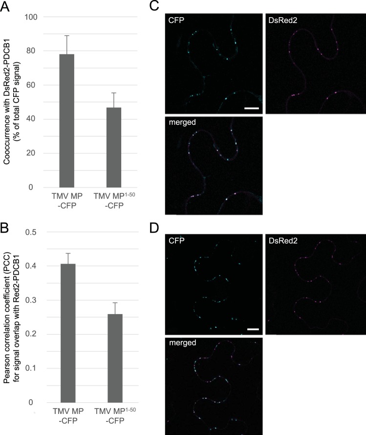
FIG 3 .
TMV MP and TMV MP1–50 localize to plasmodesmata. (A and B) TMV MP-CFP co-occurred with PDCB1-DsRed2 at plasmodesmata 78.1% ± 10.9% of the time (PCC 0.41 ± 0.03); TMV MP1–50-CFP co-occurred with PDCB1-DsRed2 at plasmodesmata 46.8% ± 8.6% of the time (PCC 0.26 ± 0.03). Data were determined on the basis of analyzing 32 expressing cells in each of three independent experiments and are expressed as means ± SE. (C) Representative single confocal sections of coexpressed TMV MP-CFP and PDCB1-DsRed2. (D) Representative single confocal sections of coexpressed TMV MP1–50-CFP and PDCB1-DsRed2. CFP signal is blue; DsRed2 signal is red; plastid autofluorescence was filtered out. Scale bars, 10 µm.

