Abstract
AIM: To demonstrate the 18F-fluorodeoxyglucose positron emission tomography (18F-FDG PET) findings in patients with non-Hodgkin’s lymphoma (NHL) involving the gastrointestinal (GI) tract and the clinical utility of modality despite of the known normal uptake of FDG in the GI tract.
METHODS: Thirty-three patients with biopsy-proven gastrointestinal NHL who had undergone FDG-PET scan were included. All the patients were injected with 10-15 mCi FDG and scanned approximately 60 min later with a CTI/Siemens HR (+) PET scanner. PET scans were reviewed and the maximum standard uptake value (SUVmax) of the lesions was measured before and after the treatment, if data were available and compared with histologic diagnoses.
RESULTS: Twenty-five patients had a high-grade lymphoma and eight had a low-grade lymphoma. The stomach was the most common site of the involvement (20 patients). In high-grade lymphoma, PET showed focal nodular or diffuse hypermetabolic activity. The average SUVmax±SD was 11.58±5.83. After the therapy, the patients whose biopsies showed no evidence of lymphoma had a lower uptake without focal lesions. The SUVmax±SD decreased from 11.58±5.83 to 2.21±0.78. In patients whose post-treatment biopsies showed lymphoma, the SUVmax±SD was 9.42±6.27. Low-grade follicular lymphomas of the colon and stomach showed diffuse hypermetabolic activity in the bowel wall (SUVmax 8.2 and 10.3, respectively). The SUVmax was 2.02-3.8 (mean 3.02) in the stomach lesions of patients with MALT lymphoma.
CONCLUSION: 18F-FDG PET contributes to the diagnosis of high-grade gastrointestinal non-Hodgkin's lymphoma, even when there is the normal background FDG activity. Furthermore, the SUV plays a role in evaluating treatment response. Low-grade NHL demonstrates FDG uptake but at a lesser intensity than seen in high-grade NHL.
Keywords: Positron emission tomography, Non-Hodgkin’s lymphoma, Gastrointestinal neoplasm
INTRODUCTION
Non-Hodgkin’s lymphoma (NHL) is known to arise from extranodal sites in 10-30% of cases[1,2]. Among the extranodal sites, the gastrointestinal tract is most frequently affected by NHL. It can involve the gastrointestinal tract partly or entirely[3]. Generally, the diagnosis of lymphomatous involvement of the gastrointestinal tract is based on clinical symptoms and imaging studies, such as double-contrast barium study, computed tomography (CT) and is confirmed by endoscopy with biopsy. CT has been widely used as the imaging modality for staging and restaging of lymphoma[4]. However, in evaluating lymphomatous involvement of the gastrointestinal tract, CT has some limitations. Examples are non-specific imaging patterns and findings that may be difficult to interpret such as wall thickening in a non-distended stomach or unopacified small bowel loops[5-7]. Gallium-67 citrate scintigraphy also plays an important role in patients with NHL for the detection of lesions, initial staging and assessment of therapeutic responses[8,9]. Gallium-67 citrate scintigraphy is known to be more sensitive for the detection of lesions in thoracic locations, but it is much less sensitive in the identification of infradiaphragmatic sites owing to physiologic hepatic and splenic uptake and excretion into the bowel[10]. Since Paul[11] first reported 18F-FDG PET imaging in lymphoma, this modality has been increasingly used to examine patients with lymphomas. Many published articles have shown that 18F-FDG PET is more sensitive in detecting disease sites than gallium-67 scintigraphy. 18F-FDG PET is at least as sensitive as CT, but more specific than CT, especially in patients undergoing restaging[12-15]. In addition, many studies have demonstrated that persistent FDG uptake after therapy may predict treatment failure or a high recurrence rate[16,17]. Most reported studies have assessed primarily patients with nodal NHL. A few studies have used 18F-FDG PET to assess NHL in the gastrointestinal tract[18-21]. Rodriguez et al[18] demonstrated that 18F-FDG PET may have a novel application in the evaluation of gastric NHL and may complement endoscopy and CT in selected patients. However, in a study by Hoffmann et al[19] in patients with mucosa-associated lymphoid tissue (MALT)-type lymphoma, no focal tracer uptake was demonstrated with 18F-FDG PET in either gastric or extragastric lesions. In addition, there is concern that normal FDG accumulation in the gastrointestinal tract or abnormal uptake in patients with inflammatory bowel disease could cause confusion in the interpretation of 18F-FDG PET images in patients with lymphoma[22-25]. Thus the clinical utility of 18F-FDG PET imaging in the evaluation of lymphomatous involvement of the gastrointestinal tract has not been clearly established. This study aimed to demonstrate the 18F-FDG PET findings in patients with NHL (high-grade vs low-grade) involving the gastrointestinal tract and the clinical utility of this modality through the normal uptake of FDG in the gastrointestinal tract was known.
MATERIALS AND METHODS
In this study, the electronic database of 907 consecutive patients with lymphoma who underwent PET imaging from October 2000 to June 2002 was retrospectively reviewed. Thirty-three patients with biopsy-proven NHL involving the gastrointestinal tract who underwent a 18F-FDG PET scan were included in this study. There were 24 men and 9 women aged 34-80 years (mean 58 years). For scanning, all patients were injected with 10-15 mCi FDG and scanned approximately 60 min later with a CTI/Siemens HR (+) PET scanner (Siemens, Knoxville, TN, USA). Each scan was performed from the head to the pelvic floor with the total time of about 60 min for image acquisition. The acquired data were reconstructed using standard vendor-provided iterative reconstruction with segmented attenuation correction. Additional transmission scanning for attenuation correction was performed.
The PET scans obtained were reviewed by an experienced nuclear physician and a radiologist who together provided the consensus reading. Reviewing was done without patient’s clinical data and the status of lymphoma. The SUVmax of the lesions was measured before and after the treatment in case the data were available. The SUVmax was measured in at least two orthogonal planes to demonstrate the best lesion appreciation. The CT scan obtained on the corresponding data was used as the guideline for demarcating those lesions when their boundaries were difficult to define. The highest SUVmax was used for each lesion. The 18F-FDG PET results and the SUV measurements at each time point were compared with the histologic data from the corresponding data. The SUVmax was compared before and after the treatment using Student’s t-test. P<0.05 was considered statistically significant.
RESULTS
Of the 33 patients with NHL involving the gastrointestinal tract, 25 had a high-grade lymphoma and 8 had a low-grade lymphoma as determined using the revised European-American classification of lymphoid neoplasm (REAL classification). The histologic subtypes included diffuse large cell lymphoma (n = 16), mantle cell lymphoma (n = 6), MALT-type lymphoma (n = 5), peripheral T-cell lymphoma (n = 3), follicular lymphoma (n = 2) and B-cell small lymphocytic lymphoma (n = 1). The stomach was the most common site of the involvement, followed (in order of decreasing prevalence) by the colon and rectum, cecum and terminal ileum, duodenum, small bowel, and esophagus (Table 1).
Table 1.
Sites of gastrointestinal involvement of NHL in 33 patients
| Sites of GI involvement | Number of patients (%) |
| Stomach | 21 (64) |
| Colon and rectum | 12 (36) |
| Terminal ileum and cecum | 9 (27) |
| Duodenum | 8 (24) |
| Small bowel | 6 (18) |
| Esophagus | 2 (6) |
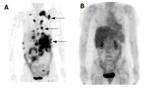
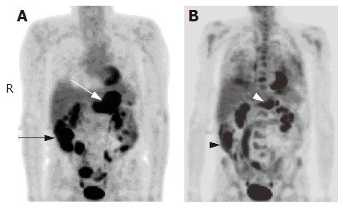
In high-grade NHL, 18F-FDG PET showed focal nodular or diffuse hypermetabolic activity, which involved mainly the wall of the gastrointestinal tract. This activity’s appearance was different from that of the normal activity in the bowel wall, which was less intense and uniform. Fifteen patients received 18F-FDG PET for staging before the treatment. In these patients, 18F-FDG PET identified 30 (100%) of 30 intestinal locations that had biopsy-proven lymphomatous involvement. These locations were the esophagus (n = 2), stomach (n = 11), duodenum (n = 5), small bowel (n = 6), cecum/terminal ileum (n = 3), and colon (n = 3). The SUVmax ranged from 3.64 to 25.10 (11.22±5.79). The SUVmax±SD in non-involved intestine was 3.09±2.34. After the therapy, uptake was absent or reduced without focal lesions in 14 patients who had complete responses and whose biopsies showed benign inflammation without evidence of lymphoma (Figure 1). The post- treatment SUVmax values were significantly lower (P<0.05), ranging from 0.89 to 4.30 (2.21±0.78). In three patients, the post-treatment biopsies still showed lymphoma (Figure 2) and the post-treatment SUVmax ranged from 3.44 to 21.90 (9.42±6.27).
Figure 1.

A 61-year-old woman with large cell lymphoma. A: Pre-treatment 18F-FDG PET scan revealed multifocal hypermetabolic activity involving the neck, chest, and abdomen (arrow). The highest SUVmax was 20.3 in the left upper abdominal area, corresponding to a positive result of a gastric biopsy; B: Follow-up 18F-FDG PET scan 5 mo later showed no evidence of residual FDG disease.
Figure 2.

A 68-year-old man with large cell lymphoma. A: Base line 18F-FDG PET scan showed hypermetabolic foci in the stomach (white arrow), cecum and terminal ileum (black arrow), and bowel loops (SUVmax = 12.7); B: 18F-FDG PET scan obtained after therapy showed partial metabolic response of the activity in the stomach (white arrowhead) and cecum (black arrowhead, SUVmax = 10.7).
In low-grade NHL, 18F-FDG PET showed diffuse hypermetabolic activity in the bowel wall in two patients with follicular lymphoma of the colon and stomach (SUVmax 8.20 and 10.30, respectively). In one patient with follicular lymphoma of the stomach, the SUVmax after 5 wk of therapy decreased from 10.30 to 2.40. In four patients with MALT-type lymphoma (Figure 3), lesions in the stomach and duodenum showed diffuse and low metabolic activity similar to that in the liver, and the SUVmax was 2.02-3.8 (average 3.02) at the time of the positive biopsy results. In another patient with MALT-type lymphoma involving the colon, the SUVmax was 6.82. However, follow-up biopsy in this patient showed high-grade transformation. The mean SUVmax in high-grade and low-grade NHL before and after the treatment is shown in Table 2. Negative post-treatment biopsy results corresponded to significantly decreased SUVmax in high-grade NHL (P<0.05). However, the SUVmax before and after the treatment was not significantly lower in low-grade NHL.
Figure 3.

An 80-year-old man with MALT-type lymphoma. A-C: 18F-FDG PET scans in transaxial and projection images showed a focal mild hypermetabolic activity (SUVmax = 3.8) in the region of stomach (black arrow); D and E: CT scans showed a bulky mass in the wall of gastric fundus and body (white arrow).
Table 2.
SUVmax in high- and low-grade NHL before and after treatment (mean±SD)
| NHL | SUVmax | SUVmax | |
| grade | before treatment |
after treatment |
|
| Biopsy positive | Biopsy negative | ||
| High grade | 11.22±5.79 | 9.42±6.27 | 2.21±0.78ac |
| (n = 15) | (n = 14) | (n = 3) | |
| Low grade | 8.57±2.47 | 3.76±2.08 | 3.03±0.89 |
| (n = 2) | (n = 5) | (n = 2) | |
n, number of patients.
P<0.05 vs SUVmax in high grade NHL before treatment;
P<0.05 vs SUVmax with biopsy positive after treatment.
DISCUSSION
The gastrointestinal tract is the most common extranodal site of NHL[1,3]. The stomach is most frequently involved (60-74% of cases), followed by the duodenum and small bowel (10-20%), ileocecal region (7-10%) and large bowel (<10%)[26,27]. Our results are consistent with these reports, except that the large bowel was involved in a greater percentage of patients in our study than the previous studies. Lymphomatous involvement of the gastrointestinal tract may be primary or secondary. The gastrointestinal tract is involved at autopsy in as many as 50% of patients with secondary lymphoma. However, most of these patients have subclinical disease while patients who have gastrointestinal symptoms are found to have primary gastrointestinal lymphoma[6,28].
Diagnostic imaging studies play an important role in documenting lymphoma, staging and re-staging the disease, evaluating treatment response and performing follow-up evaluations. Anatomical imaging modalities including computed tomography (CT) and magnetic resonance (MR) imaging have some limitations, especially when defining the viability of the residual mass, treatment response or both[4,29]. Gallium-67 scintigraphy has been proposed as a functional imaging modality to assess remission and to evaluate the nature of residual masses in patients with lymphoma. However, gallium-67 scintigraphy is of little use in the abdomen because of the high hepatic uptake and excretion into the bowel and should be performed before treatment to determine whether the patient has a gallium-fixing tumor and whether the absence of fixation after treatment corresponds to a residual mass[8,9,30]. 18F-FDG PET imaging has shown its clinical usefulness in patients with lymphoma. Several articles have reported that 18F-FDG PET has a higher sensitivity in detecting disease sites than gallium-67 scintigraphy[11-13]. 18F-FDG PET can also be used to assess the response after the therapy[16,17,31]. However, a limited number of studies have shown the utility of 18F-FDG PET imaging for the evaluation of lymphomatous involvement of the gastrointestinal tract[18-21,32,33].
In our study, 18F-FDG PET showed fixed focal nodular or diffuse hypermetabolic activity of all lesions in patients with high-grade non-Hodgkin's lymphoma, which was confirmed by histopathologic analysis. The appearance of focal intense hypermetabolic activity of the lesions was different from that of the normal activity in the bowel wall. Rodriguez et al[4,18] evaluated CT, MRI, and PET in eight patients with primary gastric lymphoma and showed that 18F-FDG PET can demonstrate both the presence and the extent of gastric NHL and is more accurate than endoscopy and CT for evaluating the extent of NHL in the gastric wall. In our study, 18F-FDG PET clearly demonstrated the involvement of specific sites, particularly in the stomach (e.g. the fundus, body, antrum, lesser and greater curvature of the stomach), corresponding to endoscopic biopsy sites. However, extension outside the bowel wall was better appreciated when interpreted with the guidance of a CT scan. It should be noted that an evaluation of the extent of NHL in the gastric wall and careful assessment of multiplicities affect the selection of therapy, since radical surgery with lymph node dissection seems to be needed for most patients with high-grade and even low-grade NHL[34].
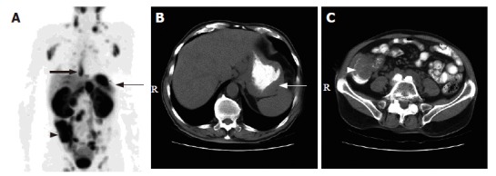
Ullerich et al[20] and Sam et al[21] showed that 18F-FDG PET could find lesions in patients with small bowel lymphoma. Najjar et al[32] also reported that 18F-FDG PET could identify lymphoma of the colon. These results are consistent with the results in our study, in which pre-treatment 18F-FDG PET showed pertinently high metabolic activity in the small bowel and colon in patients with high-grade NHL (Figure 4). It is difficult to differentiate high-grade non-Hodgkin's lymphoma from other neoplastic or inflammatory diseases. However, interpreting the results in the light of a careful clinical history, the extent of the disease and corresponding CT images, can contribute to the correct diagnosis. Moog et al[33] showed that 18F-FDG PET imaging could achieve 100% correct diagnoses of malignant lymphoma. In almost all cases of high-grade NHL in our study, CT scans also demonstrated the abnormal wall thickening or mass lesions. However, the results did not reveal the sensitivity of 18F-FDG PET vs that of CT, because both modalities were interpreted together as complementary studies.
Figure 4.

A 78-year-old man with mantle cell lymphoma. A: 18F-FDG PET scan revealed multiple hypermetabolic foci involving the lower esophagus (thick arrow), gastric fundus (thin arrow), cecum and terminal ileum (arrow head); B and C: The corresponding CT scans of the abdomen showed wall thickening at the gastric fundus (white arrow) and the cecum and terminal ileum (white arrow head)
18F-FDG PET can demonstrate diffuse hypermetabolic activity in patients with low-grade follicular NHL in the colon and stomach. The ability of gallium-67 scintigraphy and 18F-FDG PET to detect MALT-type lymphoma of the gastrointestinal tract is controversial. Hsu et al[35] showed that gallium-67 scintigraphy could not show abnormal uptake of radioactivity in patients with low-grade gastric MALT-type lymphoma. Hoffmann et al[19] studied 18F-FDG PET imaging in patients with MALT-type lymphoma and found that PET scans do not show focal tracer uptake in either gastric or extragastric lesions. Our study showed similar results of low activity in the stomach and duodenum. Even though it has been reported that CT scans may demonstrate gastric MALT-type lymphoma either as an infiltrative form or as a polypoid pattern[36,37], CT is limited in its ability to monitor the treatment response. Taken together, 18F-FDG PET imaging in low-grade NHL seems to be able to monitor the treatment response and determine high-grade transformation.
Generally, in patients with NHL, histologic grade and disease stage are clearly identified as the two major prognostic factors and therapeutic determinants. Endoscopy with biopsy and histopathologic examination are the gold standard for grading malignancies[38]. However, there have been attempts to differentiate high-grade from low-grade NHL by different methods (e.g. double-contrast radiography, CT scan and gallium-67 scintigraphy)[35,39]. 18F-FDG PET imaging in our study showed that high-grade NHL of the gastrointestinal tract had high FDG activity in the lesions as was confirmed by high SUV measurements. Though the normal background FDG activity in the gastrointestinal tract was known, abnormalities were identified in this study. In low-grade NHL of the gastrointestinal tract, it was difficult to document existing disease, especially in patients with MALT-type lymphoma. However, there was a significant difference in the SUVmax measurement between high-grade and low-grade NHL.
This study also demonstrated the clinical utility of 18F-FDG PET imaging for monitoring patients with NHL of the gastrointestinal tract especially those with high-grade lymphoma. The SUVmax measurements were significantly decreased after therapy and post-treatment biopsies were negative for lymphoma. In addition, benign inflammatory conditions such as gastritis showed significantly lower FDG activity. In patients whose post-treatment biopsies were positive for lymphoma, the SUVmax measurements were persistently high and even higher in patients with disease progression. Many studies have demonstrated that persistent FDG uptake after therapy may help to predict treatment failure or a high risk of recurrence[16,17].
In conclusion, 18F-FDG PET contributes to the diagnosis of high-grade lymphoma involving the gastrointestinal tract, even when there is the normal background FDG activity. Furthermore, the SUV plays a role in evaluating treatment response especially when the PET images are interpreted with CT scans. Low-grade lymphoma demonstrates FDG uptake, but the intensity of the uptake is lower than that in high-grade lymphoma.
ACKNOWLEDGMENTS
The authors thank Beth Wagner for manuscript preparation and literature search and Mariann Crapanzano for editorial review.
Footnotes
Science Editor Guo SY Language Editor Elsevier HK
References
- 1.Banfi A, Bonadonna G, Carnevali G, Oldini C, Salvini E. Preferential sites of involvement and spread in maignant lymphomas. Eur J Cancer. 1968;4:319–324. doi: 10.1016/0014-2964(68)90058-3. [DOI] [PubMed] [Google Scholar]
- 2.Gospodarowicz MK, Sutcliffe SB, Brown TC, Chua T, Bush RS. Patterns of disease in localized extranodal lymphomas. J Clin Oncol. 1987;5:875–880. doi: 10.1200/JCO.1987.5.6.875. [DOI] [PubMed] [Google Scholar]
- 3.Amer MH, el-Akkad S. Gastrointestinal lymphoma in adults: clinical features and management of 300 cases. Gastroenterology. 1994;106:846–858. doi: 10.1016/0016-5085(94)90742-0. [DOI] [PubMed] [Google Scholar]
- 4.Rodriguez M. Computed tomography, magnetic resonance imaging and positron emission tomography in non-Hodgkin's lymphoma. Acta Radiol Suppl. 1998;417:1–36. [PubMed] [Google Scholar]
- 5.Kaye MD, Young SW, Hayward R, Castellino RA. Gastric pseudotumor on CT scanning. AJR Am J Roentgenol. 1980;135:190–193. doi: 10.2214/ajr.135.1.190. [DOI] [PubMed] [Google Scholar]
- 6.Levine MS, Rubesin SE, Pantongrag-Brown L, Buck JL, Herlinger H. Non-Hodgkin's lymphoma of the gastrointestinal tract: radiographic findings. AJR Am J Roentgenol. 1997;168:165–172. doi: 10.2214/ajr.168.1.8976941. [DOI] [PubMed] [Google Scholar]
- 7.Komaki S. Normal or benign gastric wall thickening demonstrated by computed tomography. J Comput Assist Tomogr. 1982;6:1103–1107. doi: 10.1097/00004728-198212000-00009. [DOI] [PubMed] [Google Scholar]
- 8.Adler S, Parthasarathy KL, Bakshi SP, Stutzman L. Gallium-67-citrate scanning for the localization and staging of lymphomas. J Nucl Med. 1975;16:255–260. [PubMed] [Google Scholar]
- 9.Brown ML, O'Donnell JB, Thrall JH, Votaw ML, Keyes JW. Gallium-67 scintigraphy in untreated and treated non-hodgkin lymphomas. J Nucl Med. 1978;19:875–879. [PubMed] [Google Scholar]
- 10.Coiffier B. Positron emission tomography and gallium metabolic imaging in lymphoma. Curr Oncol Rep. 2001;3:266–270. doi: 10.1007/s11912-001-0060-1. [DOI] [PubMed] [Google Scholar]
- 11.Paul R. Comparison of fluorine-18-2-fluorodeoxyglucose and gallium-67 citrate imaging for detection of lymphoma. J Nucl Med. 1987;28:288–292. [PubMed] [Google Scholar]
- 12.Shen YY, Kao A, Yen RF. Comparison of 18F-fluoro-2-deoxyglucose positron emission tomography and gallium-67 citrate scintigraphy for detecting malignant lymphoma. Oncol Rep. 2002;9:321–325. [PubMed] [Google Scholar]
- 13.Kostakoglu L, Leonard JP, Kuji I, Coleman M, Vallabhajosula S, Goldsmith SJ. Comparison of fluorine-18 fluorodeoxyglucose positron emission tomography and Ga-67 scintigraphy in evaluation of lymphoma. Cancer. 2002;94:879–888. [PubMed] [Google Scholar]
- 14.Jerusalem G, Beguin Y, Fassotte MF, Najjar F, Paulus P, Rigo P, Fillet G. Whole-body positron emission tomography using 18F-fluorodeoxyglucose for posttreatment evaluation in Hodgkin's disease and non-Hodgkin's lymphoma has higher diagnostic and prognostic value than classical computed tomography scan imaging. Blood. 1999;94:429–433. [PubMed] [Google Scholar]
- 15.Stumpe KD, Urbinelli M, Steinert HC, Glanzmann C, Buck A, von Schulthess GK. Whole-body positron emission tomography using fluorodeoxyglucose for staging of lymphoma: effectiveness and comparison with computed tomography. Eur J Nucl Med. 1998;25:721–728. doi: 10.1007/s002590050275. [DOI] [PubMed] [Google Scholar]
- 16.Jerusalem G, Beguin Y, Fassotte MF, Najjar F, Paulus P, Rigo P, Fillet G. Persistent tumor 18F-FDG uptake after a few cycles of polychemotherapy is predictive of treatment failure in non-Hodgkin's lymphoma. Haematologica. 2000;85:613–618. [PubMed] [Google Scholar]
- 17.Spaepen K, Stroobants S, Dupont P, Van Steenweghen S, Thomas J, Vandenberghe P, Vanuytsel L, Bormans G, Balzarini J, De Wolf-Peeters C, et al. Prognostic value of positron emission tomography (PET) with fluorine-18 fluorodeoxyglucose ([18F]FDG) after first-line chemotherapy in non-Hodgkin's lymphoma: is [18F]FDG-PET a valid alternative to conventional diagnostic methods? J Clin Oncol. 2001;19:414–419. doi: 10.1200/JCO.2001.19.2.414. [DOI] [PubMed] [Google Scholar]
- 18.Rodriguez M, Ahlström H, Sundín A, Rehn S, Sundström C, Hagberg H, Glimelius B. [18F] FDG PET in gastric non-Hodgkin's lymphoma. Acta Oncol. 1997;36:577–584. doi: 10.3109/02841869709001319. [DOI] [PubMed] [Google Scholar]
- 19.Hoffmann M, Kletter K, Diemling M, Becherer A, Pfeffel F, Petkov V, Chott A, Raderer M. Positron emission tomography with fluorine-18-2-fluoro-2-deoxy-D-glucose (F18-FDG) does not visualize extranodal B-cell lymphoma of the mucosa-associated lymphoid tissue (MALT)-type. Ann Oncol. 1999;10:1185–1189. doi: 10.1023/a:1008312726163. [DOI] [PubMed] [Google Scholar]
- 20.Ullerich H, Franzius CH, Domagk D, Seidel M, Sciuk J, Schober W. 18F-Fluorodeoxyglucose PET in a patient with primary small bowel lymphoma: the only sensitive method of imaging. Am J Gastroenterol. 2001;96:2497–2499. doi: 10.1111/j.1572-0241.2001.04061.x. [DOI] [PubMed] [Google Scholar]
- 21.Sam JW, Levine MS, Farner MC, Schuster SJ, Alavi A. Detection of small bowel involvement by mantle cell lymphoma on F-18 FDG positron emission tomography. Clin Nucl Med. 2002;27:330–333. doi: 10.1097/00003072-200205000-00003. [DOI] [PubMed] [Google Scholar]
- 22.Cook GJ, Maisey MN, Fogelman I. Normal variants, artefacts and interpretative pitfalls in PET imaging with 18-fluoro-2-deoxyglucose and carbon-11 methionine. Eur J Nucl Med. 1999;26:1363–1378 DOI : 10.1007/s002590050597. doi: 10.1007/s002590050597. [DOI] [PubMed] [Google Scholar]
- 23.Shreve PD, Anzai Y, Wahl RL. Pitfalls in oncologic diagnosis with FDG PET imaging: physiologic and benign variants. Radiographics. 1999;19:61–77; quiz 150-151. doi: 10.1148/radiographics.19.1.g99ja0761. [DOI] [PubMed] [Google Scholar]
- 24.Meyer MA. Diffusely increased colonic F-18 FDG uptake in acute enterocolitis. Clin Nucl Med. 1995;20:434–435. doi: 10.1097/00003072-199505000-00012. [DOI] [PubMed] [Google Scholar]
- 25.Kresnik E, Mikosch P, Gallowitsch HJ, Heinisch M, Lind P. F-18 fluorodeoxyglucose positron emission tomography in the diagnosis of inflammatory bowel disease. Clin Nucl Med. 2001;26:867. doi: 10.1097/00003072-200110000-00015. [DOI] [PubMed] [Google Scholar]
- 26.Kolve ME, Fischbach W, Wilhelm M. Primary gastric non-Hodgkin's lymphoma: requirements for diagnosis and staging. Recent Results Cancer Res. 2000;156:63–68. doi: 10.1007/978-3-642-57054-4_8. [DOI] [PubMed] [Google Scholar]
- 27.Koch P, Grothaus-Pinke B, Hiddemann W, Willich N, Reers B, del Valle F, Bodenstein H, Pfreundschuh M, Möller E, Kocik J, et al. Primary lymphoma of the stomach: three-year results of a prospective multicenter study. The German Multicenter Study Group on GI-NHL. Ann Oncol. 1997;8 Suppl 1:85–88. [PubMed] [Google Scholar]
- 28.Ehrlich AN, Stalder G, Geller W, Sherlock P. Gastrointestinal manifestations of malignant lymphoma. Gastroenterology. 1968;54:1115–1121. [PubMed] [Google Scholar]
- 29.Coiffier B. How to interpret the radiological abnormalities that persist after treatment in non-Hodgkin's lymphoma patients? Ann Oncol. 1999;10:1141–1143. doi: 10.1023/a:1008308129857. [DOI] [PubMed] [Google Scholar]
- 30.Kaplan WD, Jochelson MS, Herman TS, Nadler LM, Stomper PC, Takvorian T, Andersen JW, Canellos GP. Gallium-67 imaging: a predictor of residual tumor viability and clinical outcome in patients with diffuse large-cell lymphoma. J Clin Oncol. 1990;8:1966–1970. doi: 10.1200/JCO.1990.8.12.1966. [DOI] [PubMed] [Google Scholar]
- 31.Zinzani PL, Magagnoli M, Chierichetti F, Zompatori M, Garraffa G, Bendandi M, Gherlinzoni F, Cellini C, Stefoni V, Ferlin G, et al. The role of positron emission tomography (PET) in the management of lymphoma patients. Ann Oncol. 1999;10:1181–1184 DOI : 10.1023/A: 1008327127033. doi: 10.1023/a:1008327127033. [DOI] [PubMed] [Google Scholar]
- 32.Najjar F, Hustinx R, Jerusalem G, Fillet G, Rigo P. Positron emission tomography (PET) for staging low-grade non-Hodgkin's lymphomas (NHL) Cancer Biother Radiopharm. 2001;16:297–304. doi: 10.1089/108497801753131372. [DOI] [PubMed] [Google Scholar]
- 33.Moog F, Bangerter M, Diederichs CG, Guhlmann A, Merkle E, Frickhofen N, Reske SN. Extranodal malignant lymphoma: detection with FDG PET versus CT. Radiology. 1998;206:475–481. doi: 10.1148/radiology.206.2.9457202. [DOI] [PubMed] [Google Scholar]
- 34.Kong SH, Kim MA, Park DJ, Lee HJ, Lee HS, Kim CW, Yang HK, Heo DS, Lee KU, Choe KJ. Clinicopathologic features of surgically resected primary gastric lymphoma. World J Gastroenterol. 2004;10:1103–1109. doi: 10.3748/wjg.v10.i8.1103. [DOI] [PMC free article] [PubMed] [Google Scholar]
- 35.Hsu CH, Sun SS, Kao CH, Lin CC, Lee CC. Differentiation of low-grade gastric MALT lymphoma and high-grade gastric MALT lymphoma: the clinical value of Ga-67 citrate scintigraphy--a pilot study. Cancer Invest. 2002;20:939–943 DOI : 10.1081/CNV-120005908. doi: 10.1081/cnv-120005908. [DOI] [PubMed] [Google Scholar]
- 36.Brown JA, Carson BW, Gascoyne RD, Cooperberg PL, Connors JM, Mason AC. Low grade gastric MALT Lymphoma: radiographic findings. Clin Radiol. 2000;55:384–389. doi: 10.1053/crad.2000.0449. [DOI] [PubMed] [Google Scholar]
- 37.Kessar P, Norton A, Rohatiner AZ, Lister TA, Reznek RH. CT appearances of mucosa-associated lymphoid tissue (MALT) lymphoma. Eur Radiol. 1999;9:693–696. doi: 10.1007/s003300050734. [DOI] [PubMed] [Google Scholar]
- 38.Skarin AT, Dorfman DM. Non-Hodgkin's lymphomas: current classification and management. CA Cancer J Clin. 1997;47:351–372. doi: 10.3322/canjclin.47.6.351. [DOI] [PubMed] [Google Scholar]
- 39.Park MS, Kim KW, Yu JS, Park C, Kim JK, Yoon SW, Lee KH, Ryu YH, Kim H, Kim MJ, et al. Radiographic findings of primary B-cell lymphoma of the stomach: low-grade versus high-grade malignancy in relation to the mucosa-associated lymphoid tissue concept. AJR Am J Roentgenol. 2002;179:1297–1304. doi: 10.2214/ajr.179.5.1791297. [DOI] [PubMed] [Google Scholar]


