Abstract
AIM: To study the relationship between the expression of human chorionic gonadotropin (HCG), CD44v6, CD44v4/5 and the infiltration, metastasis of esophageal squamous cell carcinoma.
METHODS: By labeled streptavidin-biotin technique, the expressions of HCG, CD44v6, and CD44v4/5 in 42 patients with esophageal squamous cell carcinoma were examined.
RESULTS: The positive rate of HCG expression in patients with lymph node metastasis was 85.71% (18/21), higher than that (57.14%, 12/21) in those without lymph node metastasis (P<0.05). The positive rate of CD44v6 expression was 71.43% (15/21) in lymph node metastasis group, and 38.09% (8/21) in non-metastasis group; there was a significant difference between the two groups (P<0.05). The positive rate of CD44v4/5 expression was 76.19% (16/21) in lymph node metastasis group, and 42.86% (9/21) in non-metastasis group; there was also a significant difference between them (P<0.05). From grade I to grade III in differentiation, the positive rate of HCG expression was 84.62% (11/13), 70.59% (12/17) and 58.33% (7/12), respectively; there was no significant difference among them (P>0.05). The positive rate of CD44v6 expression in grades I-III of cancer tissues was 76.92% (10/13), 52.94% (9/17), and 33.33% (4/12) respectively; there was no significant difference among them. The positive rate of CD44v4/5 expression in grades I-III of cancer tissues was 69.23% (9/13), 64.71% (11/17), and 41.67% (5/12) respectively; there was no significant difference among the three groups. There was no correlation between the positive rates of HCG and CD44v6, CD44v4/5 expression. Cancer cells in carcinomatous emboli and those infiltrating into vascular wall strongly expressed HCG, CD44v6, and CD44v4/5.
CONCLUSION: Expression of HCG, CD44v6, and CD44v4/5 in esophageal squamous cell carcinoma is related to its infiltration and metastasis. HCG, CD44v6, and CD44v4/5 have different effects on the infiltration and metastasis of esophageal squamous cell carcinoma.
Keywords: Esophageal tumor, Squamous cell carcinomas, HCG, CD44v6, CD44v4/5, Immunohistochemistry, Infiltration, Metastasis
INTRODUCTION
Human chorionic gonadotropin (HCG) is a glycoprotein molecule. Ectopic HCG produced by malignant tumors is one of the indexes of early diagnosis[1]. Researchers have studied the expression of HCG in malignant tumors, such as tumor of lung, bladder, breast[1-4], and found that HCG expression has a close correlation with the differentiation, infiltration, and metastasis of tumors. Expression of CD44 gene and its relation with metastasis of tumors are the hotspots of tumor study in recent years[5]. It has been found that expressions of mutant CD44 molecules exist in many tumors, such as stomach, lung, and cervix tumors, and have an obvious relation with metastasis of tumors[6-8]. In order to explore the relationship between the expression of HCG, CD44v6, and CD44v4/5 and the infiltration and metastasis of esophageal carcinoma, labeled streptavidin-biotin (LSAB) was used to detect the expression of these three proteins in 42 patients with esophageal squamous cell carcinoma.
MATERIALS AND METHODS
Selection of cases
Specimens were excised from 42 esophageal carcinoma patients in Tumour Hospital of Anyang city. There were 23 male patients and 19 female patients. Their age range was 33-73 years, averaged 55.7 years. None of the patients received chemotherapy, radiation therapy, or immunotherapy before surgery. All the formalin-fixed and paraffin-embedded specimens were sectioned and stained with hematoxylin-eosin. The sections were carefully diagnosed under light microscope. All the specimens excised from the patients proved to be squamous cell carcinoma by histopathology and 13 cases were grade I, 17 cases were grade II, and 12 cases were grade III. Lymph node metastasis was found in 21 cases.
Detection of HCG, CD44v6, and CD44v4/5 proteins
Immunohistochemical LSAB technique was applied. Rabbit anti-human HCG antibody was purchased from DAKO (Denmark), mouse anti-human CD44v6 and CD44v4/5 mAb was the product of R&D (USA). LSAB immunohistochemical reagent kit was purchased from Zymed (USA). Human placental chorion was taken from pregnant women as HCG positive control. Squamous basal cells were taken from normal people as CD44v6 and CD44v4/5 positive control. Antibody I was replaced with normal goat serum as negative control group. Antibody I was replaced with PBS as blank negative control group.
The criteria were established as previously described[9]. The positive cells were stained brown-yellow. The positive expression of HCG mainly displayed on cytoplasm, while CD44v6 and CD44v4/5 mainly appeared on cell membrane. The positive cells of 10 high power fields were counted and the positive expression was categorized as follows: weakly positive (+), with positive cells less than 10%; moderately positive (++), with positive cells 10-50%; strongly positive (+++), with positive cells more than 50%.
Statistical analysis
Microsoft SPSS10.0 was applied. Comparison between two and multiple specimens was made by χ2 test. Difference and correlation were analyzed by coupled χ2 test and Spearman's correlation test.
RESULTS
Distribution features of HCG and CD44v6, CD44v4/5 positive cells
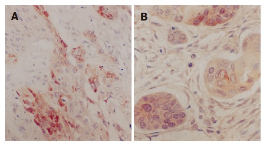
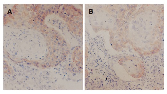
There were three patterns in the distribution of HCG and CD44v6, CD44v4/5 positive cells, namely spot, focal and diffuse distribution. In squamous cell carcinoma tissues of grade I, the distribution of HCG and CD44v6 was focal and diffuse (Figures 1A, 1B and 2A), while that of CD44v4/5 was spot, focal or diffuse. In grade II, the distribution of HCG, CD44v6, and CD44v4/5 was focal and diffuse (Figure 2B). In grade III, staining of HCG was focal or diffuse, while CD44v6 was multi-focal, CD44v4/5 was focal or spot. In carcinoma tissues with lymph node metastasis, the distribution of HCG and CD44v6, CD44v4/5 was multi-focal. The expression of keratinous pearl in grade I was negative. Tumor cells in emboli or tumor cells invading the vascular wall were frequently stained and highly positive for HCG, CD44v6, and CD44v4/5. The tumor cells at periphery of carcinoma nests with interstitial or intermuscular infiltration and mitosis were highly positive for CD44v6, CD44v4/5.
Figure 1.

Spot or focus (A) and diffuse (B) distribution of HCG positive cells.
Figure 2.

Diffuse distribution of CD44v6 (A) and CD44v4/5 (B) positive cells.
Correlation between expression of HCG, CD44v6, CD44v4/5, and differentiation degree, lymph node metastasis of esophageal squamous cell carcinoma
The correlations between expressions of HCG, CD44v6, CD44v4/5 and differentiation degree, lymph node metastasis of esophageal squamous cell carcinoma are listed in Table 1, Table 2 and Table 3.
Table 1.
Correlation between expression of HCG and differentiation degree, lymph node metastasis of esophageal squamous cell carcinoma
| Pathologic characteristics | n |
Positive expression |
Positive rates (%) | χ2 | P | |||
| - | + | ++ | +++ | |||||
| Differentiation | ||||||||
| Degree | ||||||||
| I | 13 | 2 | 3 | 3 | 5 | 84.62 | ||
| II | 17 | 5 | 4 | 2 | 6 | 70.59 | 2.122 | >0.05 |
| III | 12 | 5 | 2 | 3 | 2 | 58.33 | ||
| Lymph node metastasis | ||||||||
| Positive | 21 | 3 | 5 | 4 | 9 | 85.71 | ||
| Negative | 21 | 9 | 4 | 4 | 4 | 57.14 | 4.2 | <0.05 |
| Tumor focus | ||||||||
| Primary | 21 | 3 | 5 | 5 | 8 | 85.71 | ||
| Metastasis | 21 | 4 | 7 | 6 | 4 | 80.95 | 0.171 | >0.05 |
Table 2.
Correlation between expression of CD44v6 and differentiation degree, lymph node metastasis of esophageal squamous cell carcinoma
| Pathologic characteristics | n |
Positive expression |
Positive rates (%) | χ2 | P | |||
| - | + | ++ | +++ | |||||
| Differentiation | ||||||||
| Degree | ||||||||
| I | 13 | 3 | 3 | 4 | 3 | 76.92 | ||
| II | 17 | 8 | 5 | 1 | 3 | 52.94 | 4.788 | >0.05 |
| III | 12 | 8 | 2 | 2 | 0 | 33.33 | ||
| Lymph node metastasis | ||||||||
| Positive | 21 | 6 | 7 | 3 | 5 | 71.43 | ||
| Negative | 21 | 13 | 5 | 3 | 0 | 38.09 | 4.709 | <0.05 |
| Tumor focus | ||||||||
| Primary | 21 | 6 | 7 | 2 | 6 | 71.43 | ||
| Metastasis | 21 | 8 | 7 | 4 | 2 | 61.9 | 0.429 | >0.05 |
Table 3.
Correlation between expression of CD44v4/5 and differentiation degree, lymph node metastasis of esophageal squamous cell carcinoma
| Pathologic characteristics | n |
Positive expression |
Positive rates (%) | χ2 | P | |||
| - | + | ++ | +++ | |||||
| Differentiation | ||||||||
| Degree | ||||||||
| I | 13 | 4 | 5 | 3 | 1 | 69.23 | ||
| II | 17 | 6 | 7 | 3 | 1 | 64.71 | 2.286 | >0.25 |
| III | 12 | 7 | 4 | 1 | 0 | 41.67 | ||
| Lymph node metastasis | ||||||||
| Positive | 21 | 5 | 10 | 4 | 2 | 76.19 | ||
| Negative | 21 | 12 | 6 | 3 | 0 | 42.86 | 4.842 | <0.05 |
| Tumor focus | ||||||||
| Primary | 21 | 5 | 10 | 4 | 2 | 76.19 | ||
| Metastasis | 21 | 6 | 12 | 3 | 0 | 71.43 | 0.123 | >0.5 |
Expression of HCG, CD44v6, and CD44v4/5 in primary and metastatic carcinoma
According to Tables 1-3, if the expression of HCG, CD44v6, and CD44v4/5 was positive in primary tumor, it could be negative in metastasis tumor. If both of them were positive, the expression was similar or lower in metastasis tumor compared to primary tumor, but there was no statistically significant difference.
Positive rate of HCG, CD44v6, and CD44v4/5 expressions in esophageal squamous cell carcinoma
The positive rate of HCG, CD44v6, and CD44v4/5 expression was 71.43%, 54.76%, and 59.52% respectively. There was no significant difference among them (P>0.05), nor was there any correlation (P>0.05).
DISCUSSION
HCG is a glycoprotein hormone. Recent studies found that HCG can be expressed in many tumors, such as the tumor of lung[2], bladder[3], breast[4], HCG is related with tumor differentiation degree, infiltration and metastasis. According to our study, there is a significant difference between lymph node metastasis and non-metastasis groups in terms of the positive expression of HCG, indicating that HCG plays an important role in lymphatic metastasis of esophageal squamous cell carcinoma. The stained tumor cells in emboli or invading the vascular wall were highly positive for HCG, suggesting that HCG also has a relationship with hematogenous metastasis of esophageal squamous cell carcinoma. Most tumor cells with positive expression of HCG in esophageal squamous cell carcinoma were well differentiated and had plenty of cytoplasm. The expression of HCG was mainly in central tumor cells of carcinoma nests, and in peripheral cells. But the keratinous pearls were not stained. All of these are in accordance with the study of Li[2] on lung squamous cell carcinoma. The expression of HCG was mainly in cytoplasm, and the amount of cytoplasm of the central tumor cells was more than that in periphery of carcinoma nests, suggesting that the different expression levels of HCG of these two types of carcinoma cells result from different amounts of cytoplasm. The tumor cells with positive expression of HCG infiltrated the vascular wall, suggesting that the expression of HCG has some relationship with infiltration and metastasis of esophageal squamous cell carcinoma.
Previously it was found that the expression of HCG is negatively related with the differentiation degree of carcinoma of bladder[3] and squamous cell carcinoma of lung[2]. These discrepancies may be due to the following reasons. Types of the tumor tissues are different (the difference between squamous cell carcinoma and transitional cell carcinoma). The expression of HCG is mainly in cytoplasm, while the quantity of cytoplasm is correlated with the differentiation degree of tumor cells (there is more cytoplasm in squamous cell carcinoma of grade I, less in grade II, and the least in grade III). The positive rate of HCG in the 42 cases of esophageal squamous cell carcinoma (71.42%) was much higher than that in carcinoma of bladder[3], and also higher than that in squamous cell carcinoma of the lung (60%)[2]. All of these indicate that HCG may become a new and more sensitive biological marker.
There was a close correlation between the expression of CD44v6, CD44v4/5, and the infiltration and metastasis of tumors. The study showed that the tumor cells at the periphery of carcinoma nest with interstitial or intermuscular infiltration and the stained tumor cells with mitosis were highly positive for CD44v6 and CD44v4/5, suggesting that CD44v6 and CD44v4/5 play a role in the infiltration of esophageal squamous cell carcinoma. The results are consistent with other studies[6-8]. There was a significant difference in the positive rate of CD44v6, CD44v4/5 expression between lymph node metastasis group and non-metastasis group, indicating a correlation between the positive expression of CD44v6, CD44v4/5, and lymph node metastasis of esophageal squamous cell carcinoma. The stained tumor cells in the emboli or invading the vascular wall were highly positive for CD44v6 and CD44v4/5, suggesting that there is some relationship between the expression and risk of hematogenous metastasis of human esophageal squamous cell carcinoma.
The correlation between CD44v and metastasis of tumors was identified, while studies on relationship between CD44v and differentiation are few. This study showed that the expression rate of CD44v6, CD44v4/5 reduced with decline of the differentiation degree of esophageal squamous cell carcinoma. Yokozaki et al.[10] reported that if the differentiation of carcinoma is poor, the expression of CD44v is much lower.
There was no significant difference or any correlation among HCG, CD44v6, and CD44v4/5 in the 42 cases of esophageal squamous cell carcinoma. All these indicate that although HCG, CD44v6 and CD44v4/5 are related with the infiltration and metastasis of esophageal squamous cell carcinoma, their pathogenesis may be different.
Footnotes
Science Editor Zhu LH and Li WZ Language Editor Elsevier HK
References
- 1.Regelson W. Have we found the "definitive cancer biomarker"? The diagnostic and therapeutic implications of human chorionic gonadotropin-beta expression as a key to malignancy. Cancer. 1995;76:1299–1301. doi: 10.1002/1097-0142(19951015)76:8<1299::aid-cncr2820760802>3.0.co;2-l. [DOI] [PubMed] [Google Scholar]
- 2.Li DC, Zhou XL, Cao GQ, Li BQ. Carcinoma of lung and human chorionic gonadotropin. Zhonghua Binglixue Zazhi. 1993;22:181. [Google Scholar]
- 3.Huang JX, Zhang YH, Lin Y, Qiu QC. The expression and significance of HCG in transitional-cell carcinoma of bladder. Shantou Daxue Yixueyuan Xuebao. 2001;14:20–21. [Google Scholar]
- 4.Chi F, Sun BC, Zhao XL, Yan YH. The expression of ectopic HCG in breast cancer and its significance. Aizheng. 2000;19:493,503. [Google Scholar]
- 5.Guo LX, Xie H. CD44 and the metastasis of tumor. Shengming Kexue. 2001;13:60–63. [Google Scholar]
- 6.Zhou Y, Zong H, Xu H. Expression of CD44v6 in gastric cancer and its adjacent cancer tissues. Zhengzhou Daxue Xuebao. 2002;37:593–595. [Google Scholar]
- 7.Qiu XF, Wang XY, Zhang ZF, Zhang CP. CD44v6 expression in non-small cell lung carcinomas. Linchuang Yu Shiyan Binglixue Zazhi. 2003;19:413–415. [Google Scholar]
- 8.Chen YQ, Zheng AW, Zhang G. Expression and clinical significance of CD44v6 in cervical carcinoma. Zhongguo Zhongliu. 2002;11:485–486. [Google Scholar]
- 9.Fan ZW, Wan YX, An ZG, Zhang ZX. Study on expression of Bcl-2 and CD44 in human lung cancer. Zhongliu Fangzhi Yanjiu. 2002;29:373–375. [Google Scholar]
- 10.Yokozaki H, Ito R, Nakayama H, Kuniyasu H, Taniyama K, Tahara E. Expression of CD44 abnormal transcripts in human gastric carcinomas. Cancer Lett. 1994;83:229–234. doi: 10.1016/0304-3835(94)90324-7. [DOI] [PubMed] [Google Scholar]


