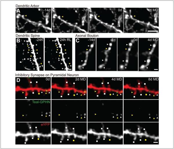
Figure 2.
Experience-dependent structural and synaptic plasticity in inhibitory circuits. Chronic two-photon in vivo images showing remodeling of (A) a dendritic branch tip, (B) dendritic spines, and (C) axonal boutons on inhibitory neurons as well as (D) inhibitory synapses and dendritic spines on excitatory pyramidal neurons in response to monocular deprivation (MD) or retinal lesion (RL) in primary visual cortex. Dynamic events (yellow) of inhibitory neurons (triangles) and pyramidal neurons (squares) with stable counterparts (white) are indicated. Scale bars: (A) 5 μm, (B-D) 2 μm. (B) from Keck and others (unpublished), with permission.

