RSNA Journals logoLink to RSNA Journals
. 2015 Jan 27;274(2):626. doi: 10.1148/radiol.14144050

Myocardial CT Perfusion Imaging and SPECT for the Diagnosis of Coronary Artery Disease: A Head-to-Head Comparison from the CORE320 Multicenter Diagnostic Performance Study

Richard T George, Vishal C Mehra, Marcus Y Chen, Kakuya Kitagawa, Armin Arbab-Zadeh, Julie M Miller, Matthew B Matheson, Andrea L Vavere, Klaus F Kofoed, Carlos E Rochitte, Marc Dewey, Tan S Yaw, Hiroyuki Niinuma, Winfried Brenner, Christopher Cox, Melvin E Clouse, João A C Lima, Marcelo Di Carli
PMCID: PMC4725290  PMID: 25625749

Originally published in:

Radiology 2014;272(2): 407–416 DOI:10.1148/ry.14140806

Erratum in:

Radiology 2015;274(2):626 DOI:10.1148/ry.14144050

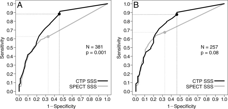
Page 411, Figure 1, The x-axis on images A and B should have read “1 – Specificity.” Corrected images are shown to the right.

Figure 1:

Figure 1:

Receiver operating characteristic curves for myocardial CT perfusion imaging (CTP) and SPECT in, A, all patients and, B, only the patients who underwent pharmacologic stress SPECT.


Articles from Radiology are provided here courtesy of Radiological Society of North America

RESOURCES