Abstract
Background
Staphylococcus aureus is a common and significant pathogen in cystic fibrosis. We sought to determine if quantitative PCR (qPCR) and 16S rRNA gene sequencing could provide a rapid, culture-independent approach to the identification of S. aureus airway infections.
Methods
We examined the sensitivity and specificity of two qPCR assays, targeting the femA and 16S rRNA gene, using culture as the gold standard. In addition, 16S rRNA gene sequencing to identify S. aureus directly from airway samples was evaluated. DNA extraction was performed with and without prior enzymatic digestion.
Results
87 samples [42 oropharyngeal (OP) and 45 expectorated sputum (ES)] were analyzed. 59 samples (68%) cultured positive for S. aureus. Using standard extraction techniques, sequencing had the highest sensitivity for S. aureus detection (85%), followed by FemA qPCR (52%) and 16SrRNA qPCR (34%). For all assays, sensitivity was higher from ES samples compared to OP swabs. Specificity of the qPCR assays was 100%, but 21.4% for sequencing due to detection of S. aureus in low relative abundance from culture negative samples. Enzymatic digestion increased the sensitivity of qPCR assays, particularly for OP swabs.
Conclusion
Sequencing had a high sensitivity for S. aureus, but low specificity. While femA qPCR had higher sensitivity than 16S qPCR for detection of S. aureus, neither assay was as sensitive as sequencing. The significance of S. aureus detection with low relative abundance by sequencing in culture-negative specimens is not clear.
Introduction
Airway infection is a significant contributor to morbidity and mortality in patients with cystic fibrosis (CF) [1]. Staphylococcus aureus is one of the predominant airway pathogens in patients with CF [2]. Culture-based techniques for identification of airway pathogens have limitations that include time needed for bacterial growth, interference by antibiotics, or overgrowth by other organisms. Molecular methods of identifying microbes may overcome some of these limitations. Improved identification of pathogens could lead to improved treatment [2].
Quantitative PCR assays targeting the small subunit rRNA (16S rRNA) gene have been used to detect bacteria from airway samples [3–6]. However, previous studies have demonstrated poor sensitivity of this qPCR assay in the identification of S. aureus in airway samples from CF subjects when compared to culture [6]. 16S rRNA qPCR assays specific for other CF pathogens were more sensitive for the identification of other important species in CF including P. aeruginosa and H. influenzae, suggesting that different approaches may be needed for detection of S. aureus [6]. Problems with staphylococcal DNA extraction may contribute to the poor sensitivity of this assay due to the rigid cell wall of S. aureus, which may limit the isolation of the DNA extraction. Modified extraction methods using lysostaphin and lysozyme may improve staphylococcal DNA extraction [7]. Another potential reason for poor sensitivity is the gene target used, alternate targets such as the femA gene may offer improved sensitivity.
FemA is an essential gene for peptidoglycan synthesis present in methicillin-susceptible S. aureus (MSSA) and -resistant S. aureus (MRSA) [8, 9]. The product of the femA gene is involved in the biosynthesis of the cell wall and is necessary for methicillin resistance [10]. Francois et al [11] developed a qPCR assay relying on the nucleic acid amplification of the femA gene for the detection and identification of S. aureus from clinical samples. This assay was shown to rapidly and accurately identify S. aureus from mixed DNA obtained from nasal, inguinal, and wound swabs.
Sequencing of the ribosomal RNA gene followed by bioinformatic identification of bacteria has also been used to identify bacteria from airway samples [3, 12, 13]. With decreasing cost of DNA sequencing and more rapid turn-around, this approach may become more practical in clinical settings.
In order for a molecular-based assay to be useful clinically, the sensitivity and specificity of the assay must be comparable to culture, the gold standard. In the current study, we assessed the sensitivity and specificity of S. aureus detection using two quantitative PCR assays and sequencing, each before and after enzymatic digestion, compared to traditional culture techniques for analyses of oropharyngeal (OP) and expectorated sputum (ES) samples. Furthermore, we correlated quantitative molecular results with quantitative culture results for sputum.
Methods
Ethics Statement
The Colorado Multiple Institutional Review Board approved the study (COMIRB 07–0835). Written informed consent and HIPAA Authorization was obtained from patients or guardians. Written informed assent was obtained for children 10–17 years of age.
Patient Demographics and Samples
Eighty-seven samples from 65 patients with cystic fibrosis ranging in age from 1.5 to 24 years were analyzed consisting of 45 ES samples and 42 OP samples (Table 1). All expectorated sputum and throat swab specimens were collected as part of standard of care for monitoring bacterial infection during routine patient visits. Clinical culture analysis included blood agar plates, MacConkey agar plates, Haemophilus isolation agar, Burkholderia cepacia selective agar plates, mannitol salt agar, and inhibitory mold agar plates. After clinical cultures were performed, excess specimen was banked and stored frozen at -80 degrees C for molecular assessment of infection. Sputum was homogenized with 10% dithiothreitol following standard CF culture guidelines. Oropharyngeal swabs were vortexed in 1 mL saline and inoculated onto culture plates [14]. 59 samples (68%) were positive and 28 (32%) were negative for S. aureus by culture.
Table 1. Subject/Sample Characteristics.
| Subject characteristics | N = 65 |
| Female, N (%) | 33 (51%) |
| Genotype, N (%) | |
| F508/F508 | 33 (51%) |
| F508/Other | 24 (37%) |
| Other | 8 (12%) |
| Non-Hispanic White, N (%) | 57 (88%) |
| # subjects w/ > 1 sample, N (%) | 16 (25%) |
| Number of samples per subject, median (range) | 1 (1–4) |
| Sample Characteristics | N = 87 |
| Age of subject at sample collection, median (range) | 12.7 (1.5–23.9) |
| Negative culture, N (%) | 8 (9%) |
| MSSA positive, N (%) | 29 (33%) |
| MRSA positive, N (%) | 27 (31%) |
DNA extraction and quantification
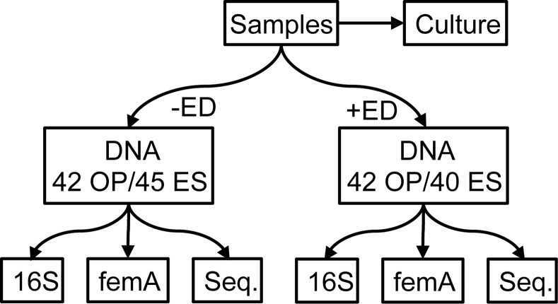
DNA extraction was performed using the manufacturer’s protocol for the Qiagen EZ1 Advanced platform. Two aliquots (200 μL) from each specimen were extracted (Fig 1). One was extracted directly without prior enzymatic digestion, and the second was pre-treated based on the protocol developed by Zhao et al [7]. Briefly, samples were mixed with lysostaphin (final concentration 0.18 mg/mL) and lysozyme (3.6 mg/mL) and incubated at 37°C for 30 minutes. Samples were then digested with proteinase K (1.4 mg/mL) and incubated at 65°C for ten minutes. Samples were then incubated at 95°C for 10 minutes. The DNA concentration of each sample was quantified using a Nanodrop Spectrophotometer.
Fig 1. Study Design.
Excess specimen collected for standard of care culture surveillance for CF pathogens were frozen at -80C. Aliquots of the thawed specimen (42 OP and 45 ES) were extracted without enzymatic digestion (-ED). A second aliquot was extracted with prior enzymatic digestion (+ED). 5 sputum samples had insufficient quantity for enzymatic digestion. The DNAs obtained were assayed using qPCR that targeted 16S and the femA gene. In addition, sequencing was performed on amplicons generated with pan-bacterial primers targeting the V1/2 region (27F/338R).
Quantitative PCR
The quantity of S. aureus was determined using qPCR assays that target the 16S rRNA gene and the S. aureus femA gene (Table 2); each assay was performed in triplicate [9, 11]. The femA qPCR assay was modified from Francois et al. to use 400 nM primer concentration (rather than 100 nm in the original description) based on titration of the primer concentration (300 nM to 800 nM) against the cloned femA standards. 400 nM was the lowest concentration where the 100 copy number S. aureus standard was amplified. Negative controls using sterile water in place of a sample were run in parallel for all experiments. All qPCR data associated with each sample is provided in S1 Table.
Table 2. qPCR Primer and Probe Sequences.
| Target | Primer | Sequence |
|---|---|---|
| Staphylococcus 16S rRNA | STPYF | ACGGTCTTGCTGTCACTTATA |
| STPYR2 | TACACATATGTTCTTCCCTAATAA | |
| femA-SA | F femA-SA | TGCCTTTACAGATAGCATGCCA |
| R femA-SA | AGTAAGTAAGCAAGCTGCAATGACC | |
| P femA-SA | TCATTTCACGCAAACTGTTGGCCACTATG |
High-throughput DNA Sequencing
16S Amplicon Library Construction: Bacterial community profiles were determined by broad-range amplification and sequence analysis of 16S rRNA genes following our previously described methods [15, 16]. In brief, amplicons were generated using primers that target approximately 300 base pairs of the V1/2 variable region of the 16S rRNA gene (27F/338R). PCR products were normalized based on agarose gel densitometry, pooled in approximately equal amounts, and the resulting mixture was gel purified and concentrated using a DNA Clean and Concentrator Kit (Zymo, Irvine, CA). Pooled amplicons were quantified using Qubit Fluorometer 2.0 (Invitrogen, Carlsbad, CA). The pool was diluted to 4nM and denatured with 0.2 N NaOH at room temperature. The denatured DNA was diluted to 15pM and spiked with 10% of the Illumina PhiX control DNA prior to loading the sequencer. Illumina paired-end sequencing was performed on the MiSeq platform using a 500 cycle version 2 reagent kit (Illumina, Inc., San Diego, CA)
Analysis of Illumina Paired-end Reads
Illumina MiSeq paired-end sequences were sorted by sample via barcodes in the paired reads with a python script. Sorted paired end sequence data were deposited in the NCBI Short Read Archive under accession number SRP043334. The sorted paired reads were assembled using phrap [17, 18]. Pairs that did not assemble were discarded. Assembled sequence ends were trimmed over a moving window of 5 nucleotides until average quality met or exceeded 20. Trimmed sequences with more than 1 ambiguity or shorter than 200 nt were discarded. Potential chimeras identified with Uchime (usearch6.0.203_i86linu32) using Schloss Silva reference sequences were removed from subsequent analyses [19, 20]. Assembled sequences were aligned and classified with SINA (1.2.11) using the 629,125 bacterial sequences in Silva 111Ref as a reference configured to yield the Silva taxonomy [21, 22]. Sequences with identical taxonomic assignments were clustered into Operational taxonomic units (OTUs). This process generated 4,397,512 sequences for 168 samples (average size: 26,176 sequences/sample; min: 7,079 max: 63,117). The median Goods coverage score was ≥99.75% at the rarefaction point of 7,079 sequences. The software package Explicet (v2.10.5, www.explicet.org) was used to extract Staphylococcus sequence counts for each library [23].
Identification of S. aureus among Staphylococcal sequences
To identify S. aureus we employed an ancillary analysis using BLAST to compare the study sequences without enzymatic digestion to the isolate sequences from Silva104 database [24, 25]. BLAST hits were required to have ≥99% sequence identity over ≥95% of the sequence in order to append the Silva binomial name to the RDP taxonomy line [26]. If any of these criteria were not met the sequence retained the genus level classification only.
Statistical Analysis
Descriptive statistics using means and standard deviations or percentages were used for continuous and categorical variables, respectively. Paired t-tests were used to evaluate whether the DNA concentration between paired samples run with and without enzymatic digestion were significantly different. Assay performance was assessed by calculating sensitivities and specificities using culture as the gold standard. Corresponding 95% confidence intervals were calculated using exact Clopper Pearson type test. Comparisons of sensitivity and specificity across OP and ES samples were performed using a Chi-square test and between methods was performed using McNemar’s test. Spearman’s correlation coefficients were used to associate continuous values for each method. Analyses were performed using SAS version 9.4 software (SAS Institute Inc.: Cary, NC, 2014).
Results
DNA Concentration
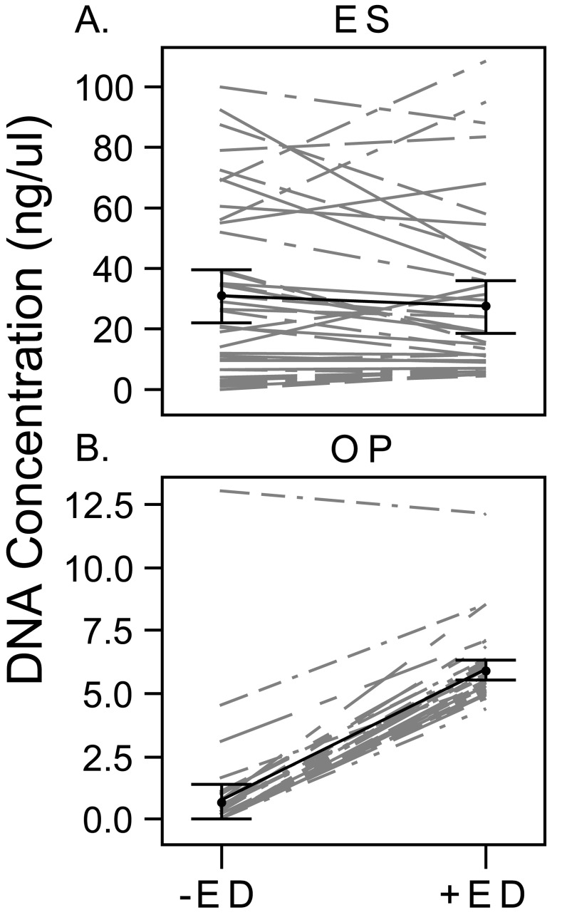
For OP samples, DNA concentrations were consistently higher using enzymatic digestion [mean difference in DNA concentration 5.2 ng/μl (SE = 0.2); p<0.01]. For ES samples, there was no significant difference in DNA concentration with or without enzymatic digestion [mean difference in DNA concentration -3.6 ng/μl (SE = 2.6); p = 0.18] (Fig 2).
Fig 2. Comparison of DNA Concentration with and without Enzymatic Digestion.
DNA concentration is shown for each paired sample of DNA extractions with and without enzymatic digestion for ES (A) and OP (B). The average concentration for all samples with each extraction approach is shown in black with 95% confidence intervals indicated by the whiskers.
Sensitivity and specificity of qPCR assays and sequencing compared to culture
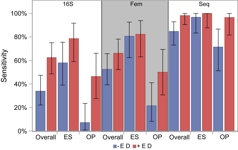
For both the 16S rRNA assay and the femA assay without enzymatic digestion, OP samples were less sensitive compared to ES samples [16S rRNA: 7.1% (95% CI: 0.1–23.5%) versus 58.1% (95% CI: 39.1–75.5%) femA: 21.4% (95% CI: 8.3–41.0%) versus 80.6% (95% CI: 62.5–92.5%), p < 0.01 for both] (Fig 3). Specificity was 100% for both qPCR assays. Sequencing had higher sensitivity, although similar to qPCR sensitivity from OP samples was lower compared to ES [71.4% (95% CI: 51.3–86.8%) versus 96.8% (95% CI: 83.3–99.9%), p = 0.01]. Specificity of sequencing was low (21.4% (95% CI: 8.3–41.0%) due to detection of S. aureus from culture negative samples. Culture negative samples had a low median relative abundance of Staphylococcus [0.04% (range: 0–0.3%)]. No Staphylococcus was detected in negative sterile water controls.
Fig 3. Sensitivity of each molecular assay with and without enzymatic digestion.
Sensitivity of qPCR assays compared to standard culture results are given for each assay based on all samples (overall), and by specimen type (ES and OP). Error bars represent 95% confidence intervals. Blue bars are sensitivity without enzymatic digestion, and red bars are with enzymatic digestion.
We compared 82 paired samples processed with and without enzymatic digestion. The addition of enzymatic digestion significantly improved the sensitivity of the 16S rRNA assay with OP samples from 7% to 46% (p < 0.01) and ES samples from 58% to 79% (p = 0.01). For the femA assay and sequencing, the addition of the enzymatic digestion significantly improved the sensitivity with OP samples from 21% to 50% and from 71% to 96% (p = 0.01 for both) but not ES samples from 81% to 82% and from 97% to 100% (p = 0.32 and p = 0.99 respectively), although particularly for sequencing, the sensitivity from ES samples was already high with standard processing. Unlike with standard DNA extraction, the sensitivity of the femA assay did not differ from the 16S rRNA assay using enzymatic digestion (p = 0.48). The sensitivity for sequencing from both ES and OP samples was high and did not differ significantly [100% (95% CI: 87.7–100%) versus 96.4% (95% CI: 81.7–99.9%) (Fig 3, p = 0.3)]. The specificity for qPCR assays did not change with enzymatic digestion (100%), and remained low with enzymatic digestion for sequencing [7.7% (95% CI: 0.1–25.1%)].
Quantitative Comparison of qPCR and sequencing results to culture
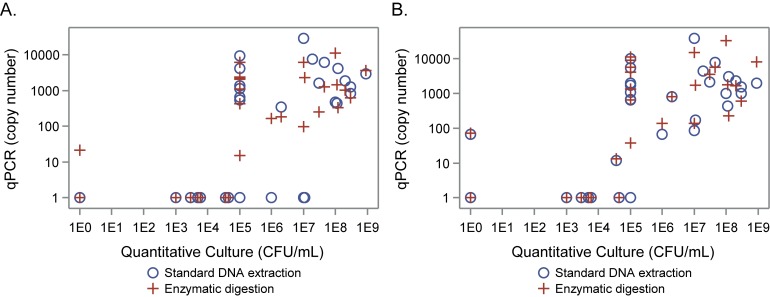
The 16S rRNA qPCR assay with enzymatic digestion and the femA assay regardless of enzymatic digestion detected S. aureus from all samples with quantities greater than 1 x 10⁵ CFU/mL by culture (Fig 4). Conversely, the 16S qPCR rRNA assay without enzymatic digestion only consistently detected S. aureus in quantities greater than 1 x 10⁷ CFU/mL by culture. In the 29 ES samples with S. aureus detected by culture, the correlations between the quantitative culture results (CFU/ml) and the qPCR results (copy number, both 16S and femA with and without enzymatic digestion) ranged from 0.54 to 0.57, p < 0.01 for all. The same comparison for the sequencing data resulted in a correlation of 0.46 for standard DNA extraction and 0.50 with enzymatic digestion (p = 0.01 for both).
Fig 4. Quantitative Culture vs. qPCR copy number in expectorated sputum samples.
qPCR copy numbers for 16S rRNA (A) and femA qPCR (B) assays with (circles) and without enzymatic digestion (plus symbol) plotted vs. CFU/mL from quantitative culture of expectorated sputum samples. Both axes are plotted on a logarithmic scale.
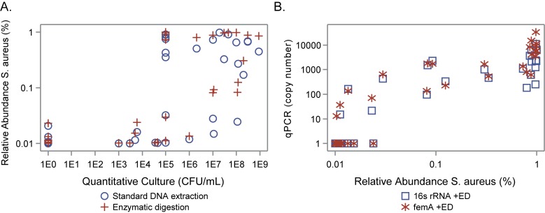
The relative abundance measured by sequencing was correlated with both the quantitative culture results and copy numbers detected with qPCR (Fig 5). Sequencing with and without enzymatic digestion consistently detected Staphylococcus when present in quantities greater than 1 x 10³ CFU/mL by culture. Sequencing also identified Staphylococcus in 22 culture negative samples all of which had relative abundance levels of Staphylococcus less than 0.3% (Fig 5). There were also 20 culture positive samples with relative abundance levels less than 0.1%. The majority of these were OP samples. The 4 ES samples had quantitative culture growth above 103 CFU/mL. Based on our quantitative comparison of culture and sequencing, we estimate that relative abundance levels less than 0.1% correspond to quantitative culture levels at or below 1 x 10³ CFU/mL (Fig 5).
Fig 5. Comparison of species specific data for S. aureus in ES samples.
(A) Quantitative culture versus sequencing. Relative abundance determined with sequencing plotted versus CFU/mL from quantitative culture. (B) Sequencing versus femA qPCR. femA copy number versus relative abundance of S. aureus.
Results of Blast analysis of Staphylococcus
To ensure the sequence comparison to culture was appropriate, we further analyzed the Staphylococcus sequences extracted without enzymatic digestion to obtain species level taxonomy where possible. Staphylococcus sequences in this study were predominated by S. aureus (90%). All samples with Staphylococcus sequences contained sequence identified as S. aureus; sensitivity and specificity compared to culture did not change with species level information.
Discussion
Sequencing demonstrated improved sensitivity and lower detection limits compared to the qPCR assays regardless of inclusion of enzymatic digestion in the DNA extraction protocol. Furthermore, the enzymatic digestion process improved the sensitivity of sequencing with the greatest gain seen with the OP samples. Optimal sensitivity (100%) was achieved with sequencing with enzymatic digestion in ES samples. Sequencing detected S. aureus in multiple culture negative samples but not in negative controls and thus may even be more sensitive than culture. With a detection limit below 1 x 10³ CFU/mL (the limit for culture), sequencing has the potential to identify additional infections. The clinical relevance of the presence of S. aureus in densities below 1 x 10³ requires further investigation. Many of the practical applications of the qPCR assays were overcome with the use of the sequencing. The main limitation of the sequencing as currently performed is its difficulty to distinguish S. aureus from other staphylococcal species. As with the qPCR assays employed in this study, sequencing is also unable to distinguish MSSA and MRSA. The mecA qPCR assay is a molecular assay that has the potential to distinguish between MSSA and MRSA.
From this study, the sensitivity of the qPCR assays does not appear sufficient to be clinically applicable. While the sensitivity of the femA assay is better compared to the 16S rRNA assay, the femA assay is still not as sensitive as culture. In particular, it is limited in its ability to detect S. aureus in oropharyngeal swab samples. We suspect that the OP swabs have lower bacterial biomass, based on lower DNA yield, which is why they behave differently than sputum samples. The addition of enzymatic digestion did appear to improve detection in the OP samples, along with increasing DNA yield. There are multiple ways in which the addition of enzymatic digestion may have increased the DNA yield. First, the goal of the enzymatic digestion was to specifically aid in the digestion of the S. aureus cell wall. In this way, the enzymatic digestion may help free the DNA for detection, particularly where bacterial biomass is lower. Additionally, the DNA extraction method with enzymatic digestion also included an additional freeze-thaw cycle when the repeat extractions were performed. The freeze-thaw cycle may also increase the amount of DNA extracted irrespective of enzymatic digestion, but we did not test this step. While the rapid quantitative detection of S. aureus using qPCR in patients with CF would be useful in guiding immediate antibiotic therapy before the culture results can be obtained, the low sensitivity of this assay limits its possible use clinically.
Methods relying on DNA sequencing techniques are becoming less expensive and increasingly prevalent in the clinical environment. 16S rRNA gene sequencing has recently been used as an alternative method for molecular detection. 16S rRNA gene sequencing is useful because the 16S rRNA gene is present in all bacteria, is well conserved, and is large enough for informatics purposes [27]. Thus, sequencing could be reasonably applied in the clinical setting, but is currently limited by cost and time to results. However, as the cost and time to results of sequencing continue to decrease, sequencing may soon be a practical alternative to culture in the clinical setting. Similar to previous work, our results demonstrated the detection of S. aureus in culture negative samples, suggesting that sequencing has the potential to be more sensitive than culture [28]. Furthermore, it has the potential to provide the advantages of earlier targeting of antibiotic therapy and the ability to track the resolution of infections.
The 16S rRNA qPCR assay has been shown to be able to successfully identify multiple types of bacteria in multiple sample types [9]. Our group previously demonstrated that qPCR amplification of the 16 rRNA gene can be used to reliably detect P. aeruginosa and H. influenzae in patients with cystic fibrosis [6]. In these studies, qPCR demonstrated increased sensitivity compared to culture for P. aeruginosa (75%), and H. influenza (75%) but similar to our currently findings, decreased sensitivity compared to culture for S. aureus (35%) [6]. Francois and colleagues demonstrated the effectiveness of an alternative assay for S. aureus identification that relies on the amplification of the femA gene [11]. The sensitivity of the qPCR assay in these samples was comparable to culture in nasal, inguinal, and wound samples in hospitalized patients at risk for S. aureus infection (100%) [11]. However, the femA assay did not improve the detection of S. aureus in our samples from children with CF, suggesting that CF may provide unique challenges in the molecular identification of S. aureus. S. aureus is a prominent pathogen in patients with CF, so it would be expected to contribute significantly to the bacterial community [3]. The standard DNA extraction method may account for the decrease in sensitivity in CF samples [7]. In fact, the addition of this method did significantly improve the sensitivity of multiple molecular methods in our studies.
Methicillin resistance is an important factor in targeting antibiotic therapy in the clinical setting. The mecA gene is the gold standard for detecting methicillin resistance and can be used in conjunction with the femA assay to distinguish between MSSA and MRSA. The mecA assay demonstrates limitations as well. Francois and co-workers demonstrated a high number of false positive results with the mecA gene [11]. The mecA gene also occurs in coagulase negative staphylococci (CNS). However, MRSA can be distinguished from CNS with the combination of the femA, femB, and mecA assays [29]. Another potential limitation is that there is evidence that detecting the mecA gene decreases with storage at -80°C over two years [30]. Although distinguishing between MSSA and MRSA is very important clinically, the main aim of the study was the detection of S. aureus. The tests to assess resistance merit further study.
Our study is not without limitations. First, our sample size is relatively low, and larger studies may be required to further evaluate the effectiveness of these molecular methods. Additionally, S. aureus was detected in a large proportion of samples by sequencing. However, it is currently unclear whether the small relative abundance seen in many sample is clinically relevant. Finally, it is unclear how the enzymatic digestion step affected the detection of other organisms.
Conclusion
Our work suggests that sequencing has a detection limit that is low enough to provide useful clinical information in the setting of S. aureus infection in patients with CF. Based on our studies, sequencing can detect samples with lower quantities compared to cultures and just above the lowest reported values by culture. Quantitative PCR may be useful for rapidly detecting and quantifying S. aureus, but requires appropriate DNA extraction. However, sensitivity is lower than sequencing, particularly for OP swabs. Future work should also focus on the ability to differentiate between MSSA and MRSA. Sequencing could be a useful tool to track the effectiveness of antibiotic therapy and resolution of infection in the clinical setting.
Supporting Information
(XLSX)
Data Availability
All relevant data are within the paper and its supporting information files.
Funding Statement
This work was supported by Cystic Fibrosis Foundation Therapeutics, Inc.
References
- 1.Gibson RL, Burns JL, Ramsey BW. Pathophysiology and management of pulmonary infections in cystic fibrosis. Am J Respir Crit Care Med. 2003;168(8):918–51. Epub 2003/10/14. 10.1164/rccm.200304-505SO 168/8/918 [pii]. . [DOI] [PubMed] [Google Scholar]
- 2.Sibley CD, Rabin H, Surette MG. Cystic fibrosis: a polymicrobial infectious disease. Future Microbiol. 2006;1:53–61. Epub 2007/07/31. 10.2217/17460913.1.1.53 . [DOI] [PubMed] [Google Scholar]
- 3.Cox MJ, Allgaier M, Taylor B, Baek MS, Huang YJ, Daly RA, et al. Airway microbiota and pathogen abundance in age-stratified cystic fibrosis patients. PLoS ONE. 2010;5(6):e11044 Epub 2010/06/30. 10.1371/journal.pone.0011044 [DOI] [PMC free article] [PubMed] [Google Scholar]
- 4.Deschaght P, De Baere T, Van Simaey L, Van Daele S, De Baets F, De Vos D, et al. Comparison of the sensitivity of culture, PCR and quantitative real-time PCR for the detection of Pseudomonas aeruginosa in sputum of cystic fibrosis patients. BMC microbiology. 2009;9:244 10.1186/1471-2180-9-244 [DOI] [PMC free article] [PubMed] [Google Scholar]
- 5.Filkins LM, Hampton TH, Gifford AH, Gross MJ, Hogan DA, Sogin ML, et al. Prevalence of streptococci and increased polymicrobial diversity associated with cystic fibrosis patient stability. J Bacteriol. 2012;194(17):4709–17. Epub 2012/07/04. 10.1128/JB.00566-12 [DOI] [PMC free article] [PubMed] [Google Scholar]
- 6.Zemanick ET, Wagner BD, Sagel SD, Stevens MJ, Accurso FJ, Harris JK. Reliability of quantitative real-time PCR for bacterial detection in cystic fibrosis airway specimens. PLoS ONE. 2010;5(11):e15101 Epub 2010/12/15. 10.1371/journal.pone.0015101 [DOI] [PMC free article] [PubMed] [Google Scholar]
- 7.Zhao J, Carmody LA, Kalikin LM, Li J, Petrosino JF, Schloss PD, et al. Impact of enhanced Staphylococcus DNA extraction on microbial community measures in cystic fibrosis sputum. PLoS One. 2012;7(3):e33127 Epub 2012/03/14. 10.1371/journal.pone.0033127 [DOI] [PMC free article] [PubMed] [Google Scholar]
- 8.Maidhof H, Reinicke B, Blümel P, Berger-Bächi B, Labischinski H. femA, which encodes a factor essential for expression of methicillin resistance, affects glycine content of peptidoglycan in methicillin-resistant and methicillin-susceptible Staphylococcus aureus strains. J Bacteriol. 1991;173(11):3507–13. [DOI] [PMC free article] [PubMed] [Google Scholar]
- 9.Matsuda K, Tsuji H, Asahara T, Kado Y, Nomoto K. Sensitive quantitative detection of commensal bacteria by rRNA-targeted reverse transcription-PCR. Appl Environ Microbiol. 2007;73(1):32–9. Epub 2006/10/31. doi: AEM.01224-06 [pii] 10.1128/AEM.01224-06 [DOI] [PMC free article] [PubMed] [Google Scholar]
- 10.Johnson S, Kruger D, Labischinski H. FemA of Staphylococcus aureus: isolation and immunodetection. FEMS Microbiol Lett. 1995;132(3):221–8. . [DOI] [PubMed] [Google Scholar]
- 11.Francois P, Pittet D, Bento M, Pepey B, Vaudaux P, Lew D, et al. Rapid detection of methicillin-resistant Staphylococcus aureus directly from sterile or nonsterile clinical samples by a new molecular assay. J Clin Microbiol. 2003;41(1):254–60. [DOI] [PMC free article] [PubMed] [Google Scholar]
- 12.Lynch SV, Bruce KD. The cystic fibrosis airway microbiome. Cold Spring Harbor perspectives in medicine. 2013;3(3):a009738 Epub 2013/03/05. 10.1101/cshperspect.a009738 . [DOI] [PMC free article] [PubMed] [Google Scholar]
- 13.Caverly LJ, Zhao J, LiPuma JJ. Cystic fibrosis lung microbiome: Opportunities to reconsider management of airway infection. Pediatr Pulmonol. 2015;50 Suppl 40:S31–8. 10.1002/ppul.23243 . [DOI] [PubMed] [Google Scholar]
- 14.Burns JL, Emerson J, Stapp JR, Yim DL, Krzewinski J, Louden L, et al. Microbiology of sputum from patients at cystic fibrosis centers in the United States. Clin Infect Dis. 1998;27(1):158–63. Epub 1998/07/24. . [DOI] [PubMed] [Google Scholar]
- 15.Hara N, Alkanani AK, Ir D, Robertson CE, Wagner BD, Frank DN, et al. Prevention of virus-induced type 1 diabetes with antibiotic therapy. J Immunol. 2012;189(8):3805–14. Epub 2012/09/19. 10.4049/jimmunol.1201257 . [DOI] [PubMed] [Google Scholar]
- 16.Markle JG, Frank DN, Mortin-Toth S, Robertson CE, Feazel LM, Rolle-Kampczyk U, et al. Sex differences in the gut microbiome drive hormone-dependent regulation of autoimmunity. Science. 2013;339(6123):1084–8. Epub 2013/01/19. 10.1126/science.1233521 . [DOI] [PubMed] [Google Scholar]
- 17.Ewing B, Green P. Base-calling of automated sequencer traces using phred. II. Error probabilities. Genome Res. 1998;8(3):186–94. Epub 1998/05/16. . [PubMed] [Google Scholar]
- 18.Ewing B, Hillier L, Wendl MC, Green P. Base-calling of automated sequencer traces using phred. I. Accuracy assessment. Genome Res. 1998;8(3):175–85. . [DOI] [PubMed] [Google Scholar]
- 19.Edgar RC, Haas BJ, Clemente JC, Quince C, Knight R. UCHIME improves sensitivity and speed of chimera detection. Bioinformatics. 2011;27(16):2194–200. Epub 2011/06/28. 10.1093/bioinformatics/btr381 [DOI] [PMC free article] [PubMed] [Google Scholar]
- 20.Schloss PD, Westcott SL. Assessing and improving methods used in operational taxonomic unit-based approaches for 16S rRNA gene sequence analysis. Appl Environ Microbiol. 2011;77(10):3219–26. 10.1128/AEM.02810-10 [DOI] [PMC free article] [PubMed] [Google Scholar]
- 21.Pruesse E, Peplies J, Glockner FO. SINA: accurate high-throughput multiple sequence alignment of ribosomal RNA genes. Bioinformatics. 2012;28(14):1823–9. Epub 2012/05/05. 10.1093/bioinformatics/bts252 [DOI] [PMC free article] [PubMed] [Google Scholar]
- 22.Quast C, Pruesse E, Yilmaz P, Gerken J, Schweer T, Yarza P, et al. The SILVA ribosomal RNA gene database project: improved data processing and web-based tools. Nucleic Acids Res. 2013;41(Database issue):D590–6. Epub 2012/11/30. 10.1093/nar/gks1219 [DOI] [PMC free article] [PubMed] [Google Scholar]
- 23.Robertson CE, Harris JK, Wagner BD, Granger D, Browne K, Tatem B, et al. Explicet: Graphical user interface software for the management, analysis and visualization of microbial ecology data and metadata. Bioinformatics. 2013. [DOI] [PMC free article] [PubMed] [Google Scholar]
- 24.Altschul SF, Gish W, Miller W, Myers EW, Lipman DJ. Basic local alignment search tool. J Mol Biol. 1990;215(3):403–10. Epub 1990/10/05. 10.1006/jmbi.1990.9999 S0022283680799990 [pii]. 2231712. [DOI] [PubMed] [Google Scholar]
- 25.Pruesse E, Quast C, Knittel K, Fuchs BM, Ludwig W, Peplies J, et al. SILVA: a comprehensive online resource for quality checked and aligned ribosomal RNA sequence data compatible with ARB. Nucleic Acids Res. 2007;35(21):7188–96. Epub 2007/10/20. doi: gkm864 [pii] 10.1093/nar/gkm864 [DOI] [PMC free article] [PubMed] [Google Scholar]
- 26.Zemanick ET, Harris JK, Wagner BD, Robertson CE, Sagel SD, Stevens MJ, et al. Inflammation and airway microbiota during cystic fibrosis pulmonary exacerbations. PLoS ONE. 2013;8(4):e62917 Epub 2013/05/07. 10.1371/journal.pone.0062917 [DOI] [PMC free article] [PubMed] [Google Scholar]
- 27.Janda JM, Abbott SL. 16S rRNA gene sequencing for bacterial identification in the diagnostic laboratory: pluses, perils, and pitfalls. J Clin Microbiol. 2007;45(9):2761–4. 10.1128/JCM.01228-07 [DOI] [PMC free article] [PubMed] [Google Scholar]
- 28.Mahboubi MA, Carmody LA, Foster BK, Kalikin LM, VanDevanter DR, LiPuma JJ. Culture-based and culture-independent bacteriologic analysis of cystic fibrosis respiratory specimens. J Clin Microbiol. 2015. 10.1128/JCM.02299-15 . [DOI] [PMC free article] [PubMed] [Google Scholar]
- 29.Kobayashi N, Wu H, Kojima K, Taniguchi K, Urasawa S, Uehara N, et al. Detection of mecA, femA, and femB genes in clinical strains of staphylococci using polymerase chain reaction. Epidemiol Infect. 1994;113(2):259–66. [DOI] [PMC free article] [PubMed] [Google Scholar]
- 30.van Griethuysen A, van Loo I, van Belkum A, Vandenbroucke-Grauls C, Wannet W, van Keulen P, et al. Loss of the mecA gene during storage of methicillin-resistant Staphylococcus aureus strains. J Clin Microbiol. 2005;43(3):1361–5. 10.1128/JCM.43.3.1361-1365.2005 [DOI] [PMC free article] [PubMed] [Google Scholar]
Associated Data
This section collects any data citations, data availability statements, or supplementary materials included in this article.
Supplementary Materials
(XLSX)
Data Availability Statement
All relevant data are within the paper and its supporting information files.


