Fig 2 and Fig 4 are swapped. The image in Fig 2 should be the image in Fig 4 and the image in Fig 4 should be the image in Fig 2. The captions remain unchanged. The publisher apologizes for the error. Please see the corrected Fig 2 and Fig 4 here.
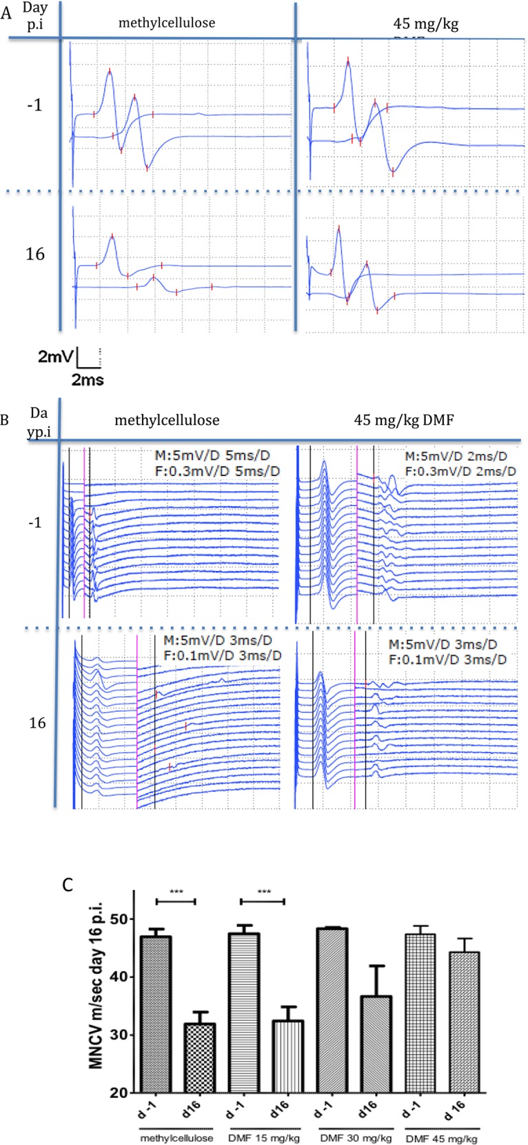
Fig 2. Dimethyl fumarate improved proximal and distal nerve conduction.

(A) Representative CMAP (compound motor action potentials) traces during EAN course at days −1 and 16 p.i. showing a conduction block for methylcellulose-treated rats at day 16 p.i. whereas for 45 mg/kg DMF-treated rats no conduction block was recorded. (B) Representative F-wave traces after distal stimulation showing prolonged F-waves latencies only for the methylcellulose-treated group at day 16 p.i. in comparison to day -1. Rats treated with 45mg/kg did not show any significant differences in the F-wave latencies between day -1 and 16 p.i. The black vertical line defines the motor (M) response and the F (F-wave) response latency. On the left of the red vertical line applies the M response regarding distance (horizontally, ms) and vertically (mV) and on the right of the red vertical line applies the F response data (ms, mV), (M: M response, F: F response, D: distance of one side of the dotted lined squares). (C) After proximal and distal stimulation of the sciatic nerve the conduction velocity was calculated. A statistical significant reduction of the MNCV (motor nerve conduction velocity) appeared for the control group and the 15mg/kg group (p<0,0001 ***, n = 10), but no difference in the MNCV was seen for the 45mg/kg DMF treated group indicating a protective role of DMF against demyelination. Mean values and SEM are depicted.
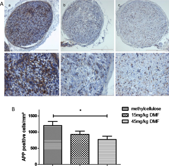
Fig 4. Dimethyl fumarate reduced early axonal damage at the peak of EAN course.
(A) Representative photos of APP (amyloid precursor protein) staining for sciatic nerve transverse sections of rats (n = 6/group) treated with DMF 15mg/kg (b, e), 45mg/kg (c, f) and methylcellulose-treated animals (a, d), showing an reduction of APP positive cells for DMF-treated rats. Scale bars indicate 100μm for a-c and 50μm for d-f. (B) Mean numbers of APP positive cells per mm2 sciatic nerve sections as calculated by immunohistochemistry on day 16 p.i. from EAN rats (n = 6/group) receiving orally DMF at different doses (15mg/kg, 45mg/kg/day) and methylcellulose-treated rats. Mean values and SEM are depicted (*p<0,05). The experiment was repeated 2 times with similar results.
Reference
- 1.Pitarokoili K, Ambrosius B, Meyer D, Schrewe L, Gold R (2015) Dimethyl Fumarate Ameliorates Lewis Rat Experimental Autoimmune Neuritis and Mediates Axonal Protection. PLoS ONE 10(11): e0143416 doi:10.1371/journal.pone.0143416 [DOI] [PMC free article] [PubMed] [Google Scholar]


