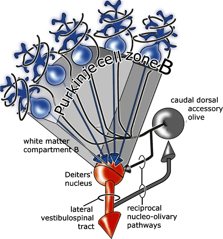
Fig. 1.
Diagram of the B module. It consists of the lateral vermal Purkinje cell zone B, Deiters’ nucleus as its target nucleus, a climbing fiber projection from the contralateral caudal dorsal accessory olive, reciprocally organized nucleo-olivary pathways, and the lateral vestibulospinal tract as its efferent pathway. The Purkinje cell axons and the olivocerebellar fibers are contained in the white matter compartment B (Fig. 7b)

